Jump to content
In this section we have spot diagnoses posted on a daily basis since June 2010, now over 4000! You can review the archived cases and read the suggested diagnoses by users and the final comment by the contributors.
Case are uploaded each week day by 10 am UK time with the correct diagnosis will generally be posted at 8 pm UK time. Why not view the most recent spot diagnosis and proffer a diagnosis?

Case Number : Case 1094 - 2nd September Posted By: Guest

Please read the clinical history and view the images by clicking on them before you proffer your diagnosis.
Submitted Date :
   (0 reviews)

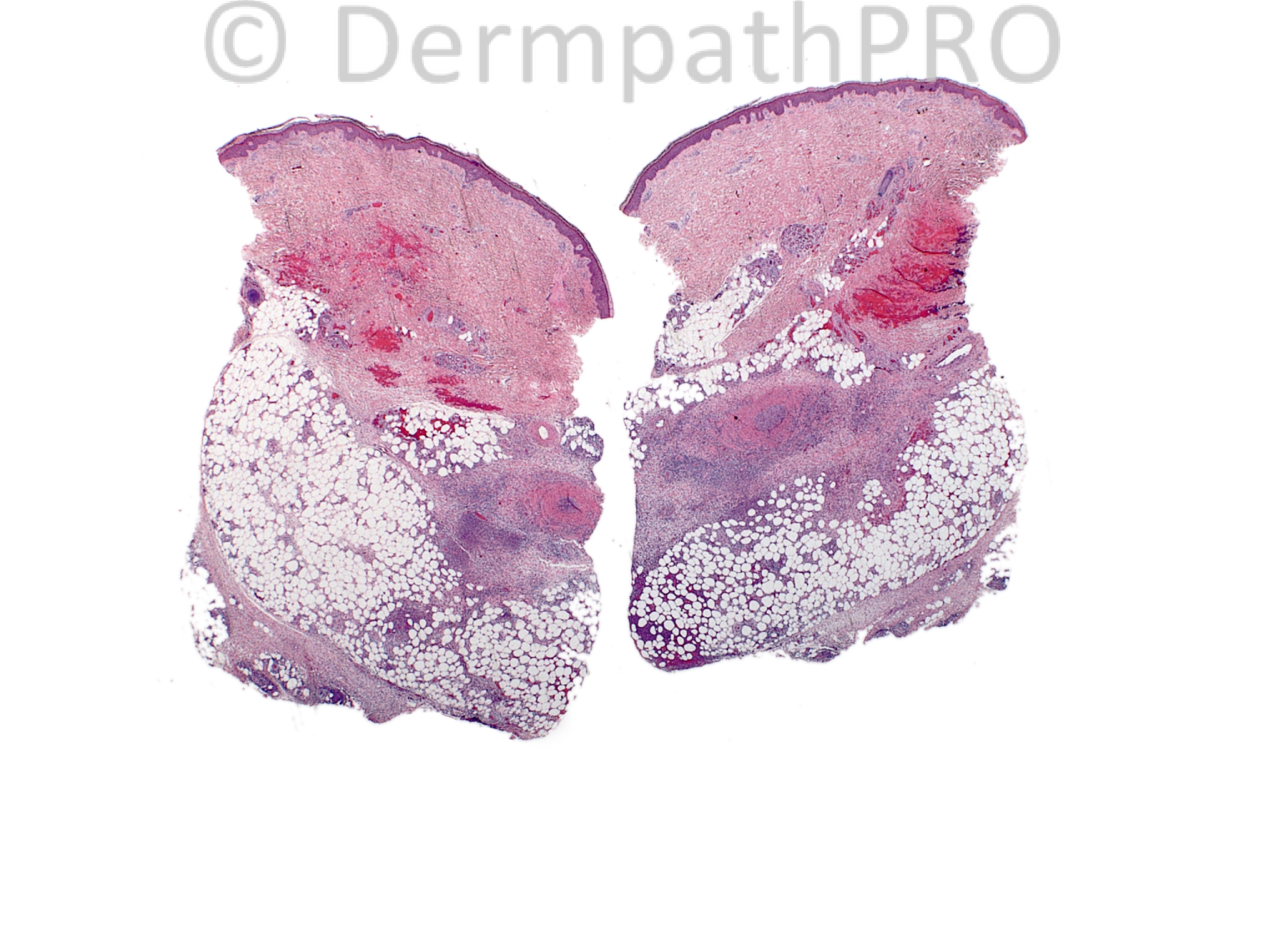
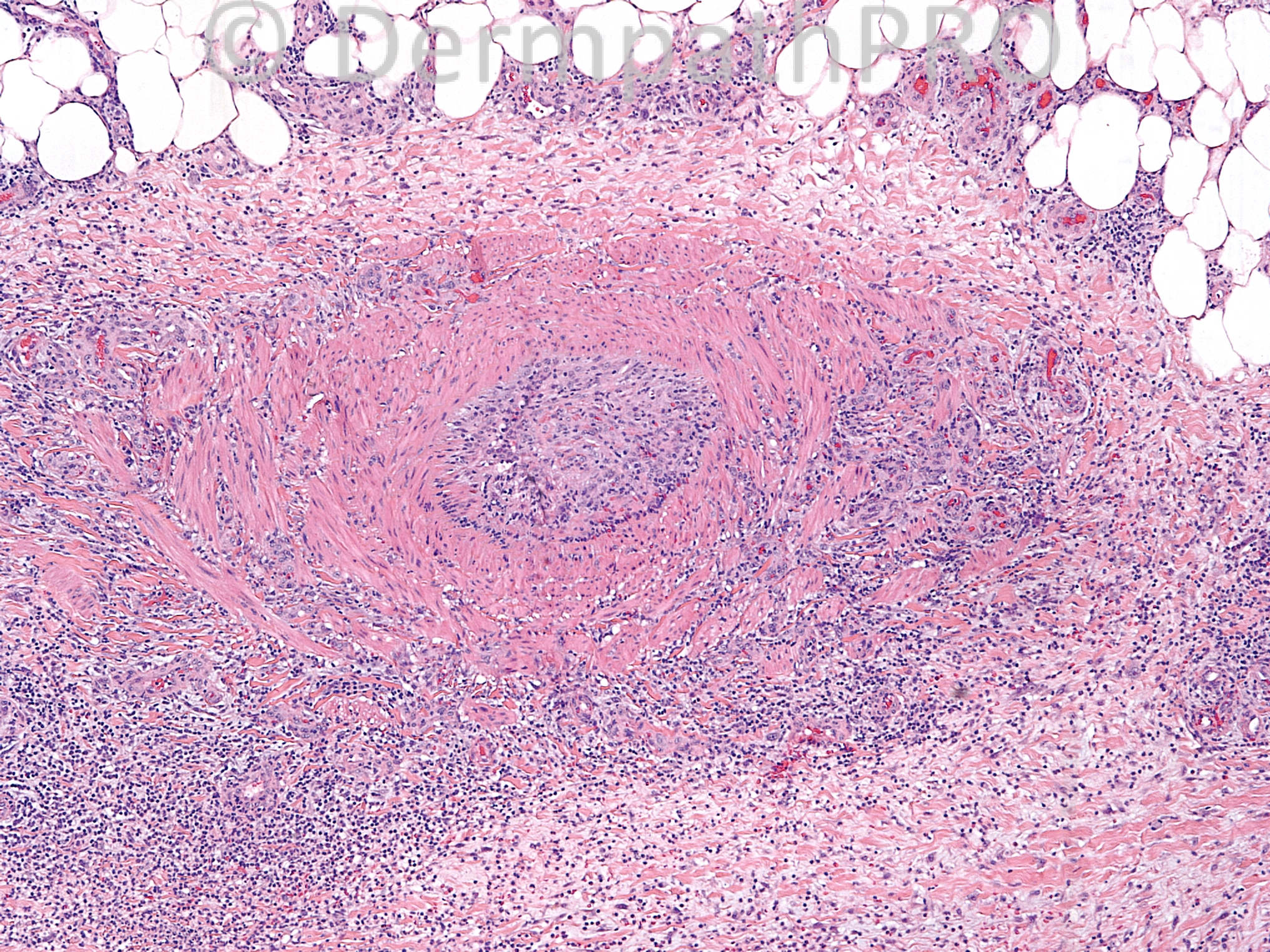
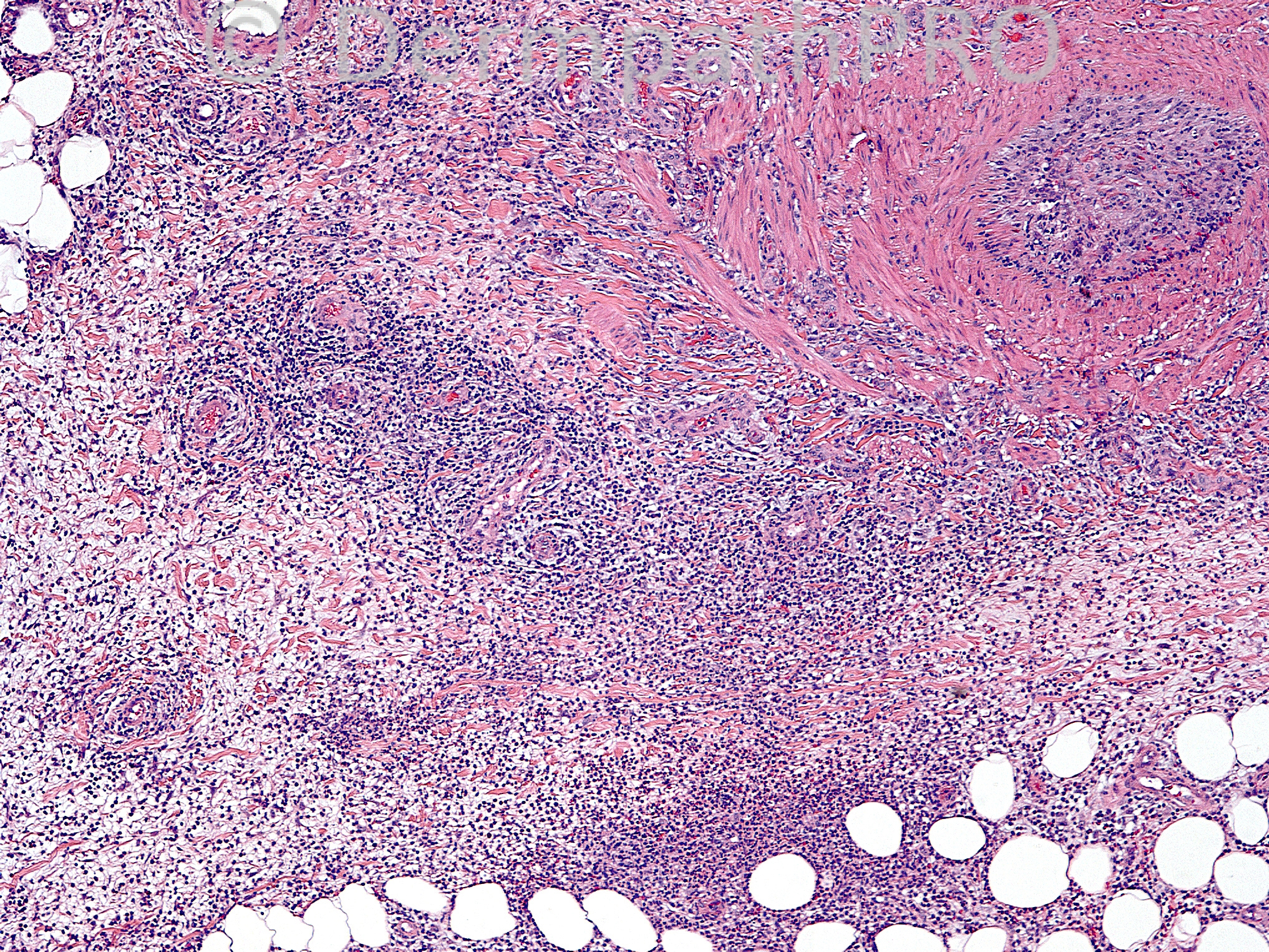
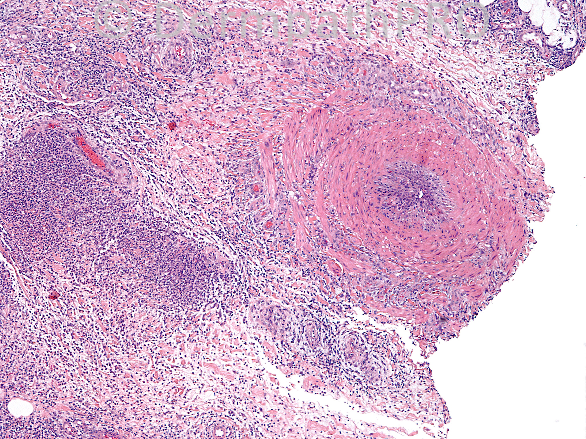
A 34-year-old male with violaceous plaques and nodules on bilateral lower extremities. Tender, come and go, patient otherwise in good health (no fever or chills, no weight loss, no fatigue). Duration approximately one year and intermittent. Biopsy of right thigh.

Case posted by Dr.Uma Sundram.


  Report Record

User Feedback


Arti Bakshi

Posted

The inflammation appears to be centered on a medium sized vessel on low power (not sure if artery or vein).
The fat lobules away from this vessel are not affected. ?Superficial thrombophlebitis ?PAN

Share this comment


Link to comment
share_externally

Dr. Mona Abdel-Halim

Posted

The inflammation is limited to the vicinity of the vessel, no extension to the whole fat lobules, no extensive fat necrosis. I think this speaks against nodular vasculitis. I agree with Arti that this could be superficial thrombophlebitis or PAN. Favour superficial thrombophlebitis based on clinical history.

Share this comment


Link to comment
share_externally

I also do not see extensive lobular necrosis, a find so common in Nodular vasculitis. I don´t like to decide between PAN and superficial trombophlebitis without an elastic stain, but since there´s no big destruction of the vessel wall and the lumen is obstructed I think this case is probably superficial trombophlebitis.

Share this comment


Link to comment
share_externally

Uma Sundram

Posted

This is quite a difficult case; thank you for all of your comments. I agree that the elastin stain is the best way to distinguish between superficial thrombophlebitis and PAN; however, in inflammatory situations the elastin can be destroyed (see recent paper by Hall LD, Elston DM, et al.). A relatively reliable H+E method is to look for intervening collagen fibers in the vessel wall. Veins have these; arteries do not. The paper by Hall et al has some nice visuals of these two settings (PAN and ST) comparing the smooth muscle patterns. I called this superficial thrombophlebitis based on the clinical setting and the smooth muscle patterns of the vessels. Nodular vasculitis is also an excellent thought, and a differential diagnostic consideration. I favored this less as the infiltrate seems relatively localized (although there is some spillover into the lobules) and not as diffuse as cases of NV that I have previously encountered.

Share this comment


Link to comment
share_externally

Dr. Richard Carr

Posted

Agree this vessel is a vein but I was favouring nodular vasculitis too. Uma - what in the clinical setting was more typical of thrombophlebitis?

Share this comment


Link to comment
share_externally

Uma Sundram

Posted

Richard, the lesions were smaller than those typically found in panniculitides, had a linear, rope like pattern in the overlying skin, and 'came and went', according to the patient. Based on the clinical appearance, the clinicians favored superficial migratory thrombophlebitis.

Share this comment


Link to comment
share_externally


Join the conversation

You can post now and register later. If you have an account, sign in now to post with your account.

Guest
Add a comment...

×   Pasted as rich text.   Paste as plain text instead

  Only 75 emoji are allowed.

×   Your link has been automatically embedded.   Display as a link instead

×   Your previous content has been restored.   Clear editor

×   You cannot paste images directly. Upload or insert images from URL.

Loading...

×
×
  • Create New...