Jump to content
In this section we have spot diagnoses posted on a daily basis since June 2010, now over 4000! You can review the archived cases and read the suggested diagnoses by users and the final comment by the contributors.
Case are uploaded each week day by 10 am UK time with the correct diagnosis will generally be posted at 8 pm UK time. Why not view the most recent spot diagnosis and proffer a diagnosis?

Case Number : Case 1100 - 10th September Posted By: Guest

Please read the clinical history and view the images by clicking on them before you proffer your diagnosis.
Submitted Date :
   (0 reviews)

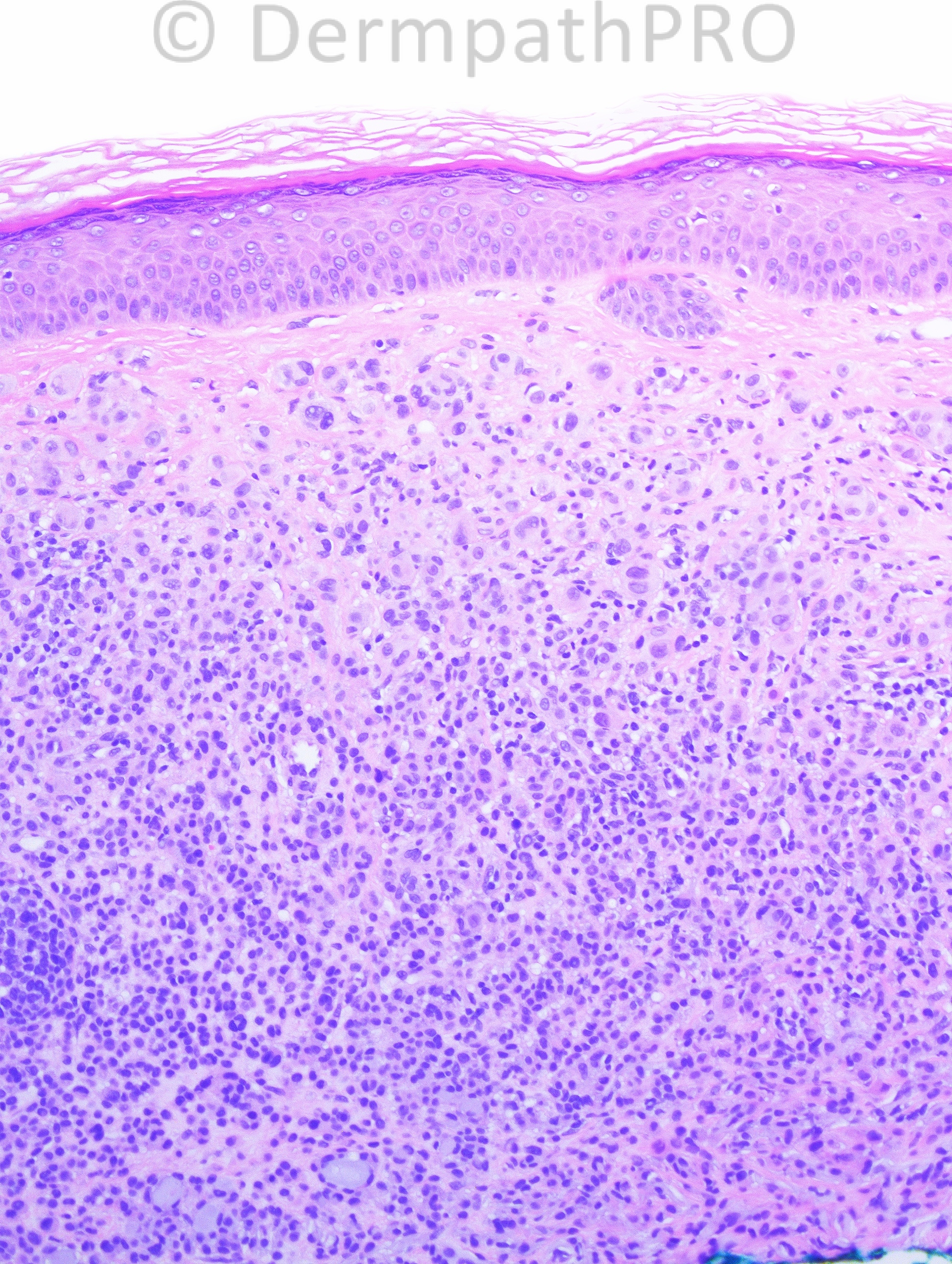
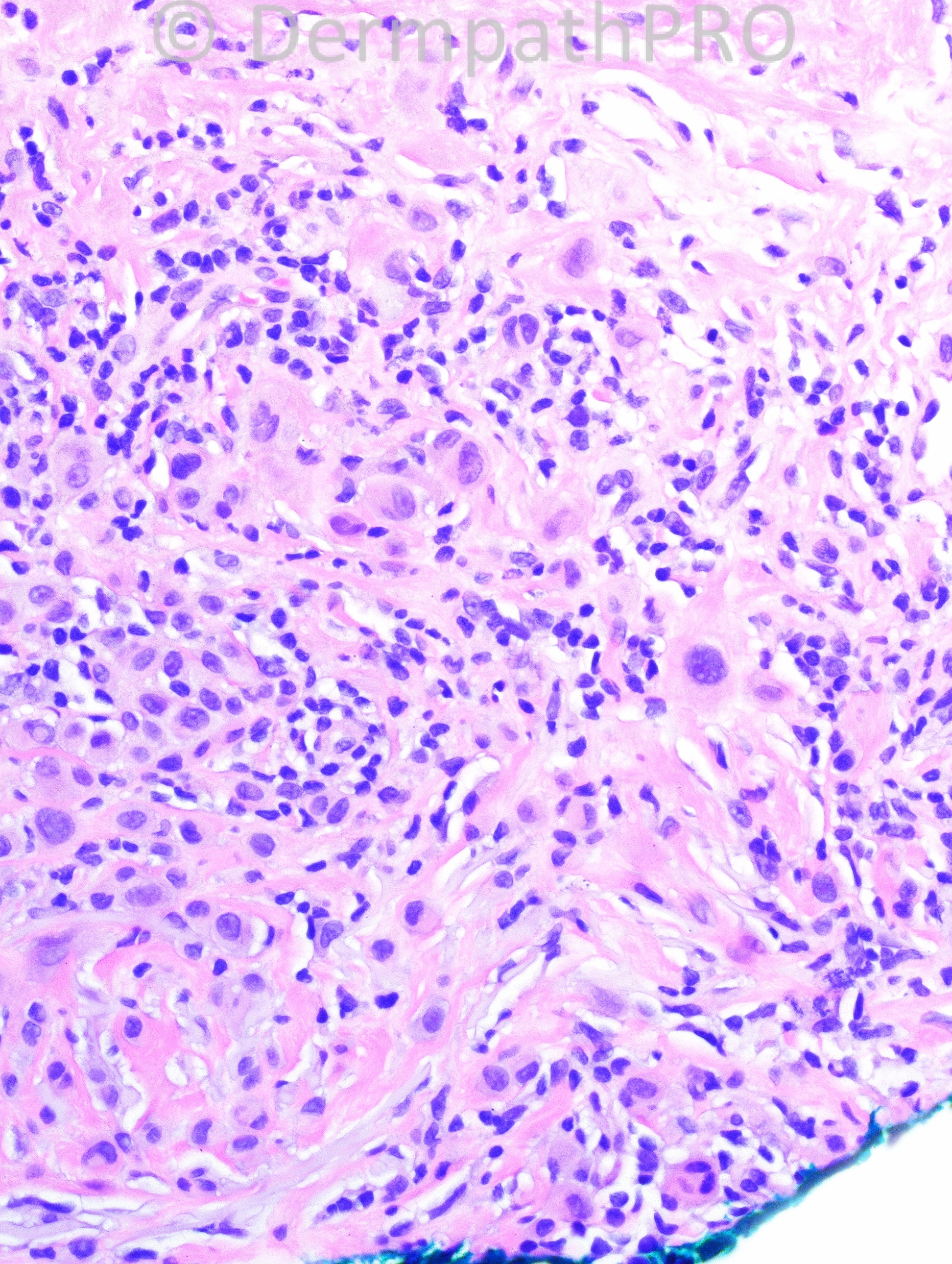
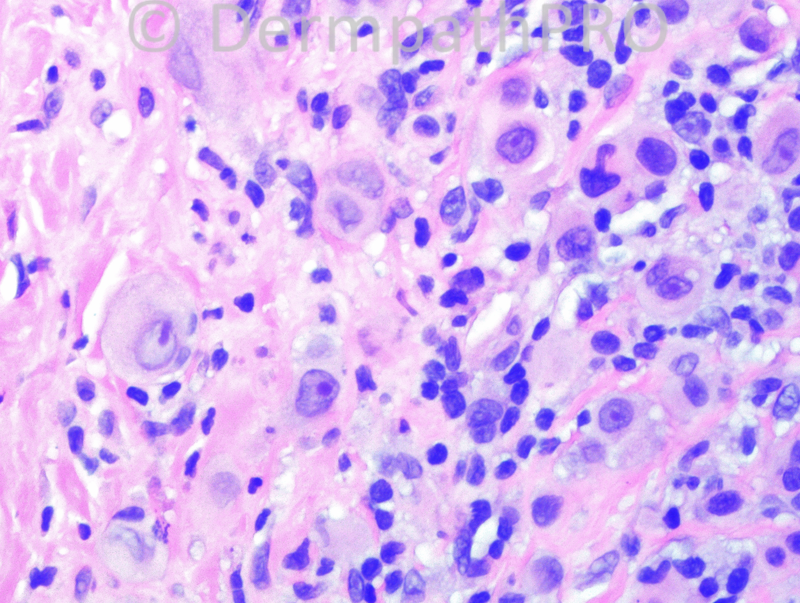
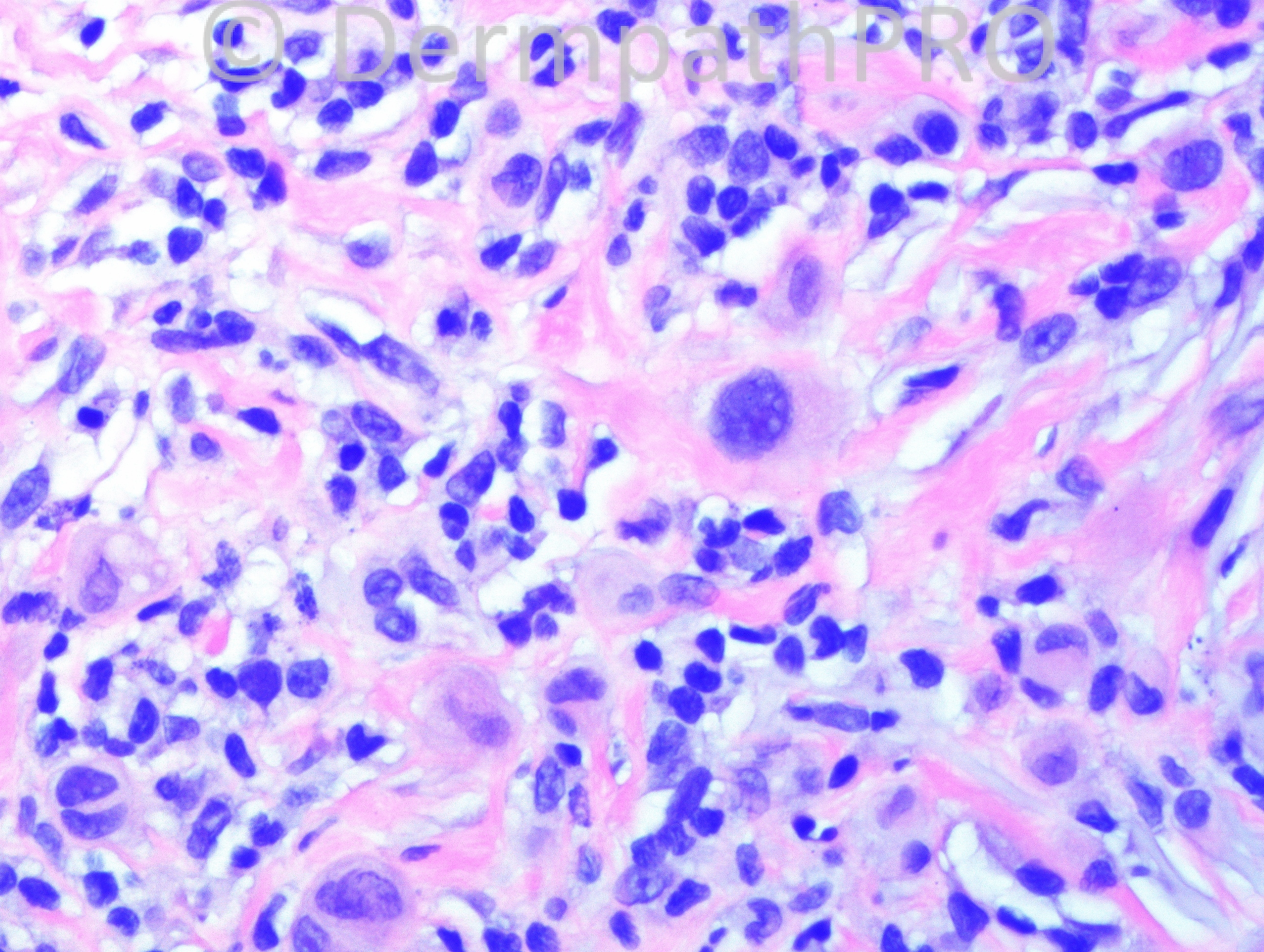
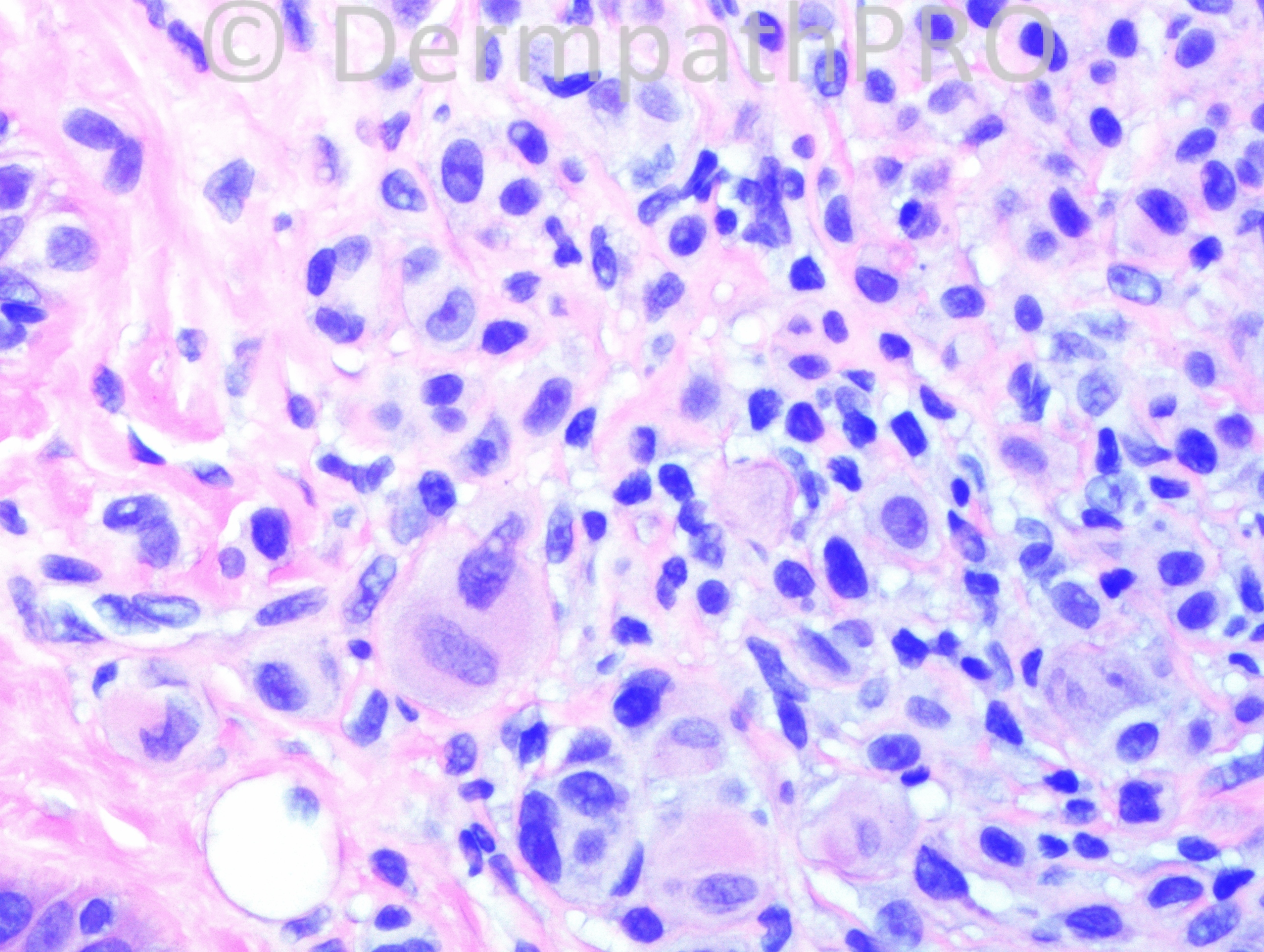
44 year-old female with right arm lesion. The clinical differential is: BCC vs neurofibroma.

Case posted by Dr. Hafeez Diwan.

  Report Record

User Feedback


Dr. Mona Abdel-Halim

Posted

Very difficult !!! Could it be Spitzoid lesion for further work up as in this age it is not easy to go with a diagnosis of Spitz nevus. Epithelioid shwannoma is a good possibility but my perception of shwannomas is that they r capsulated lesions?

Share this comment


Link to comment
share_externally

?Epithelioid benign fibrous histiocytoma. As far as I know, epithelioid schwannoma is a very rare lesion with less than 50 cases reported.

Share this comment


Link to comment
share_externally

Guest Romualdo

Posted

Well, let's try again! Intradermal melanocytic nevus with halo type reaction.

Share this comment


Link to comment
share_externally

Robledo F. Rocha

Posted

According to Guido Massi and Philip LeBoit in their excellent book about melanocytic pathology, as a rule, halo nevi do not occur on extremities.
The microscopic picture of Spitz-like epithelioid cells with ground glass cytoplasm accompanied by a lymphocytic infiltrate suggests me solitary reticulohistiocytoma.

Share this comment


Link to comment
share_externally

Epitheliod histoocytoma vs spitz nevus for histochemical stain

Share this comment


Link to comment
share_externally

Guest Giovanni Falconieri

Posted

Quite difficult. Cannot make a firm choice. The confluent pattern and the scattered larger cells might suggest Bapoma, and a work up including assay for the Bap1 loss (Wiesner nevus) [font=Helvetica][size=1] [/size][/font]might be worth in a case like this, of course in addition to the stains entailed by the broader differential above.

Share this comment


Link to comment
share_externally

Dr. Hafeez Diwan

Posted

There was loss of BAP1, so this is a Wiesner nevus. Only a few cells were Ki67 positive (using a double pan-melanocytic marker + Ki67 stain).

Share this comment


Link to comment
share_externally

Dr. Hafeez Diwan

Posted

I advised complete excision and indicated the associations of Wiesner nevus in my diagnostic comment.

Share this comment


Link to comment
share_externally


Join the conversation

You can post now and register later. If you have an account, sign in now to post with your account.

Guest
Add a comment...

×   Pasted as rich text.   Paste as plain text instead

  Only 75 emoji are allowed.

×   Your link has been automatically embedded.   Display as a link instead

×   Your previous content has been restored.   Clear editor

×   You cannot paste images directly. Upload or insert images from URL.

Loading...

×
×
  • Create New...