Jump to content
In this section we have spot diagnoses posted on a daily basis since June 2010, now over 4000! You can review the archived cases and read the suggested diagnoses by users and the final comment by the contributors.
Case are uploaded each week day by 10 am UK time with the correct diagnosis will generally be posted at 8 pm UK time. Why not view the most recent spot diagnosis and proffer a diagnosis?

Case Number : Case 1624 - 15 September Posted By: Guest

Please read the clinical history and view the images by clicking on them before you proffer your diagnosis.
Submitted Date :
   (0 reviews)

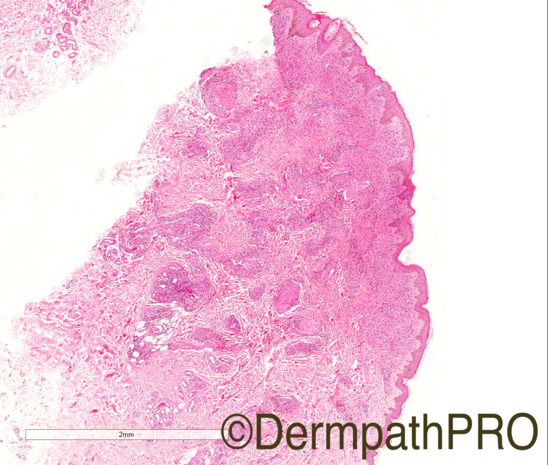
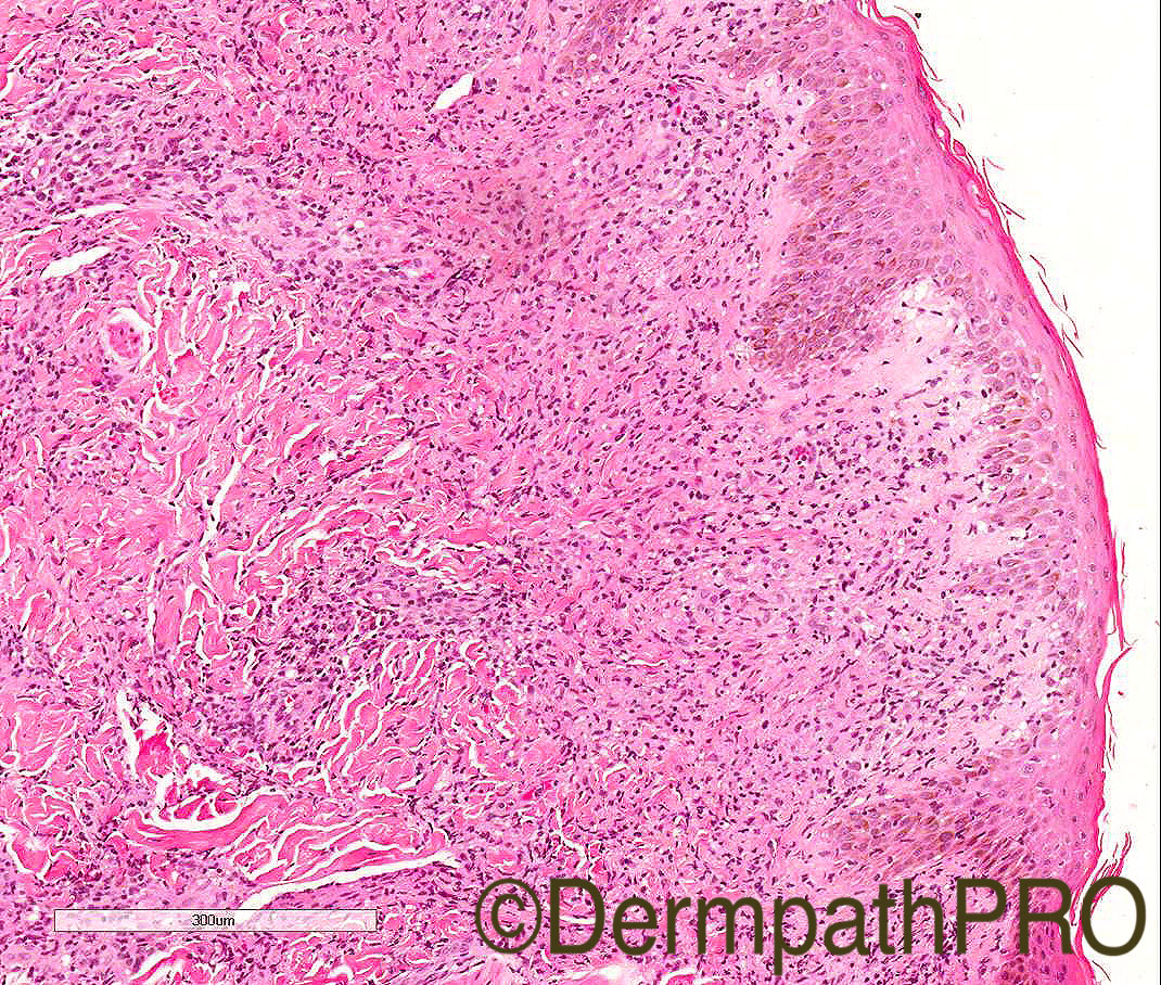
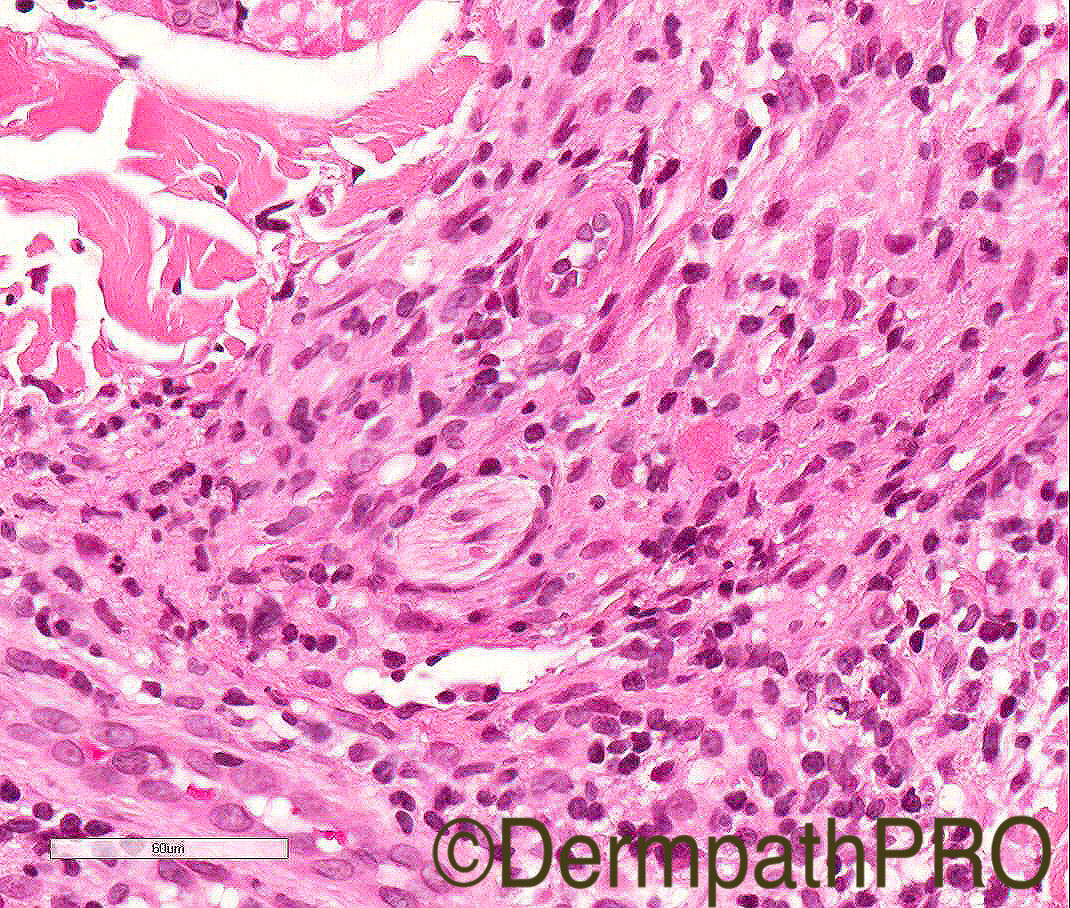
21/M, Florid rash with oedema of lower arm, nodules+

Case Posted by Dr Arti Bakshi


  Report Record

User Feedback


Raul Perret

Posted

Primary granulomatous lymphocyte poor dermal inflammation, frequently surrounding nerves. There seems to be some interface change and dermal edema in picture 2. The differentials include: Leprosy, non-tuberculoous mycobacterial infection, sarcoidosis, and less probable syphilis (due to lack of plasmocytes and mainly granulomatous infiltrate). Clinicopathological correlation is really important here as well as eventual PCR, culture. Would still perform ZN, and Warthin-starry/ IHQ for treponems. Here a nice article of entities with perineural inflammation. I think my favourite diagnosis is first leprosy and second sarcoidosis. Everything (rash and nodules) is confined to the lower arm or the rash is systemic? Because if confined to the arm then atypical mycobacterial infection is algo a good consideration.

Share this comment


Link to comment
share_externally

Guest Arash Daryakarr

Posted

Agree with Raul, i consider more leprosy and atypical mycobacterial infection.further studies are needed.

Share this comment


Link to comment
share_externally

Dr. Mona Abdel-Halim

Posted

Agree, leprosy in reaction tops the list

Share this comment


Link to comment
share_externally

vincenzo polizzi

Posted

Agree with above. Histoid Leprosy could be a good diff.  

Share this comment


Link to comment
share_externally

Arti Bakshi

Posted

Yes this is case of leprosy. 

The patient was a student from Srilanka. As this is quite an uncommon diagnosis in this part of the world, the dermatologists were not quite familiar with its cutaneous spectrum and did not suspect the diagnosis. Following the histology report, the infectious disease consultants were involved and the patient had quite typical anaeasthetic patches, ulnar nerve thickening and early clawing of hand. The AFB was negative on the histology section but a slit skin smear was positive (1+).

Cant claim to have any expertise in classifying leprosy, as this is the only case I have ever diagnosed in UK! But from my early pathology years in India where I did see quite a few cases, I classified this as a Borderline Tuberculoid leprosy. 

Share this comment


Link to comment
share_externally


Join the conversation

You can post now and register later. If you have an account, sign in now to post with your account.

Guest
Add a comment...

×   Pasted as rich text.   Paste as plain text instead

  Only 75 emoji are allowed.

×   Your link has been automatically embedded.   Display as a link instead

×   Your previous content has been restored.   Clear editor

×   You cannot paste images directly. Upload or insert images from URL.

Loading...

×
×
  • Create New...