Jump to content
In this section we have spot diagnoses posted on a daily basis since June 2010, now over 4000! You can review the archived cases and read the suggested diagnoses by users and the final comment by the contributors.
Case are uploaded each week day by 10 am UK time with the correct diagnosis will generally be posted at 8 pm UK time. Why not view the most recent spot diagnosis and proffer a diagnosis?

Case Number : Case 1247 - 3 April Posted By: Guest

Please read the clinical history and view the images by clicking on them before you proffer your diagnosis.
Submitted Date :
   (0 reviews)

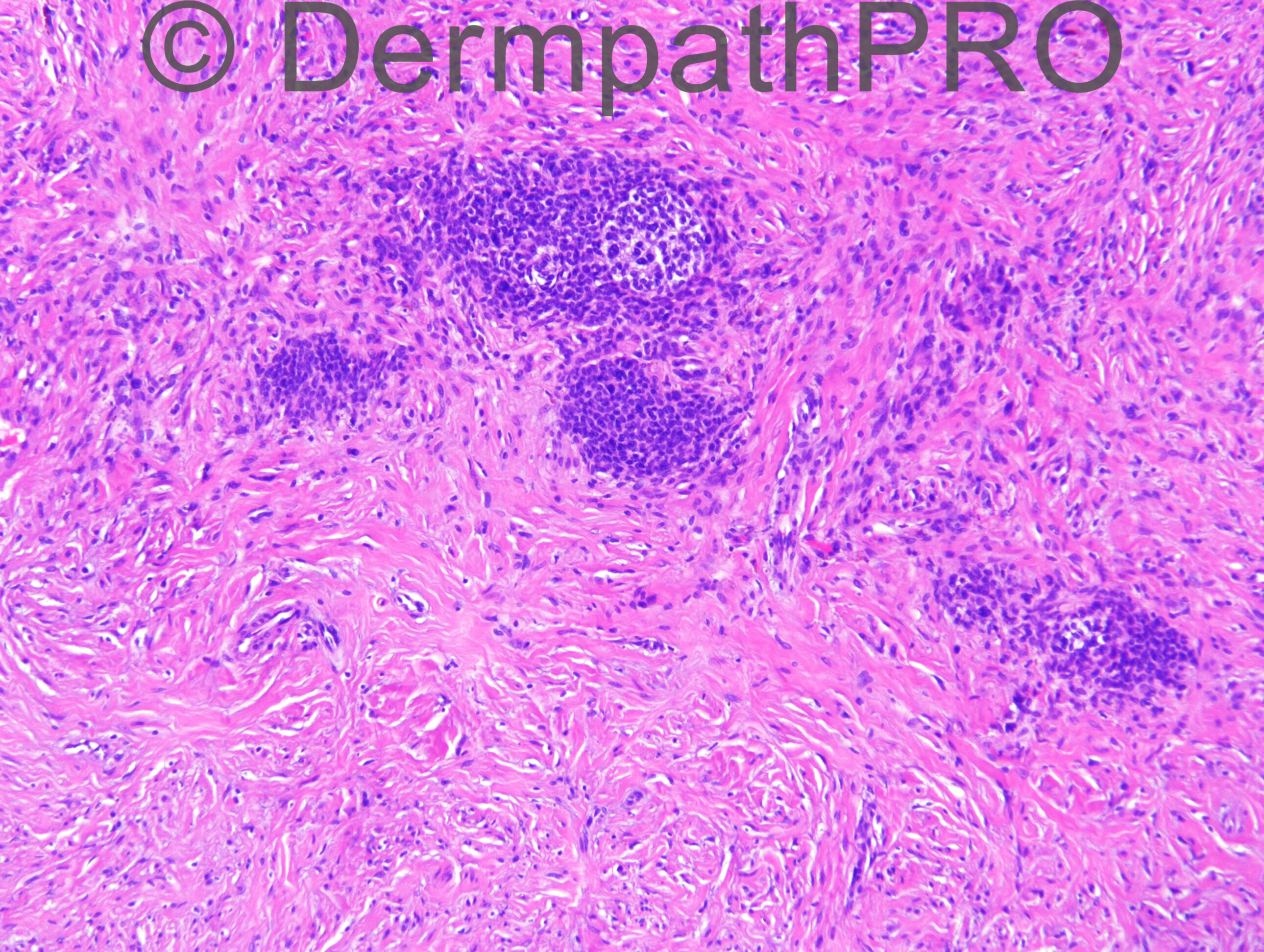
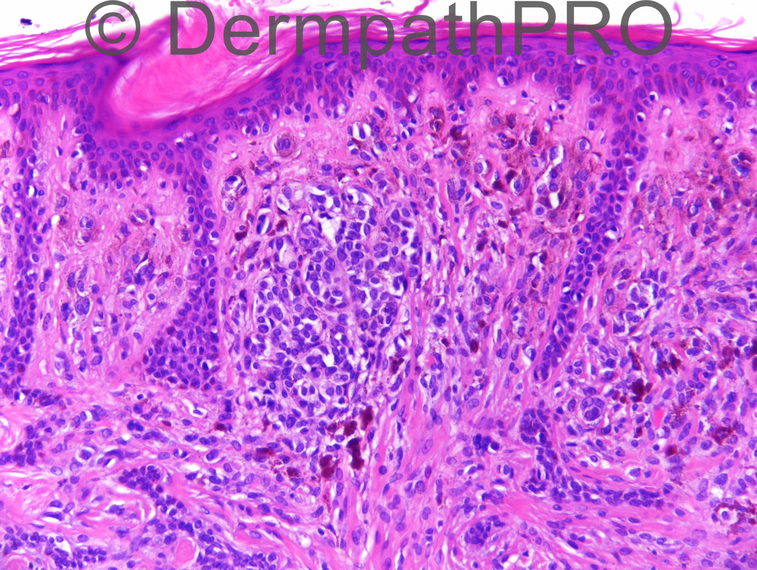
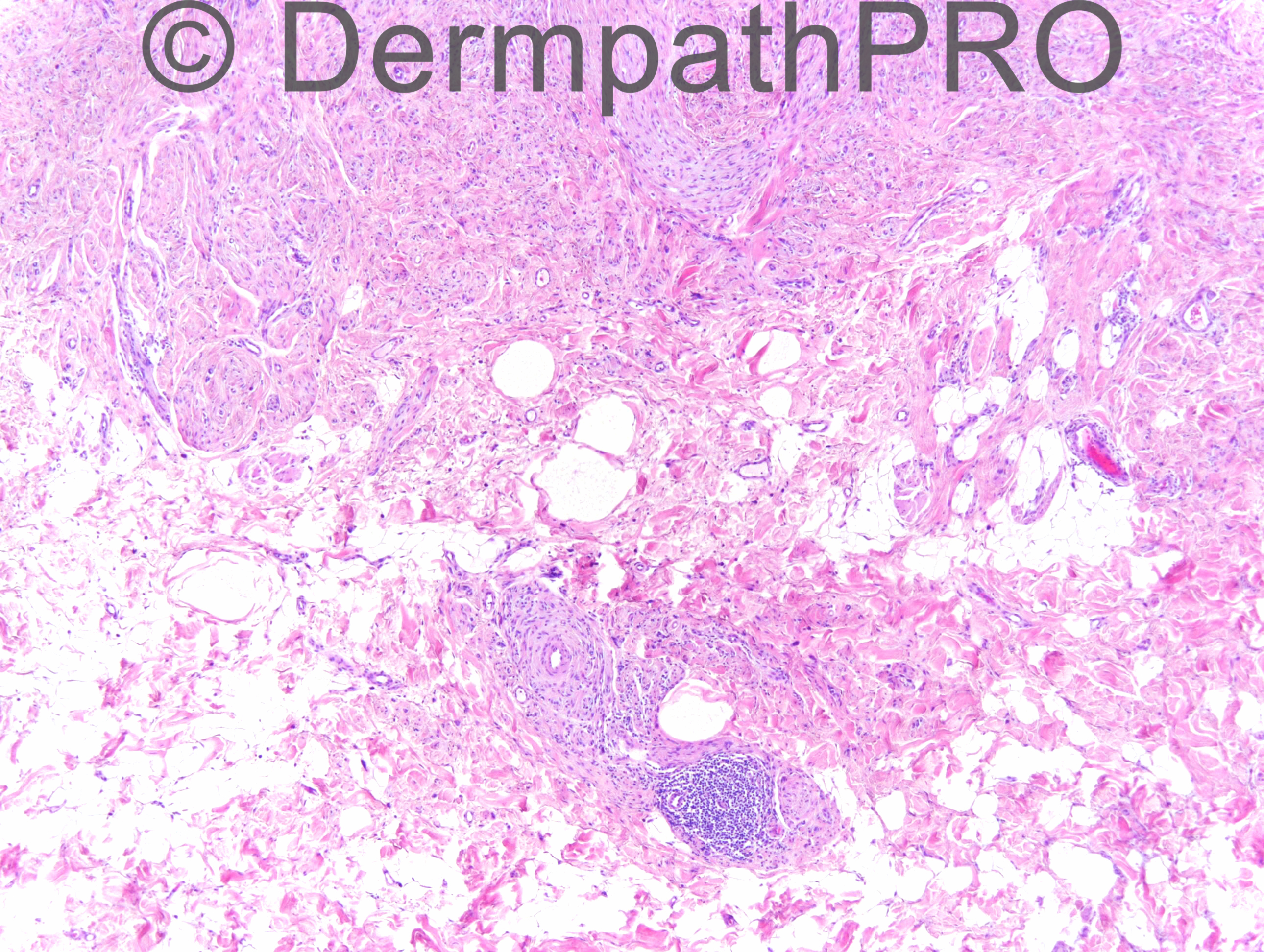
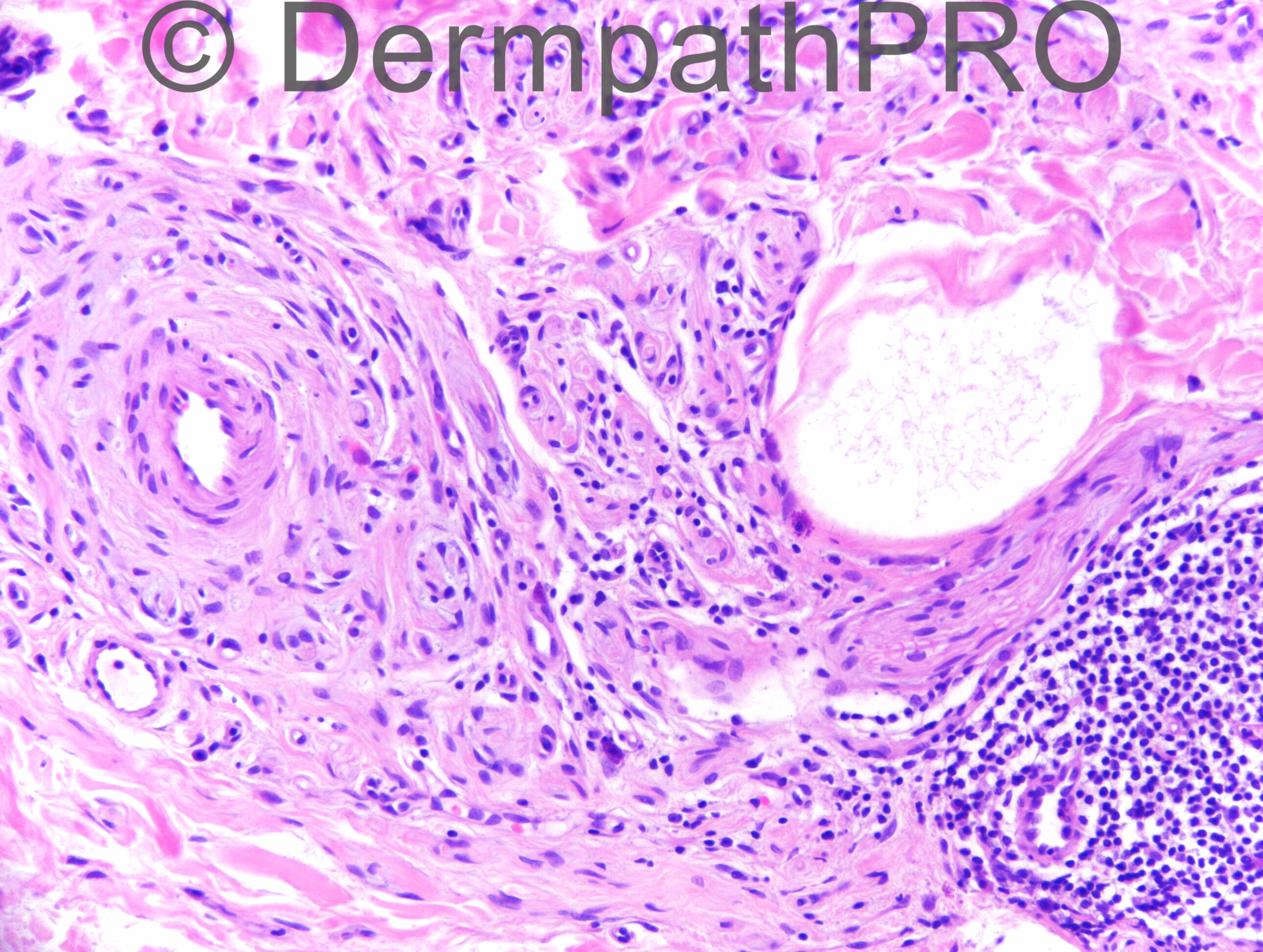
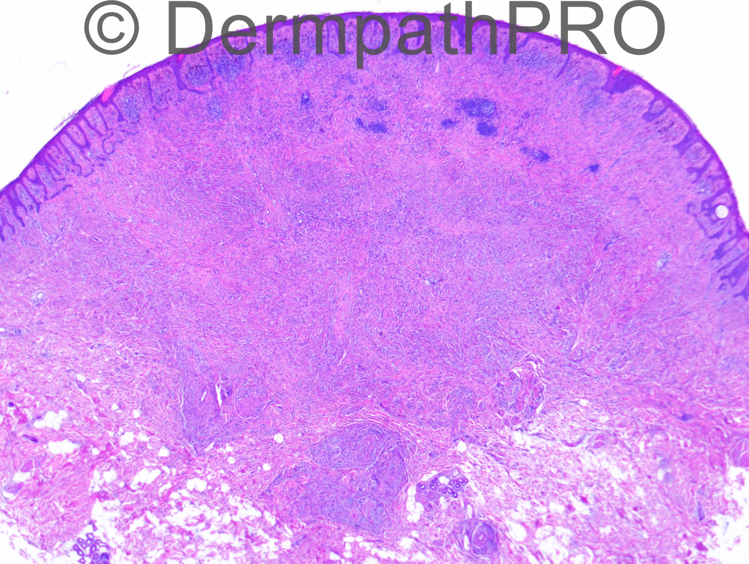
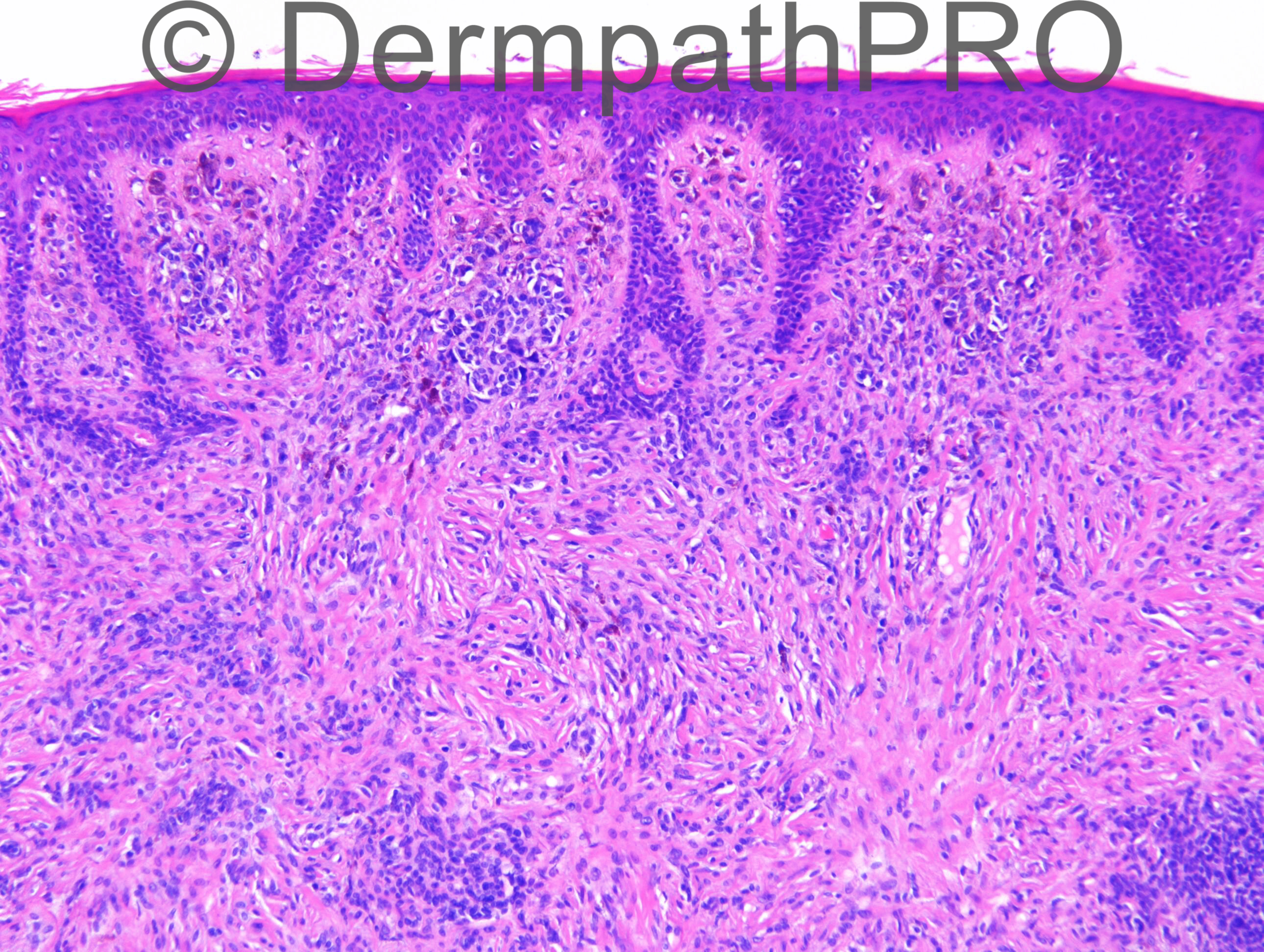
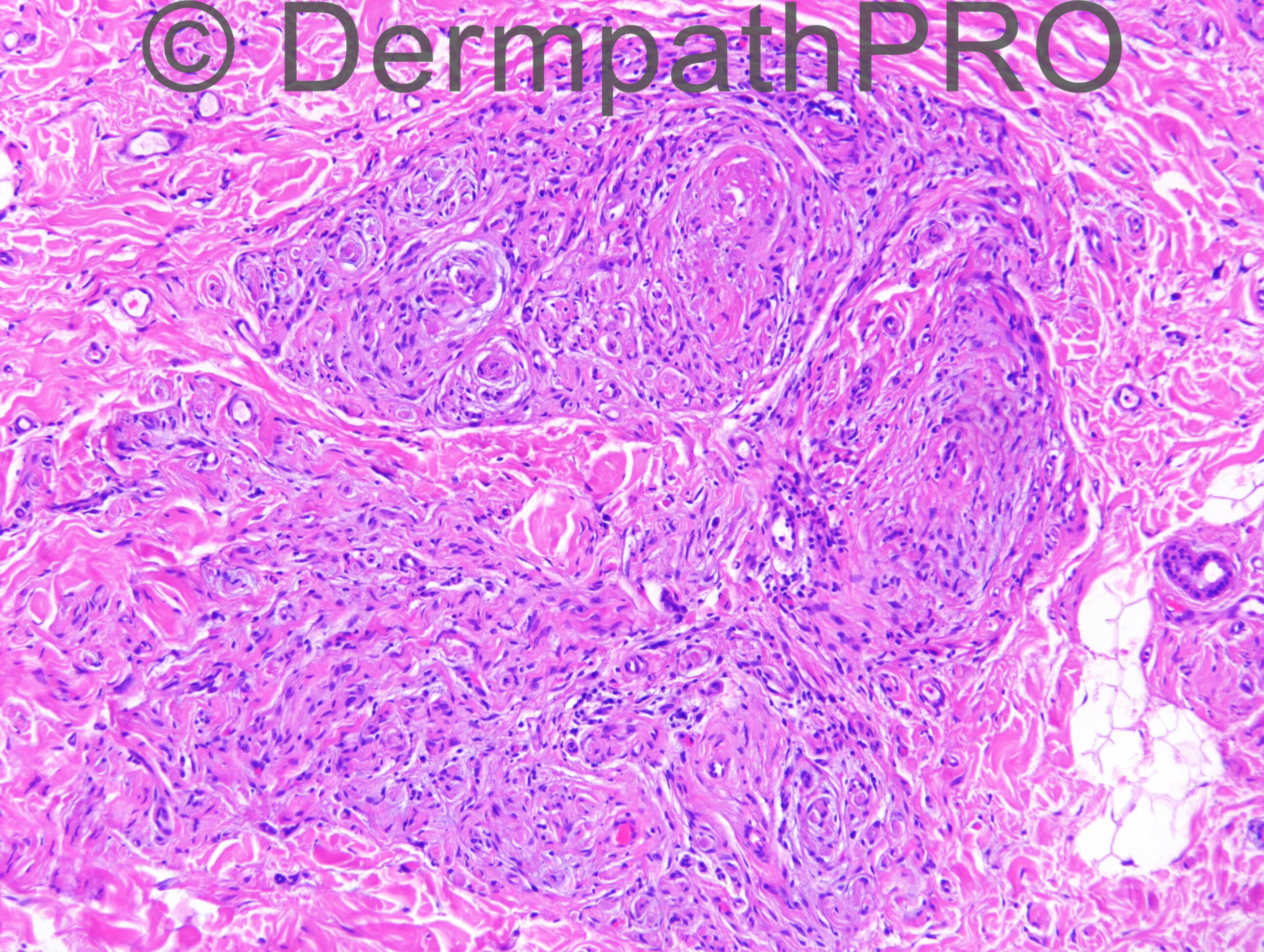
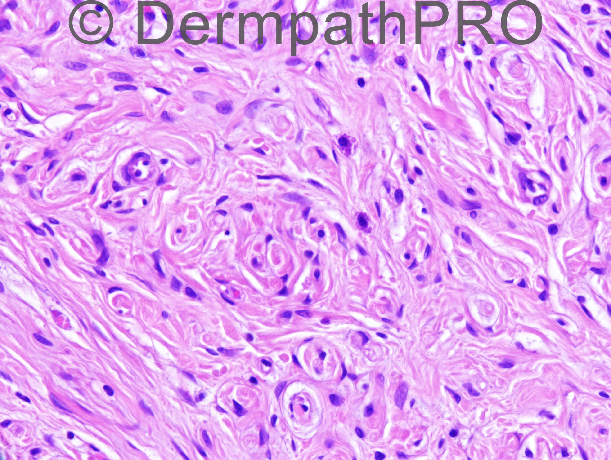
M65. Altered naevus on back.

Case posted by Dr Richard Carr

  Report Record

User Feedback


Dr. Mona Abdel-Halim

Posted

I will consider this a spindle cell/neurotropic melanoma developing in a nevus.

Share this comment


Link to comment
share_externally

Guest Romualdo

Posted

Desmoplastic (sclerotic) nevus with lymphocytic aggregates?

Share this comment


Link to comment
share_externally

Guest Jim Davie MD

Posted

Agree most likely desmoplastic neurotropic melanoma, developing in pre-existing nevus.
- Clinical is supportive (older male, changing lesion).
- Targeted lymphoplasmacytic infiltrate surrounding nodules of moderately atypical pale nevoid cells in the upper dermis (images 1,5) and also adjacent to a nerve with spindle-cell desmoplastic focus in the deeper dermis (images 3, 4).
- Junction has a subtle lentiginous proliferation with pagetoid component.

S100 or SOX10 might be useful to confirm spindle cells; Ki67 may show proliferative activity in the spindle/nevoid dermal components that (if present) would be supportive of DM over nevus. Absence of MelanA/MART1 staining would help support DM over differential consideration of a desmoplastic nevus /hypopigmented blue nevus variant.

Share this comment


Link to comment
share_externally

Dr. Richard Carr

Posted

This is a referred case was initially out-sourced but the local pathologist on reviewing the slides (reported by a known UK expert) asked for a further opinion. I shared the case with my colleague who is also well known in the field and reported the case as follows:

Many thanks for the opportunity to review this case, which has also been seen by Dr XXXX and by one of our visiting Diploma Dermopath Candidates, Dr XXXX.

In our opinion, this is a benign lesion showing residual naevocellular neavus superficially and dramatically neurotised naevus in the deeper part; closely resembling neurofibroma. We were not worried for melanoma in this case and the leave and wait policy could be adopted.

I understand the case had been reported initially by Dr XXXX who is a national expert on melanocytic tumours [and raised a possibility of desmoplastic/neurotrophic melanoma] but as you know, experts can vary in opinions for difficult melanocytic cases.

Comments: It is interesting how we can see the same case but come up with such different [favoured] diagnoses or raise serious concerns. It is understandable why patients must be non-plussed with medico-legal implications. The only points I will dissagree on with Jim is that the [purple] aggregates in the upper field are most likely naevocellular [compare with blue dots in lower field of a genuine lymphoid aggregate] and the slight prominence of non-atypical, non-confluent melanocytes over a naevus are quite regular for these lesions. Having said that there was a lymphoid aggregate in the mid/upper dermis in another image not presented (no plasma cells though). I am guessing in the future nearly all cases will be diagnosed on digitised slides - but I hope this will only happen when the technology is as fast as a microscope and equal in quality of image and of proven saftey and equally if not lower in litigation! This case illustrates there is a fine line between certain diagnoses and I remember well Dr Mckee saying at the start of every sign out say "desmoplastic melanoma" to yourself!!! It is however possible we can be too sensitive to certain diagnoses the more knowledgeable/experienced we become sometimes the more uncertain (I know this is true of Spitz lesions for me). If you have a doubt then my advice is to show someone else although it does not necessarily mean you are right if both agree it does provide some measure of protection to criticsm if somone else dissagrees.

Share this comment


Link to comment
share_externally


Join the conversation

You can post now and register later. If you have an account, sign in now to post with your account.

Guest
Add a comment...

×   Pasted as rich text.   Paste as plain text instead

  Only 75 emoji are allowed.

×   Your link has been automatically embedded.   Display as a link instead

×   Your previous content has been restored.   Clear editor

×   You cannot paste images directly. Upload or insert images from URL.

Loading...

×
×
  • Create New...