Jump to content
In this section we have spot diagnoses posted on a daily basis since June 2010, now over 4000! You can review the archived cases and read the suggested diagnoses by users and the final comment by the contributors.
Case are uploaded each week day by 10 am UK time with the correct diagnosis will generally be posted at 8 pm UK time. Why not view the most recent spot diagnosis and proffer a diagnosis?

Case Number : Case 1248 - 6 April Posted By: Guest

Please read the clinical history and view the images by clicking on them before you proffer your diagnosis.
Submitted Date :
   (0 reviews)

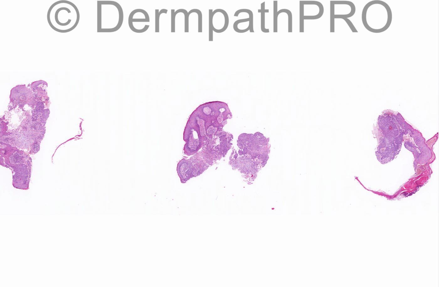
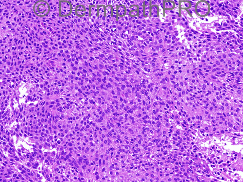
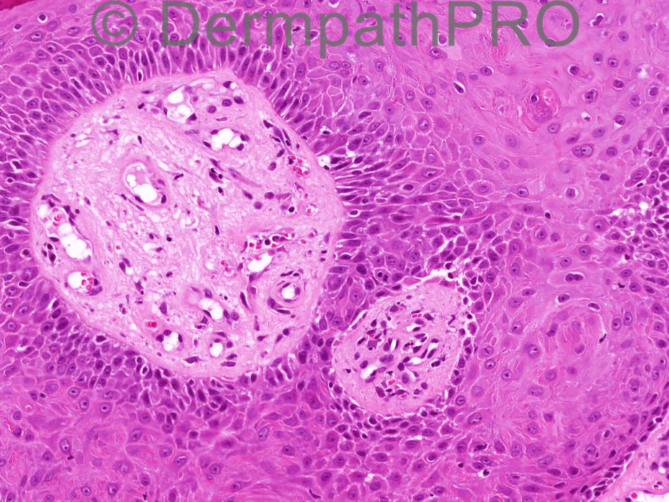
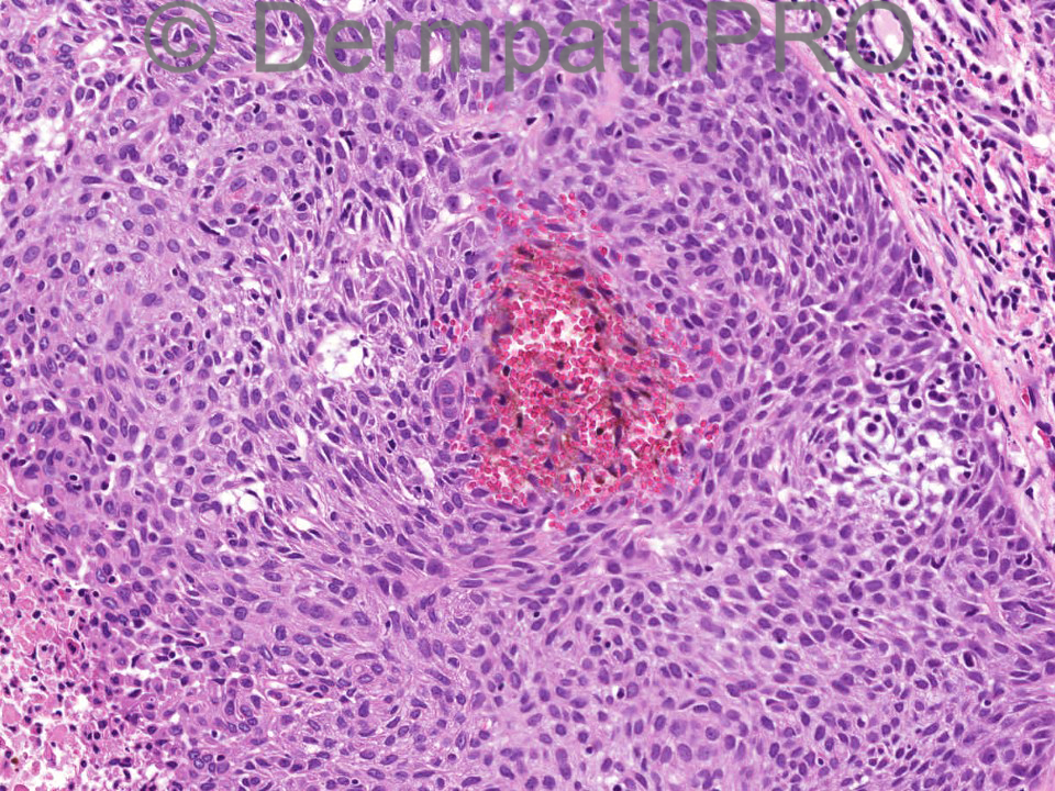
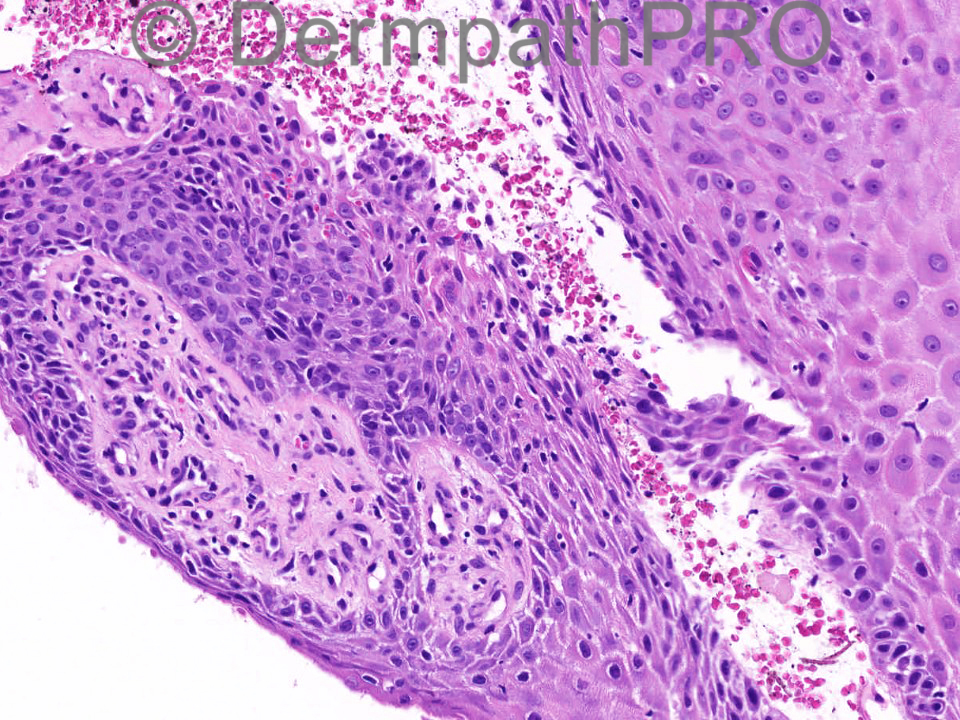
72 years male; benign skin growth upper lip.

Case posted by Dr Iskander Chaudhry

  Report Record

User Feedback


Dr. Mona Abdel-Halim

Posted

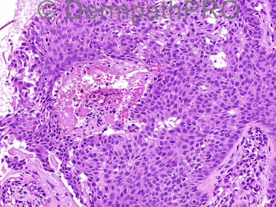
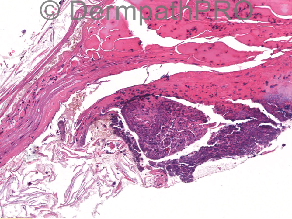
Images 1,2 and 6, to me are poroid and look like poroma. The infiltrative pattern in images 7 and 8 is worrisome. I need a scanning view of the lesion !!!!

Share this comment


Link to comment
share_externally

Guest Romualdo

Posted

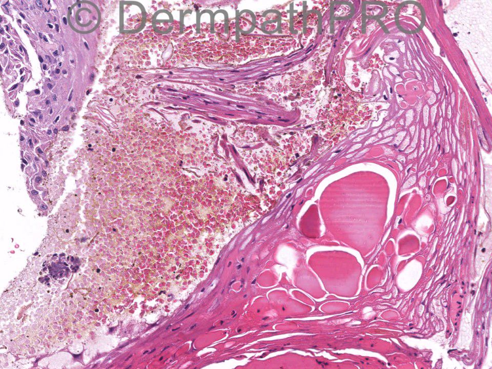
In image 1 I see a large epithelial lobule with smooth contour and thick basement membrane. Images 7 and 8 show what I think are pseudoinfiltrative features. In images 9, 10 and 11 there are dilated capillaries within dermal papillae and maybe other features suggestive of common warts. I think this is desmoplastic trichilemmoma. As a second possibility, depending on a complete examination of the lesion, an old verruca vulgaris with trichilemmoma-like areas.

Share this comment


Link to comment
share_externally

I agree with the diagnosis of Desmoplastic trichilemmoma.

Share this comment


Link to comment
share_externally

Eman El-Nabarawy

Posted

I thought of desmoplastic seborrheic keratosis.

Share this comment


Link to comment
share_externally

I agree With Dr Abdel Halim about the need of an ovaerall view. These close-up snapshots show a squamous lesion, without definite evidence of malignancy, but I can't go any further. This may well be a tricholemmoma, but I am far from convinced.

Share this comment


Link to comment
share_externally

Dr. Richard Carr

Posted

Can't see significant cellular atypia. Given the anatomic location traumatised (focally desmoplastic) tricholemmoma would be my favoured diagnosis. CD34 and PAS (highlights BM) should be helpful.

Share this comment


Link to comment
share_externally

Iskander H. Chaudhry

Posted

Thank you all for your comments on a bank holiday here in the UK. The final diagnosis is desmoplastic trichilemmoma and as Richard says there is no significant atypia.

Share this comment


Link to comment
share_externally


Join the conversation

You can post now and register later. If you have an account, sign in now to post with your account.

Guest
Add a comment...

×   Pasted as rich text.   Paste as plain text instead

  Only 75 emoji are allowed.

×   Your link has been automatically embedded.   Display as a link instead

×   Your previous content has been restored.   Clear editor

×   You cannot paste images directly. Upload or insert images from URL.

Loading...

×
×
  • Create New...