Jump to content
In this section we have spot diagnoses posted on a daily basis since June 2010, now over 4000! You can review the archived cases and read the suggested diagnoses by users and the final comment by the contributors.
Case are uploaded each week day by 10 am UK time with the correct diagnosis will generally be posted at 8 pm UK time. Why not view the most recent spot diagnosis and proffer a diagnosis?

Case Number : Case 1252 - 10 April Posted By: Guest

Please read the clinical history and view the images by clicking on them before you proffer your diagnosis.
Submitted Date :
   (0 reviews)

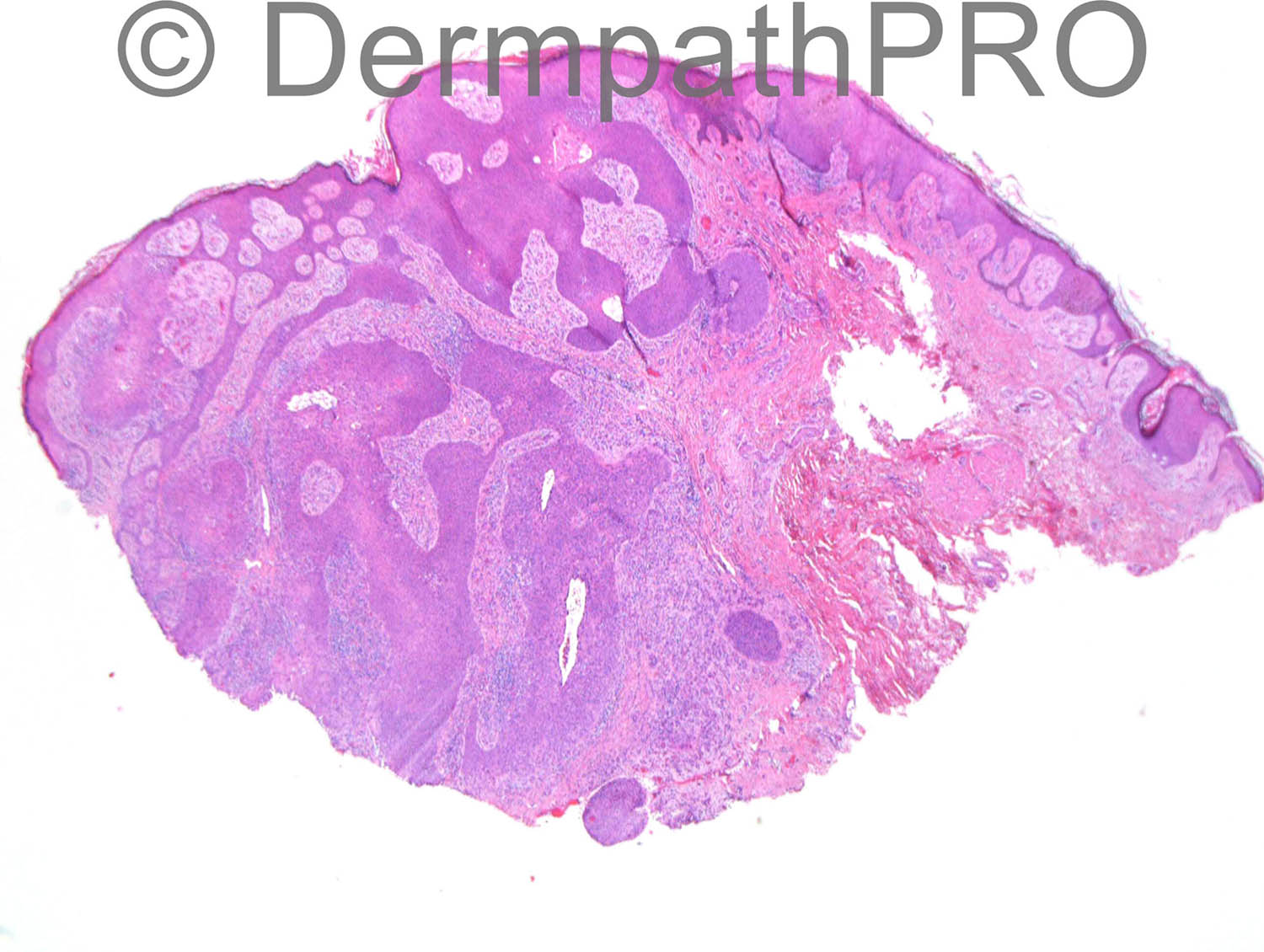
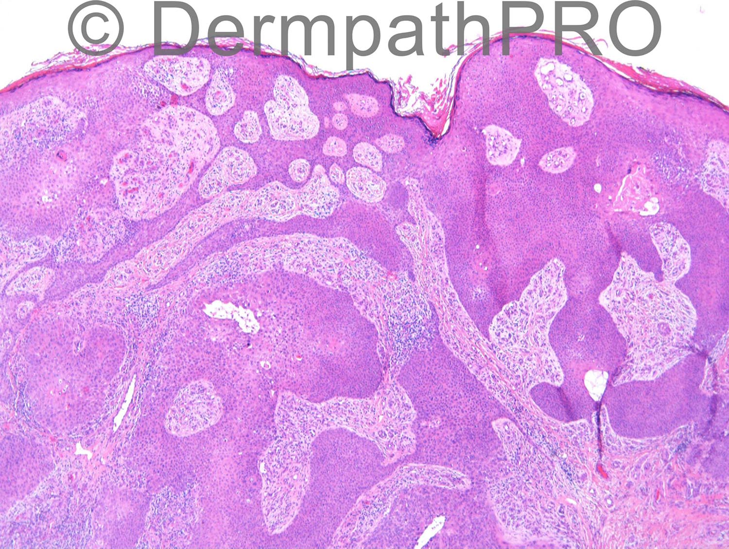
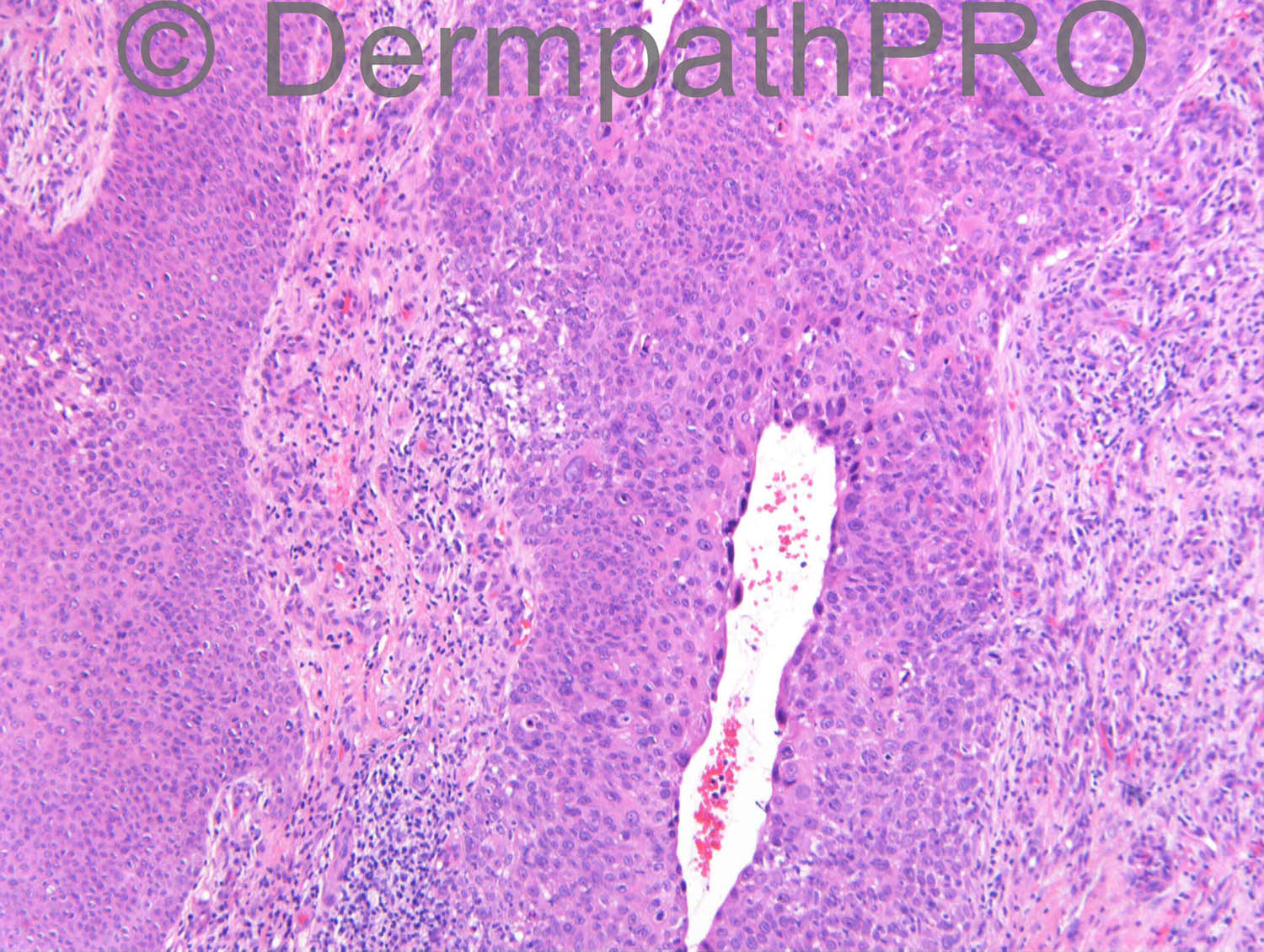
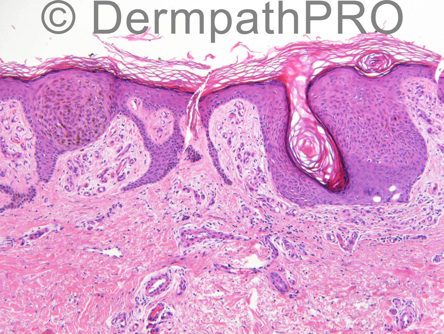
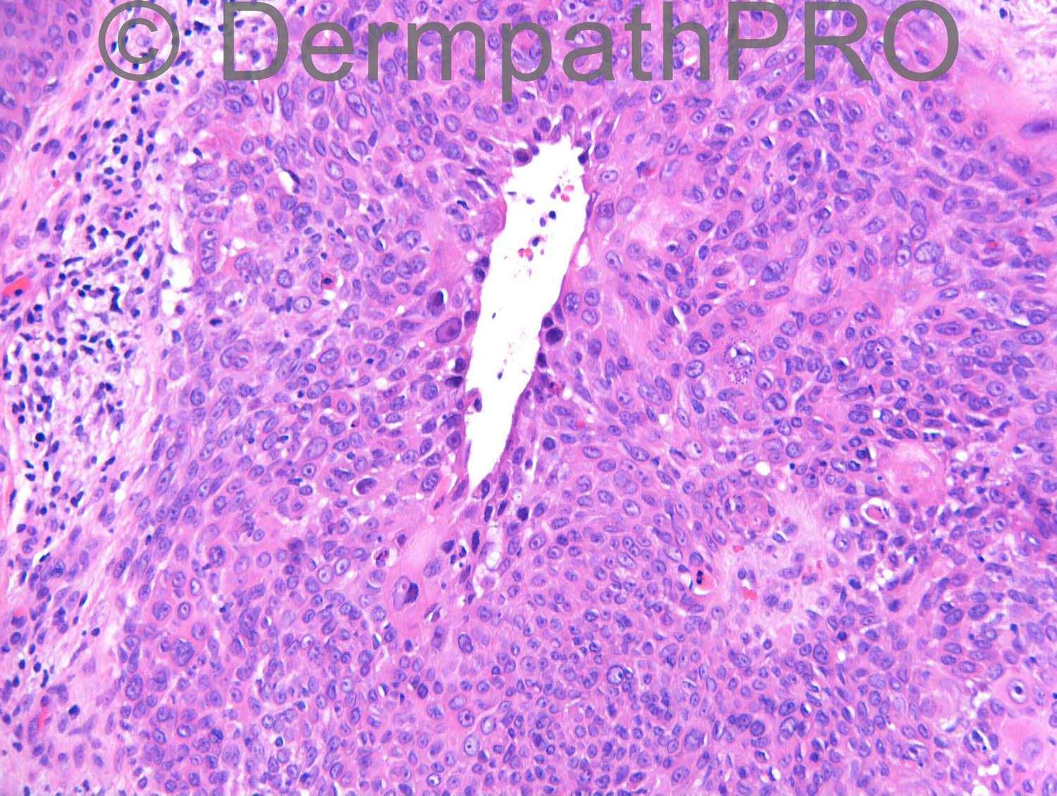
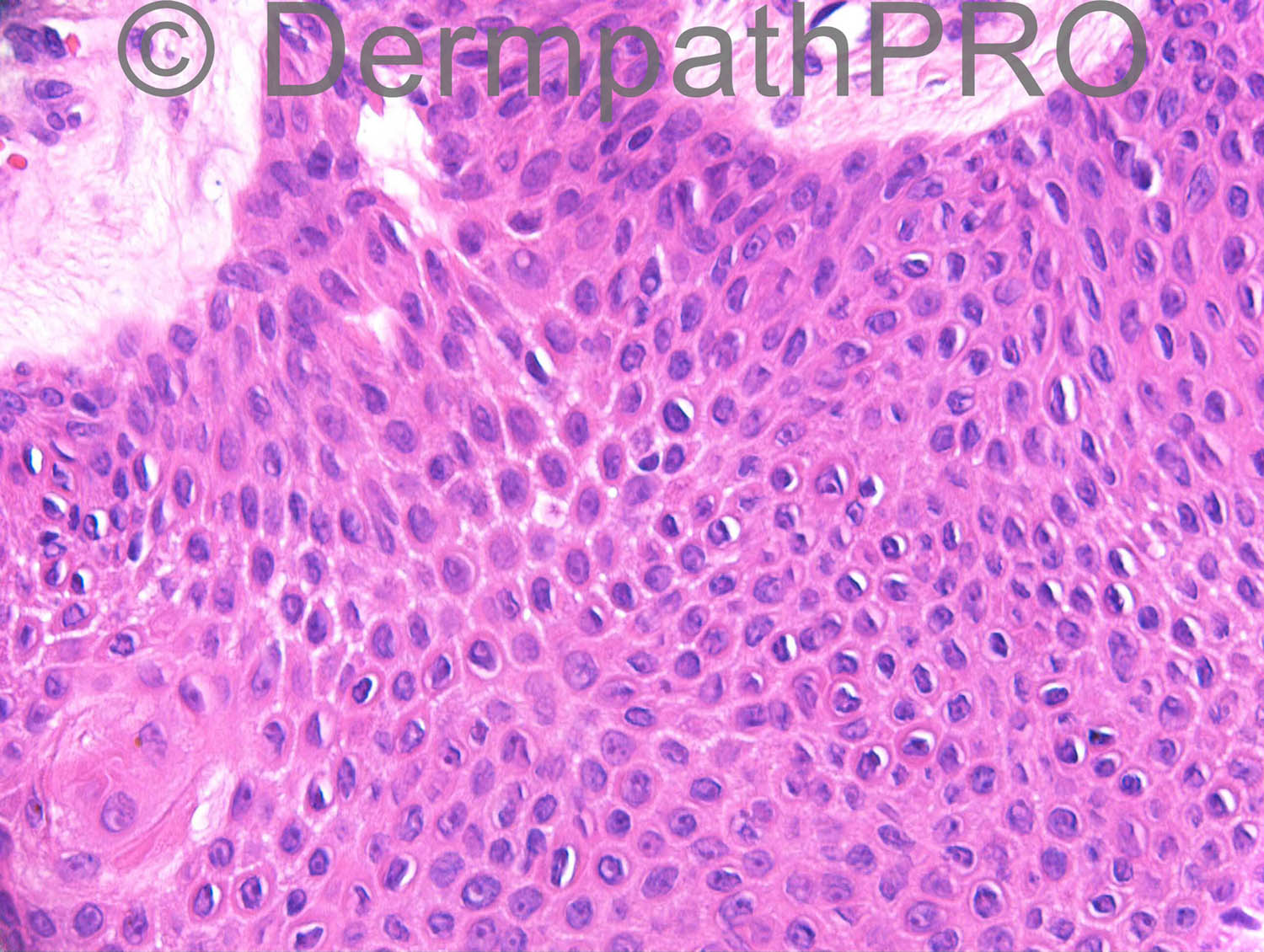
M65 lower leg lesion ?BCC ?Bowen’s

Case posted by Dr Richard Carr


  Report Record

User Feedback


Eman El-Nabarawy

Posted

Porocarcinoma (Bowenoid) arising in hidroacanthoma simplex.
DD collision of Porocarcinoma and seb k (clonal type).

Share this comment


Link to comment
share_externally

Guest Romualdo

Posted

Agree, eccrine porocarcinoma. Some figures show coexisting hidroacanthoma simplex and eccrine poroma.

Share this comment


Link to comment
share_externally

Dr. Mona Abdel-Halim

Posted

Yes, my first impression is porocarcinoma, arising from a precursor hidroacanthoma simplex manifesting focal pigmentation and borst jaddason phenomenon (image 4).

Share this comment


Link to comment
share_externally

Agree with Porocarcinoma and the hidroacanthoma simplex component.

Share this comment


Link to comment
share_externally

Guest Romualdo

Posted

Dr. Mark Hurt is probably correct. There is no ductal differentiation in figures 2 and 4, that probably correspond to seborrheic keratosis, clonal in figure 2. Presence of melanin and transitional cells at the border of abnormal nests also favor clonal seborrheic keratosis.The large lumens seen in several figures may represent residual eccrine duct lumens. Now, porocarcinoma as a second possibility.

Share this comment


Link to comment
share_externally

Dr. Richard Carr

Posted

I reported this as poroma, porocarcinoma and background clonal seborrhoeic keratosis. The top two images show the typical raining down pattern of poroma (benign on upper right) and porocarcinoma (in situ) pushing borders lower and left. There is clear cut ductal differentiation in the images on the left side (and in other images I took - not shown). Ki67 was markedly increased in areas of porocarinoma in situ and p53 was null in these areas (wild type in the poroma and adjacent seborrhoeic keratosis. Hopefully the dermpathpro team will post the IHC which should have been done at 4pm on friday. As a general comment it can be difficult at times to distinguish poroid SEBK from poroma and indeed Bowen's from bowenoid porocarcinoma and arguably the latter is academic. I have seen a number of SEBK in association with both bowen's and porocarcinoma and indeed one rare case of all three with additional sebaceous differentiation (bowenoid sebaceous carcinoma)!! As an aside I have noticed that a majority of clonal lesions (benign and malignant) arise on the legs. Hope you all enjoyed your weekends.

Share this comment


Link to comment
share_externally

Dr. Richard Carr

Posted

[img]https://dermpathpro.com/uploads/spot_diagnosis_comment_img/Case%201252_RAC6901x20c_p53_Label.jpg[/img]

[img]https://dermpathpro.com/uploads/spot_diagnosis_comment_img/Case%201252_RAC6901x10b_Ki67_Label.jpg[/img]

[img]https://dermpathpro.com/uploads/spot_diagnosis_comment_img/Case%201252_RAC6901x20b_p53_Label.jpg[/img]

[img]https://dermpathpro.com/uploads/spot_diagnosis_comment_img/Case%201252_RAC6901x20b_Ki67_Label.jpg[/img]

[img]https://dermpathpro.com/uploads/spot_diagnosis_comment_img/Case%201252_RAC6901x20_Ki67_Label.jpg[/img]

[img]https://dermpathpro.com/uploads/spot_diagnosis_comment_img/Case%201252_RAC6901x10b_p53_Label.jpg[/img]

Share this comment


Link to comment
share_externally


Join the conversation

You can post now and register later. If you have an account, sign in now to post with your account.

Guest
Add a comment...

×   Pasted as rich text.   Paste as plain text instead

  Only 75 emoji are allowed.

×   Your link has been automatically embedded.   Display as a link instead

×   Your previous content has been restored.   Clear editor

×   You cannot paste images directly. Upload or insert images from URL.

Loading...

×
×
  • Create New...