Jump to content
In this section we have spot diagnoses posted on a daily basis since June 2010, now over 4000! You can review the archived cases and read the suggested diagnoses by users and the final comment by the contributors.
Case are uploaded each week day by 10 am UK time with the correct diagnosis will generally be posted at 8 pm UK time. Why not view the most recent spot diagnosis and proffer a diagnosis?

Case Number : Case 1262 - 24 April Posted By: Guest

Please read the clinical history and view the images by clicking on them before you proffer your diagnosis.
Submitted Date :
   (0 reviews)

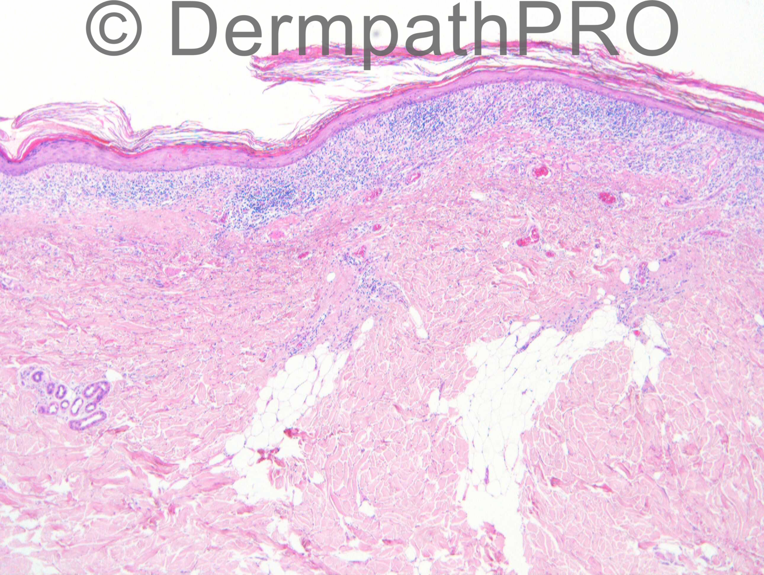
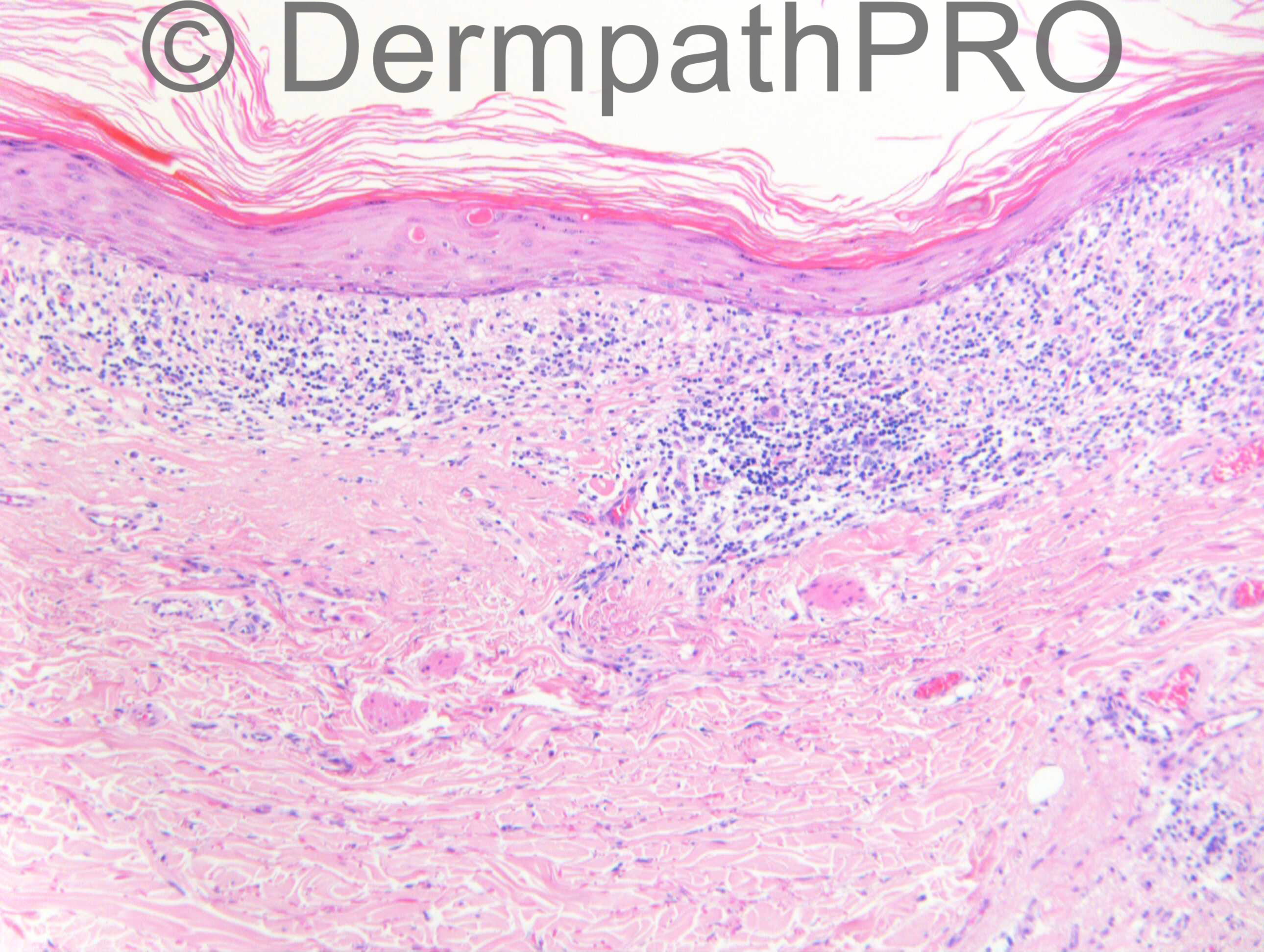
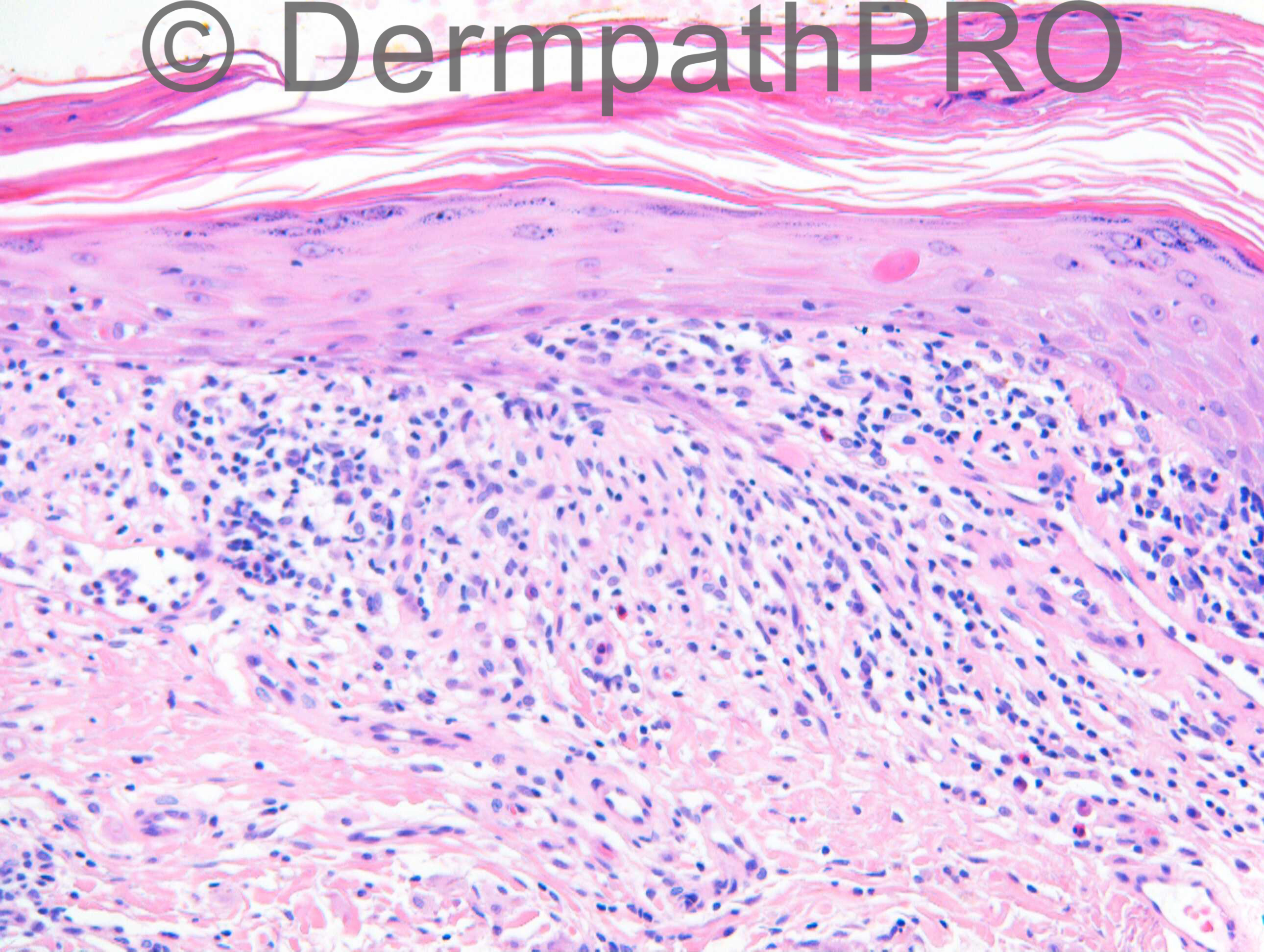
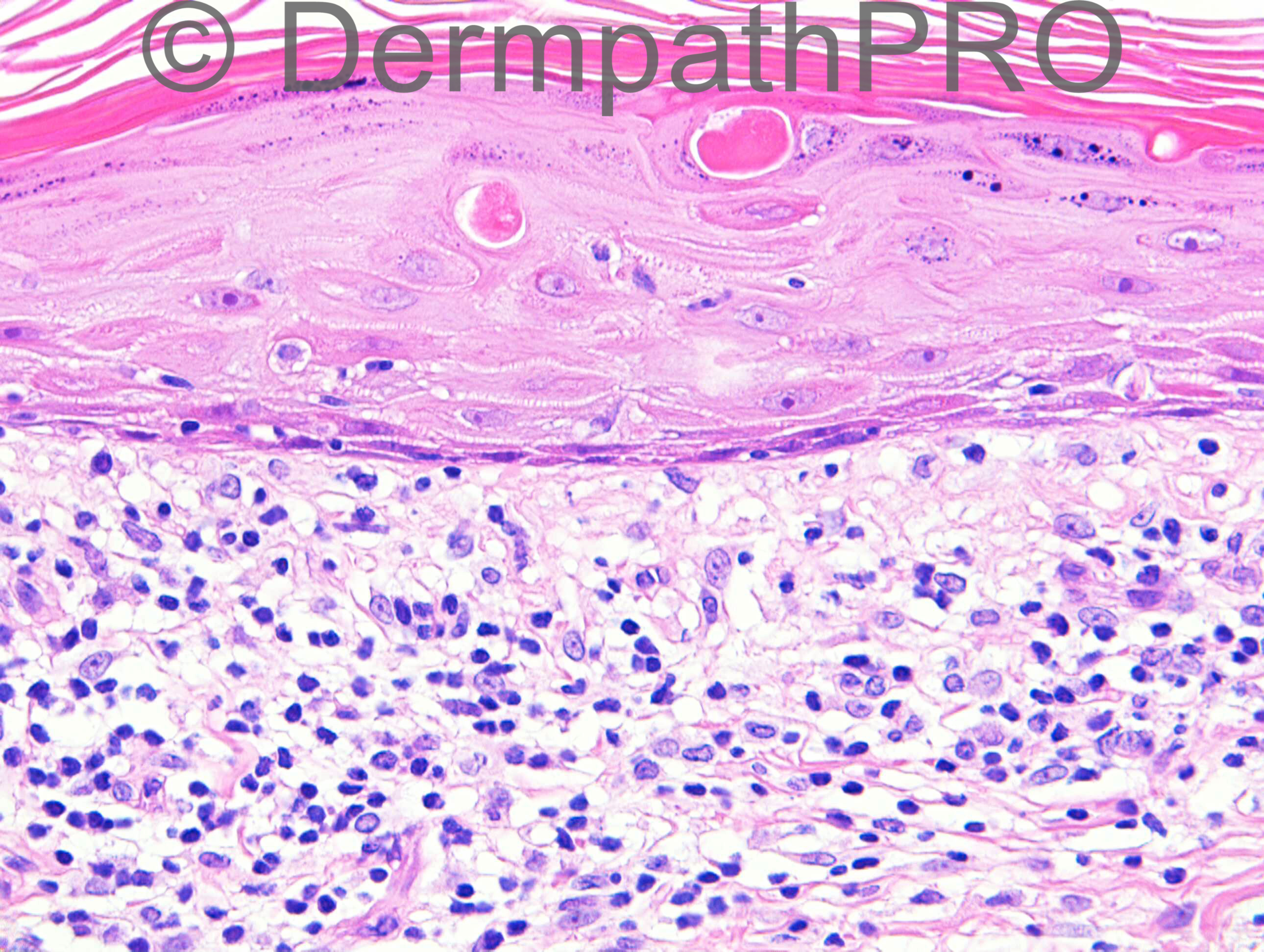
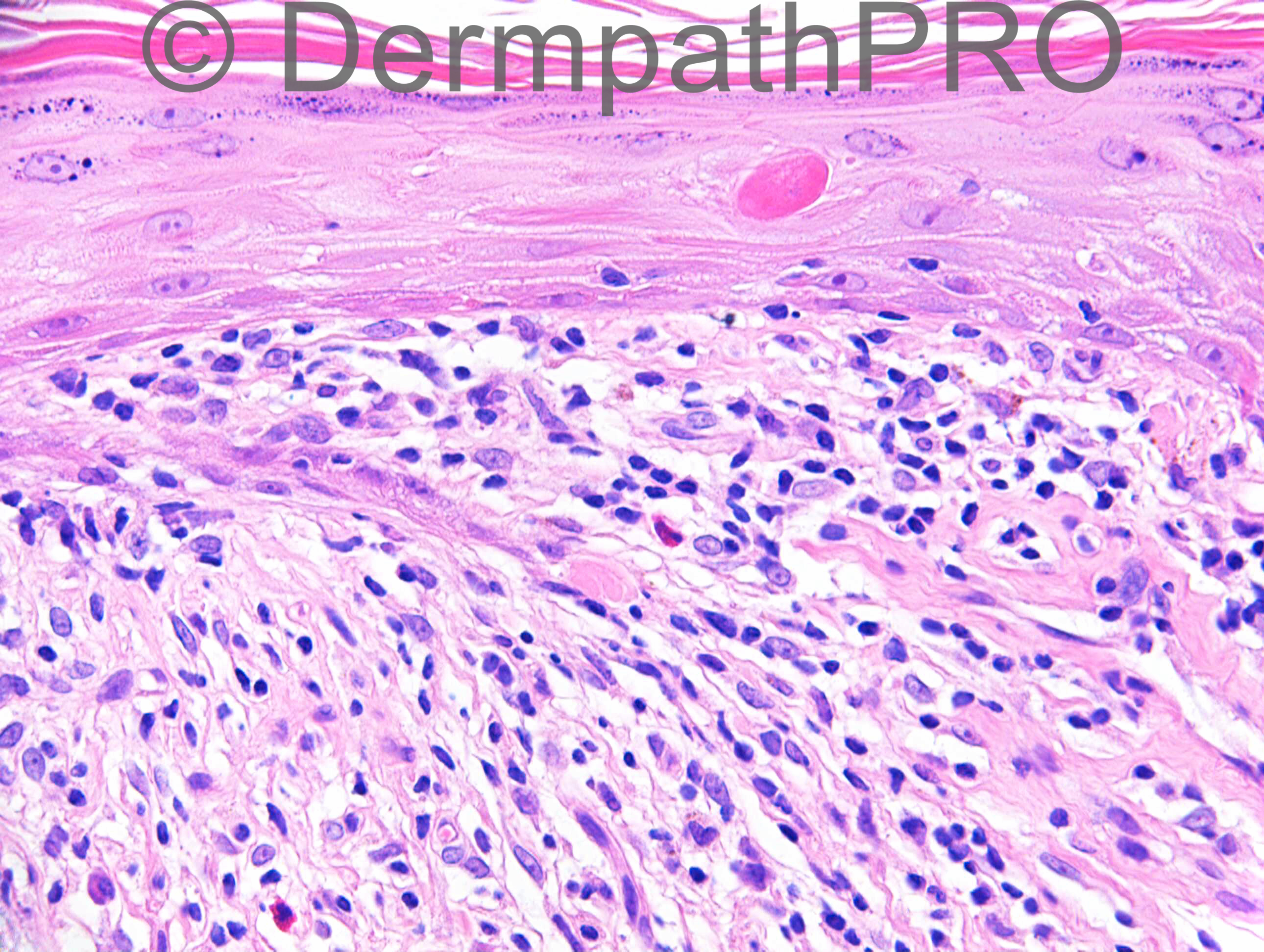
M60. Left shoulder lesion. ?Psoriasis, ?GVHD

Case posted by Dr Richard Carr


  Report Record

User Feedback


Eman El-Nabarawy

Posted

Atrophic type of lichen planus-like keratosis.

Share this comment


Link to comment
share_externally

Dr. Richard Carr

Posted

Eman the history should have read "Left shoulder biopsy. ?Psoriasis ?GVHD"

Share this comment


Link to comment
share_externally

Guest Romualdo

Posted

[u]I think this is compatible with late chronic GVHD disease.[/u]

Share this comment


Link to comment
share_externally

Arti Bakshi

Posted

This is a lichenoid inflammatory reaction with many high dyskeratotic keratinocytes.
Presumably there is a history of organ transplantation, in which case GVHD is likely. Howevere, a drug reaction will show similar features and cannot be reliably distinguished.

Share this comment


Link to comment
share_externally

I believe the main point here is trying to differentiate between chronic GVHD x Lichen planus. Although eosinophils are common in Hypertrophic lichen planus this is not the case. Plasma cells are also conspicuous in this case. So I think this is early chronic GVHD disease.

Share this comment


Link to comment
share_externally

Lichenoid GVHD (versus lichenoid drug reaction).

Share this comment


Link to comment
share_externally

Dr. Hafeez Diwan

Posted

Lichenoid GVHD, given the clinical.

Share this comment


Link to comment
share_externally

Dr. Richard Carr

Posted

I agree and favoured early chronic GVHD.
My report as follows.
Marked band-like lichenoid inflammation with complete loss of the basal layer (squamatisation) and some Civatte bodies (necrotic intra-epideral keratinocytes) above the basal layer. Sparse eosinophils only.
Histologically there is some resemblance to lichen planus but Weedon's text states that early chronic GVHD can have these appearaces. The histological differential includes a lichenoid drug eruption and clinical correlation is clearly essential.
Enjoy your weekends.

Share this comment


Link to comment
share_externally


Join the conversation

You can post now and register later. If you have an account, sign in now to post with your account.

Guest
Add a comment...

×   Pasted as rich text.   Paste as plain text instead

  Only 75 emoji are allowed.

×   Your link has been automatically embedded.   Display as a link instead

×   Your previous content has been restored.   Clear editor

×   You cannot paste images directly. Upload or insert images from URL.

Loading...

×
×
  • Create New...