Jump to content
In this section we have spot diagnoses posted on a daily basis since June 2010, now over 4000! You can review the archived cases and read the suggested diagnoses by users and the final comment by the contributors.
Case are uploaded each week day by 10 am UK time with the correct diagnosis will generally be posted at 8 pm UK time. Why not view the most recent spot diagnosis and proffer a diagnosis?

Case Number : Case 1264 - 28 April Posted By: Guest

Please read the clinical history and view the images by clicking on them before you proffer your diagnosis.
Submitted Date :
   (0 reviews)

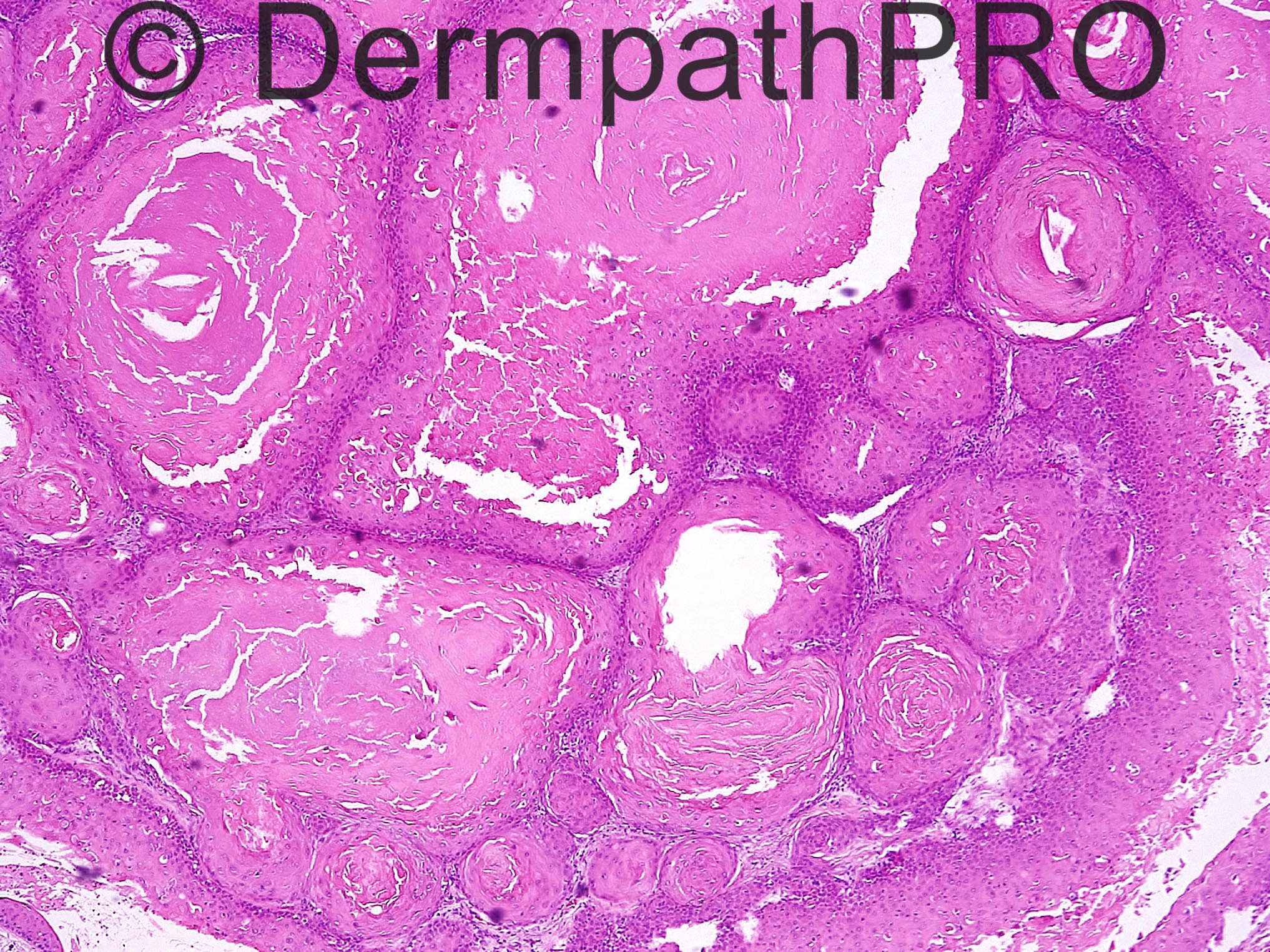
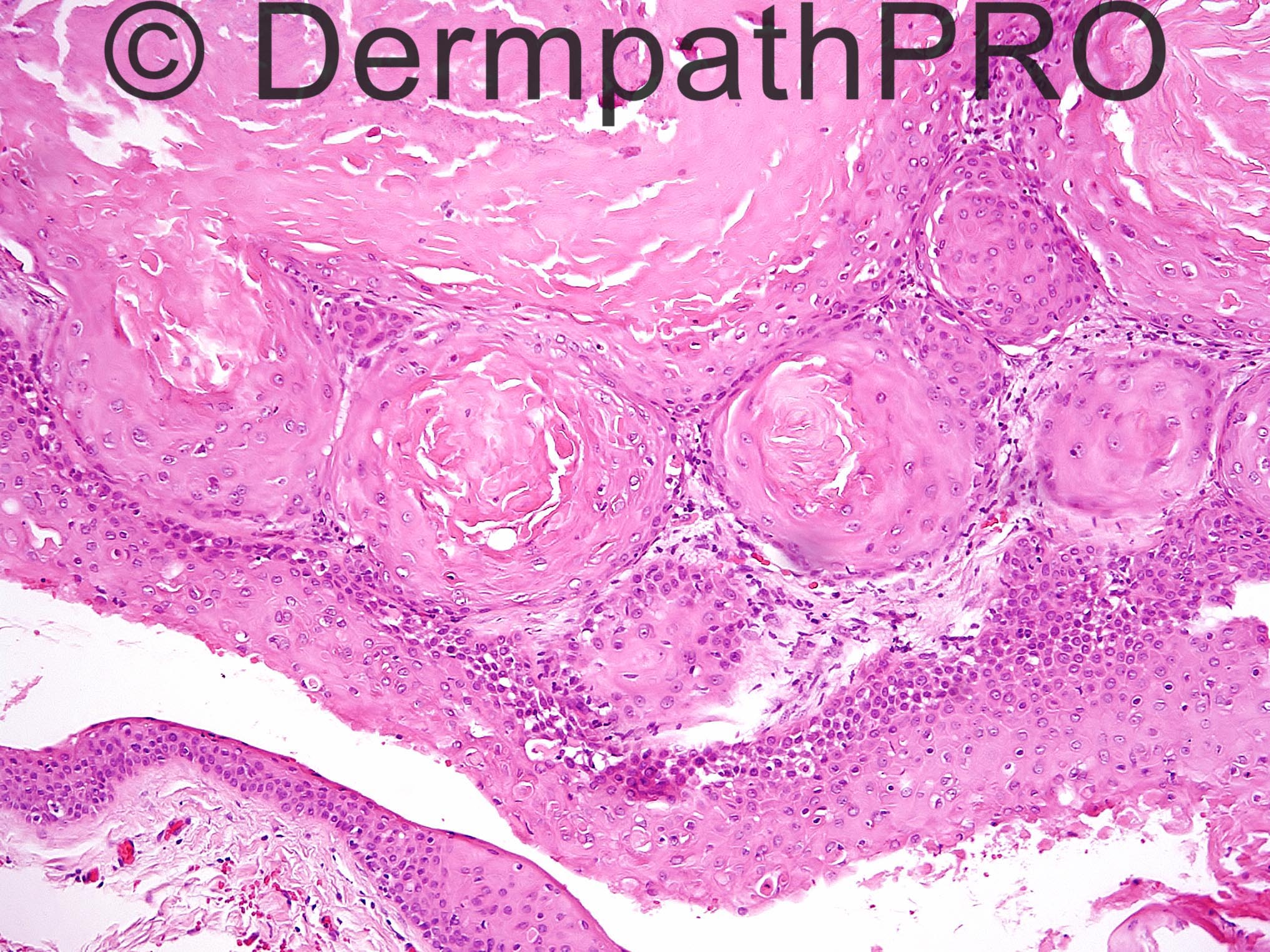
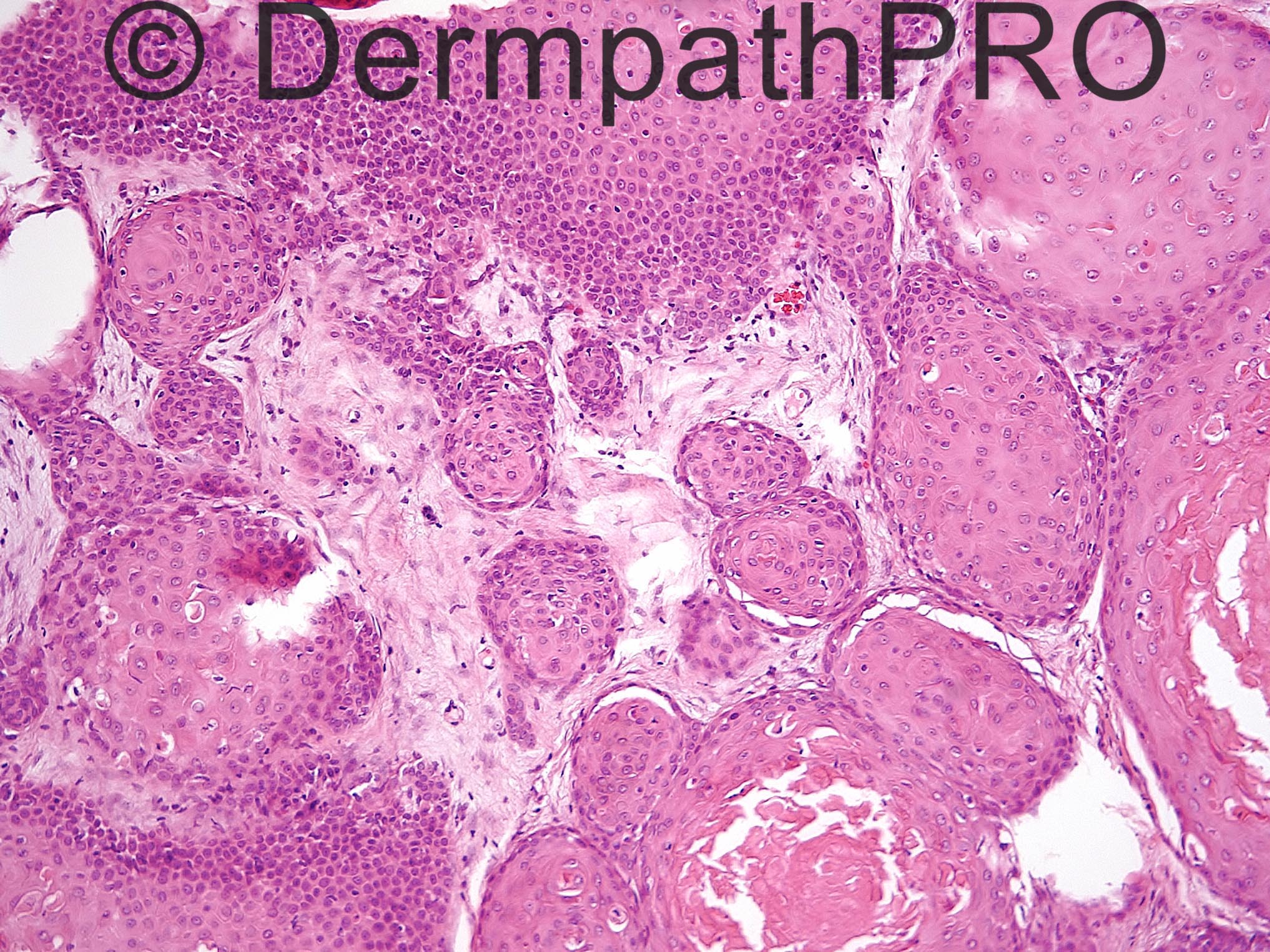
60 year old man with long standing scalp tumor and sudden recent growth.

Case posted by Dr Uma Sundram


  Report Record

User Feedback


Guest Romualdo

Posted

This tumor shows trichilemmal (pilar) type of keratinization so my diagnosis is proliferating trichilemmal (pilar) cyst/ tumor.

Share this comment


Link to comment
share_externally

Dr. Mona Abdel-Halim

Posted

Proliferating trichilemmal cyst

Share this comment


Link to comment
share_externally

Uma Sundram

Posted

I called this a proliferating tricholemmal tumor with the caveat that morphology is not an accurate predictor of clinical outcome and a recommendation that this be treated as if it were a carcinoma. I completely agree with Dr. Hurt that calling this tumor a carcinoma is a perfectly valid approach as well.

Share this comment


Link to comment
share_externally

Dr. Richard Carr

Posted

Not surprisingly in this case I would suggest it looks benign and it will almost certainly behave so but local recurrence is a possibility. In the exceedingly unlikely event of a metastasis from this lesion it would not prove malignancy as the operator may have pushed tumour it into vessels during the procedure (assuming the lesion has not regrown and then undergone malignant transformation). Not sure I would be advising a re-excision as it may be shelled-out completely. I confess I have not read the original papers regarding malignant behavior or proliferating pilar tumours that are nicely circumscribed, appear cytologically bland and were adequately sampled (I do note the history might be a concern in this case but surely we should see some morphological evidence to support a diagnosis of carcinoma).

Share this comment


Link to comment
share_externally


Join the conversation

You can post now and register later. If you have an account, sign in now to post with your account.

Guest
Add a comment...

×   Pasted as rich text.   Paste as plain text instead

  Only 75 emoji are allowed.

×   Your link has been automatically embedded.   Display as a link instead

×   Your previous content has been restored.   Clear editor

×   You cannot paste images directly. Upload or insert images from URL.

Loading...

×
×
  • Create New...