Jump to content
In this section we have spot diagnoses posted on a daily basis since June 2010, now over 4000! You can review the archived cases and read the suggested diagnoses by users and the final comment by the contributors.
Case are uploaded each week day by 10 am UK time with the correct diagnosis will generally be posted at 8 pm UK time. Why not view the most recent spot diagnosis and proffer a diagnosis?

Case Number : Case 1343 - 17 August Posted By: Guest

Please read the clinical history and view the images by clicking on them before you proffer your diagnosis.
Submitted Date :
   (0 reviews)

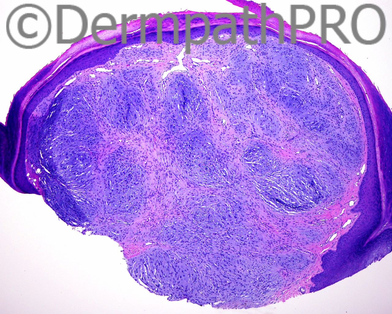
The patient is a 32 year old woman with a shave biopsy of a changing lesion on the left shoulder. Clinical Diagnosis: melanocytic nevus

Case posted by Dr Mark Hurt


  Report Record

User Feedback


Arti Bakshi

Posted

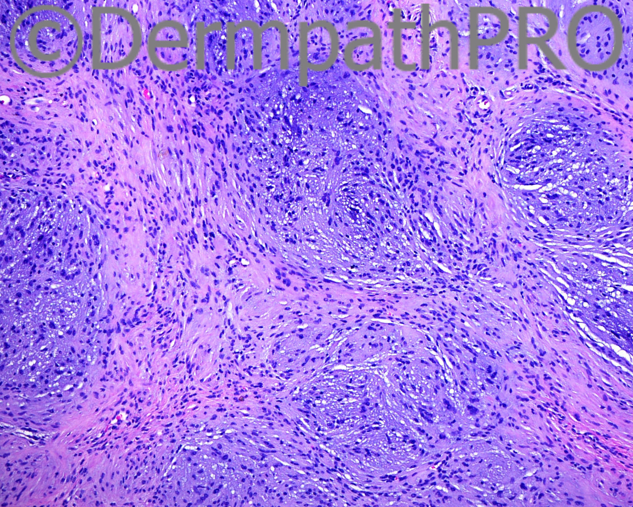
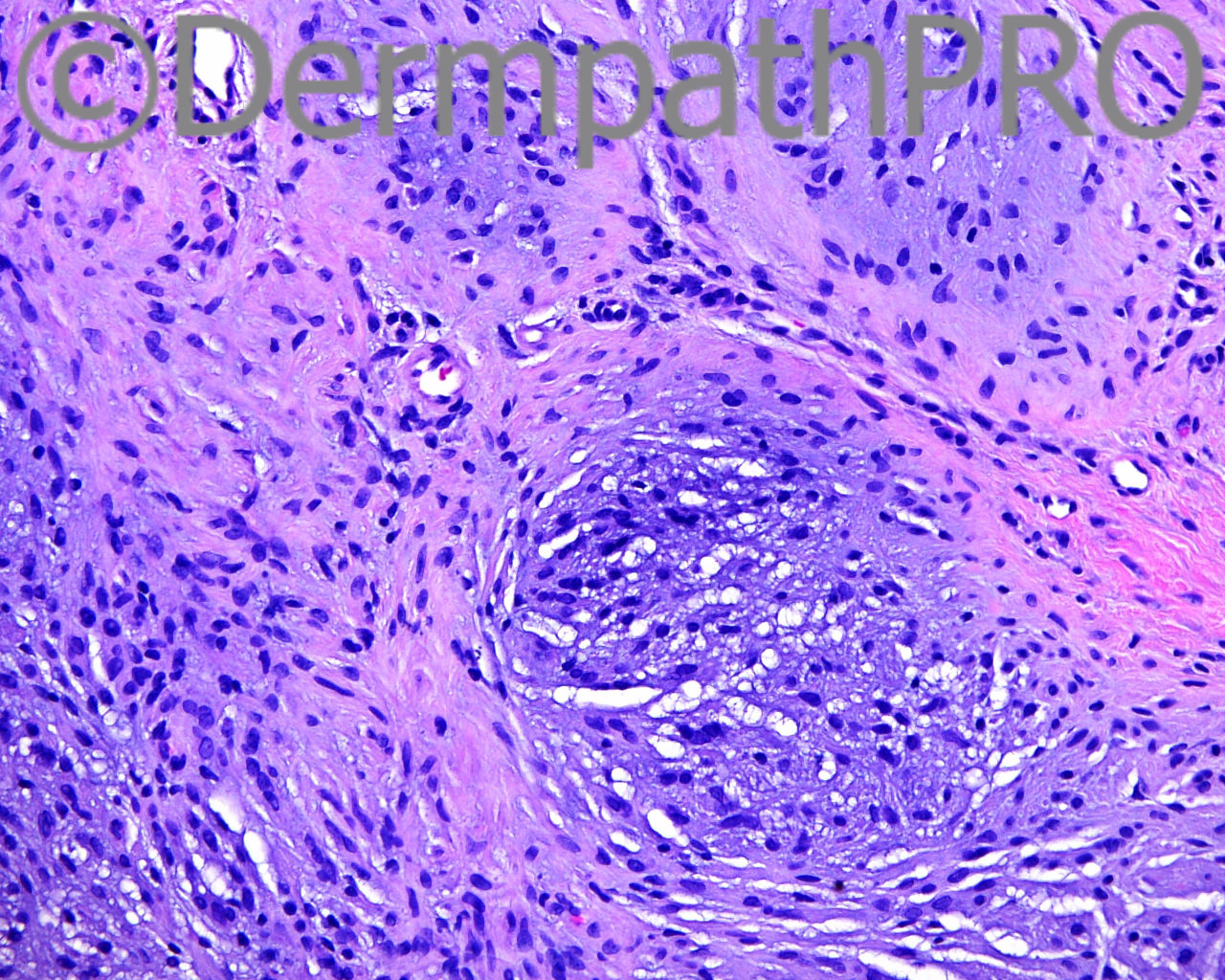
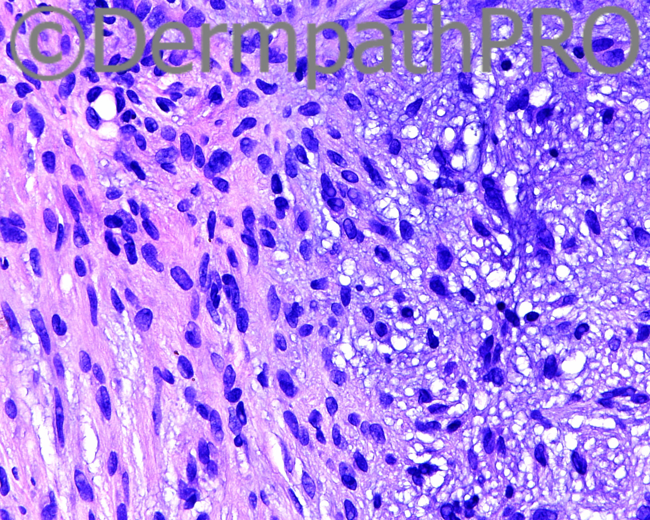
This lesion has a biphasic appearance with peripheral spindle cells with eosinophilic cytoplasm and central nodular areas with basophilic cells in a myxoid/?chondroid matrx.
?Solitary Myofibroma

Share this comment


Link to comment
share_externally

Dr. Mona Abdel-Halim

Posted

Nice suggestion of myofibroma. I did not think of it, I thought of cellular neurothekoma with prominent myxoid stroma. The clinical impression of a melanocytic nevus does not fit with the usual clinical presentation of solitary myofibromas. I am not expert in this area and I am looking forward to the correct diagnosis.

Share this comment


Link to comment
share_externally

Mark A. Hurt MD

Posted

Here is my microscopic description:

There is a circumscribed lesion that contains small epicenters of somewhat myxoid areas that have epithelioid cells within them, and in the "fibrous" zones between the myxoid areas, the cells are slightly smaller. They are all uniform throughout the entire field however. There is no nuclear pleomorphism in the lesion. Immunostains show that this lesion is strongly positive with S100 protein in nuclei and cytoplasm of the small cluster-like zones, while CD63 is weakly positive. Melan-A, neuron specific enolase, D2-40, EMA, actin, CD10, and CD68 are all negative or very diminished. Ki67 (clone 30-9) has an index of less than 1% positive in my opinion.

Share this comment


Link to comment
share_externally

Mark A. Hurt MD

Posted

And here is my diagnosis:

[b]-- NERVE SHEATH MYXOMA[/b]

Share this comment


Link to comment
share_externally

Arti Bakshi

Posted

Very difficult case today! Diffuse S100 obviously points to a neural tumour and rules out the other possibilities considered. But Dr Hurt, isn't the tumour too cellular for a nerve sheath myxoma? Would you consider a variant of dermal schwannoma (plexiform/epithelioid) in the differential ??

Share this comment


Link to comment
share_externally

Mark A. Hurt MD

Posted

Arti, I see your point, and perhaps you're right.

Share this comment


Link to comment
share_externally


Join the conversation

You can post now and register later. If you have an account, sign in now to post with your account.

Guest
Add a comment...

×   Pasted as rich text.   Paste as plain text instead

  Only 75 emoji are allowed.

×   Your link has been automatically embedded.   Display as a link instead

×   Your previous content has been restored.   Clear editor

×   You cannot paste images directly. Upload or insert images from URL.

Loading...

×
×
  • Create New...