Jump to content
In this section we have spot diagnoses posted on a daily basis since June 2010, now over 4000! You can review the archived cases and read the suggested diagnoses by users and the final comment by the contributors.
Case are uploaded each week day by 10 am UK time with the correct diagnosis will generally be posted at 8 pm UK time. Why not view the most recent spot diagnosis and proffer a diagnosis?

Case Number : Case 1427- 11 December Posted By: Guest

Please read the clinical history and view the images by clicking on them before you proffer your diagnosis.
Submitted Date :
   (0 reviews)

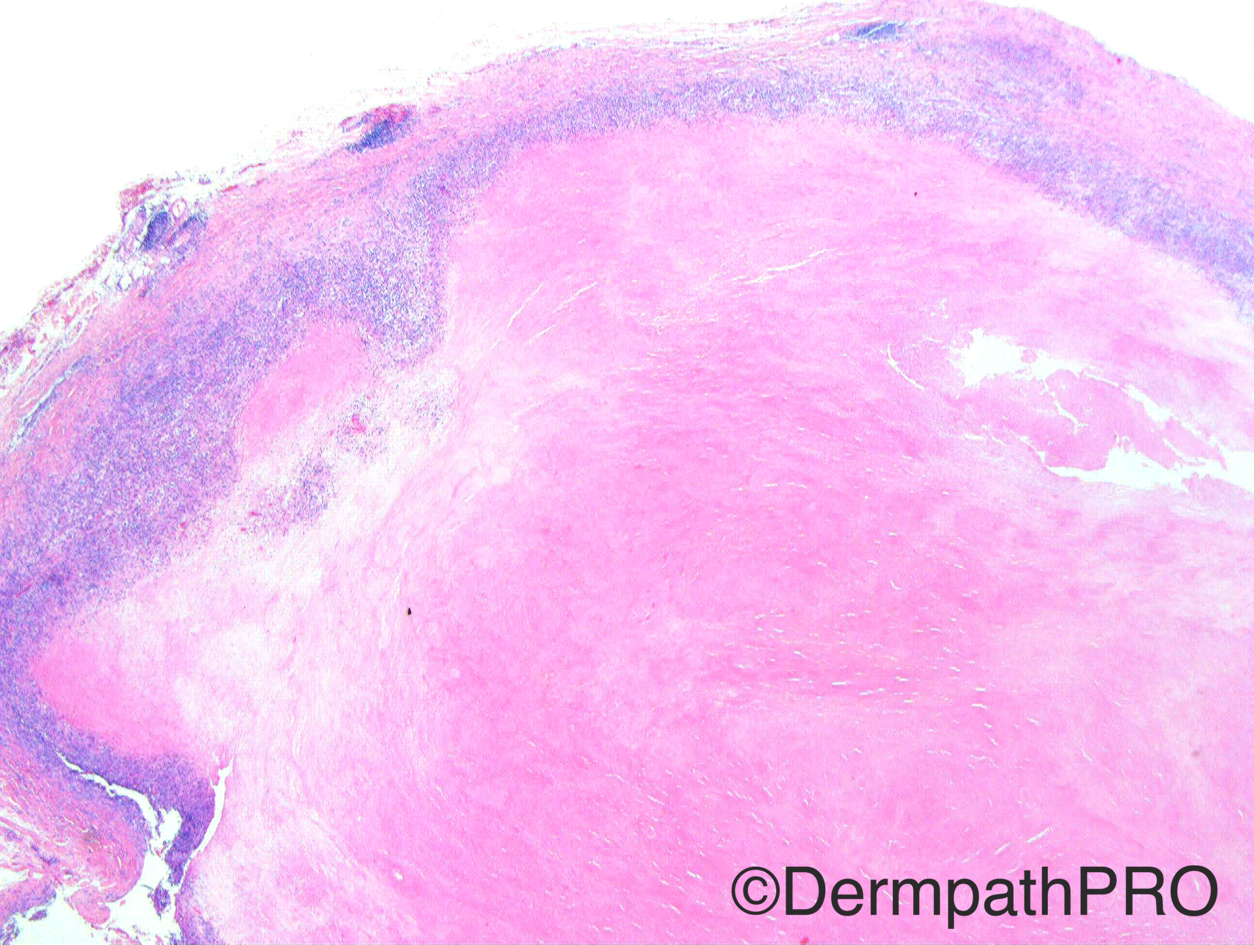
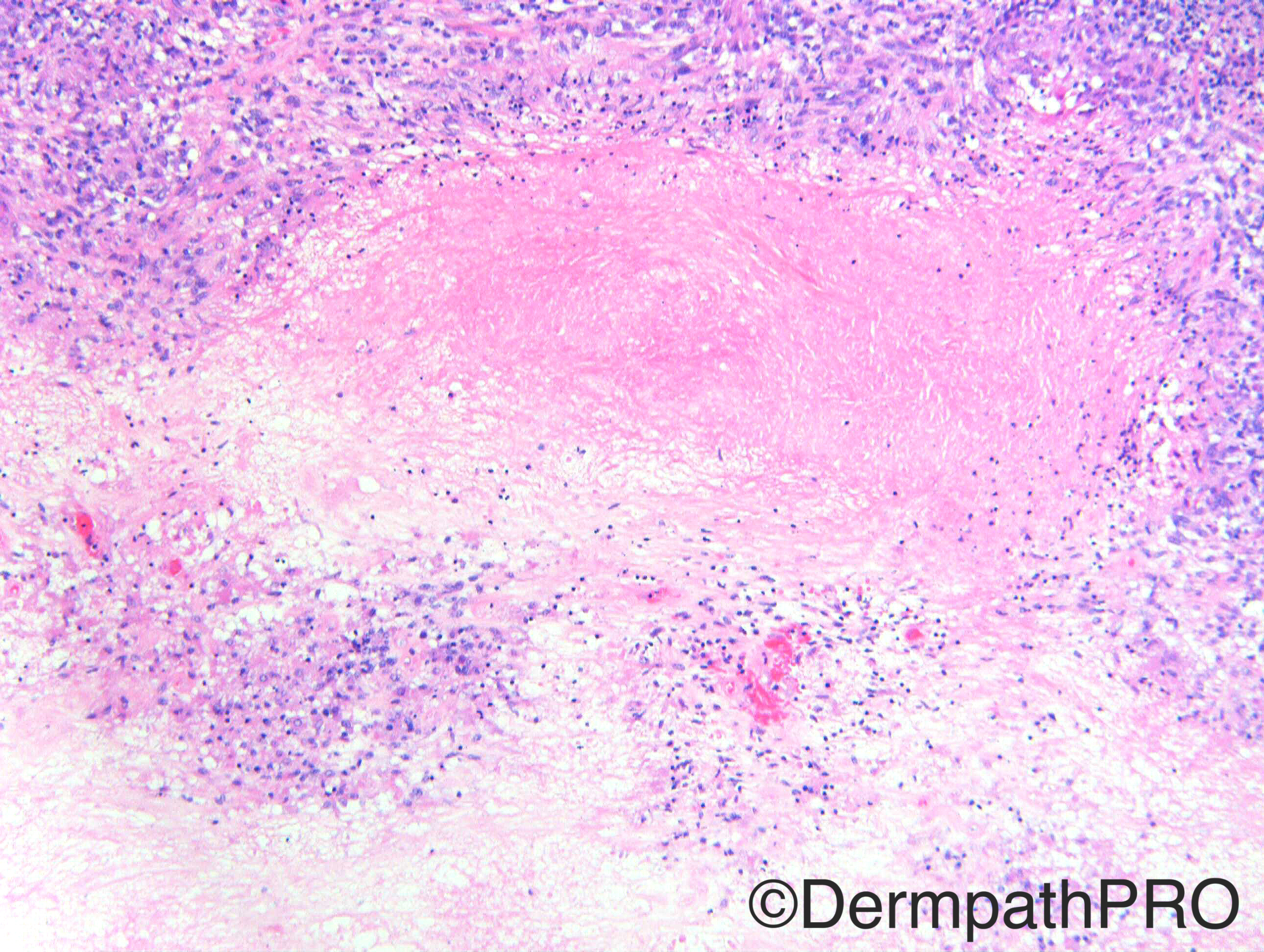
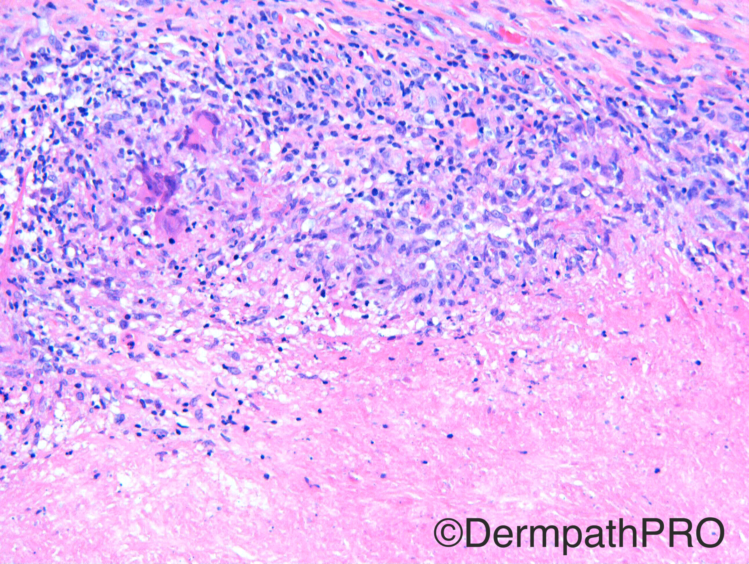
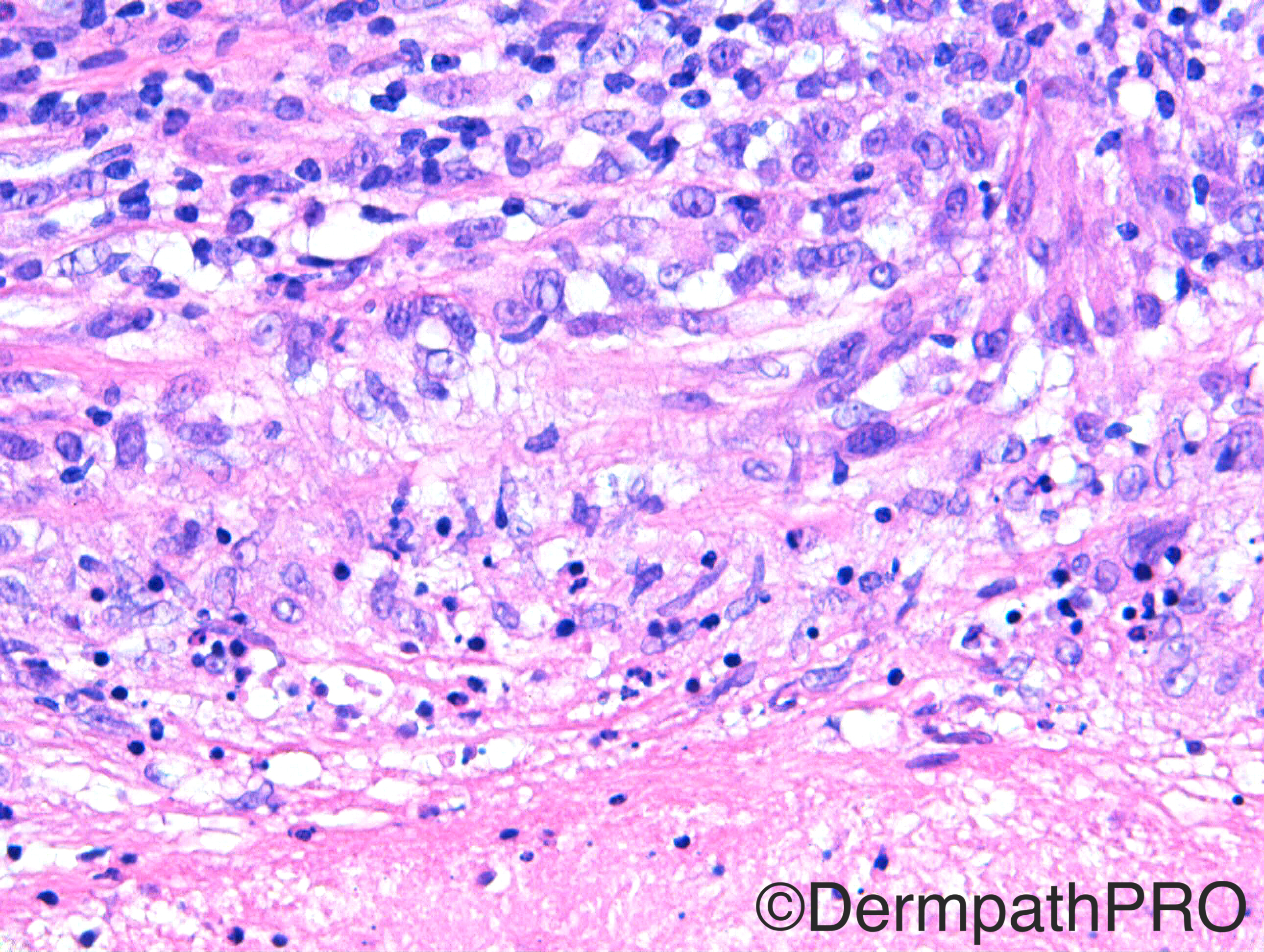
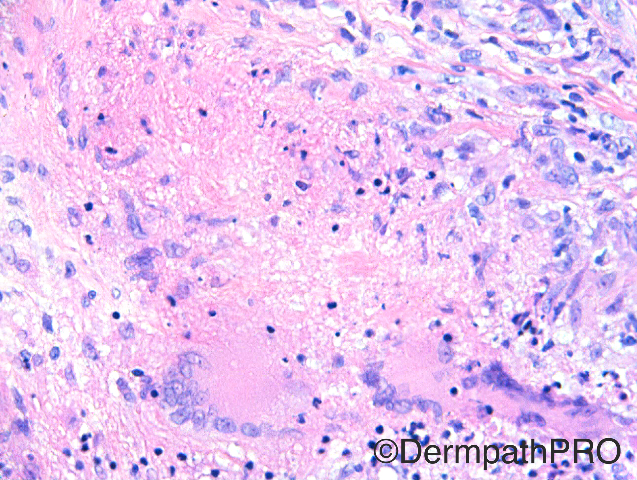
Case History: F45. Elbow. Hard slow growing lump ?Lymph node, ?cyst, ?granuloma ?neoplastic

Case posted by Dr Richard Carr


  Report Record

User Feedback


I see a deep granuloma composed by histiocytes with some palisading and giant cells. The necrotic center in some pictures, like picture 3, reminds me the fibrinoid type. My first hypothesis is Rheumatoid nodule. Special stains for microorganisms are important.

Share this comment


Link to comment
share_externally

Dr. Mona Abdel-Halim

Posted

As Igor said, my first thought is rheumatoid nodule

Share this comment


Link to comment
share_externally

vincenzo polizzi

Posted

Extensive fibrinoid necrosis with peripheral lymphocytic, histolytic and giant cell infiltration. In second image there is a necrotising vasculitis. I need some clinical correlation, thinking, first of all, to a Wegener Granulomatosis...

Share this comment


Link to comment
share_externally

Arti Bakshi

Posted

Agree with rheumatoid nodule (wonder whats the catch??!!)

Share this comment


Link to comment
share_externally

Sasi Attili

Posted

Loong time since I have managed to visit the site. Hopefully will be more regular from now. R.Nodule is my first thought too!

Share this comment


Link to comment
share_externally

Raul Perret

Posted

agree with rheumatoid nodule but as Igor said would make special stains before signing out

Share this comment


Link to comment
share_externally

Guest Romualdo

Posted

Agree with rheumatoid nodule, after exclusion of infections.

Share this comment


Link to comment
share_externally

Dr. Richard Carr

Posted

Seems I got you all again!!


I had met the patient at a regional meeting in 2009 long-standing history of nodules with crusts, unilateral overlying elbow.  She was otherwise well and the case had been presented for ?diagnosis.  ?GA, ?Atypical mycobacterial, ?fungal, ?Other.  Based on images presented at the meeting I had suggested perforating granuloma annulare and heard no more until...


She was re-biopsied in 2014 with the history supplied to you. 

The lesions have persisted and due to clinical concern (for a neoplasm etc) I was invited to see the sections this year. 


My report as follows:

Reviewing the original histology, this does have features of circumscript areas of distinct collagen necrobiosis with palisading granulomatous reaction and a surface transepidermal elimination via follicles.  All these features are typical of granuloma annulare. 

The more recent histology is a rather striking deep, dermal / subcutaneous necrobiotic nodule, in keeping with the subcutaneous variant of granuloma annulare (pseudo-rheumatoid nodule).  The lack of neutrophilic infiltrate is certainly against any infective condition.  We ascertained that there was a case series of adults with juxta-articular pseudo-rheumatoid nodules, these usually are in relation to the joints of the hands.  I am uncertain if cases have been reported related to the elbow.


This case prompted me to review the prior dermpathpro cases:

https://dermpathpro.com/spot-diagnosis-1/_/2015-spot-diagnoses/august-2015/case-1334-04-august-r1388


https://dermpathpro.com/spot-diagnosis-1/_/2014-spot-diagnoses/november-2014/case-1150-19th-november-r1194


https://dermpathpro.com/spot-diagnosis-1/_/2014-spot-diagnoses/january-2014/case-925-8th-january-r963


And also to review cases from my slide collection and photographed cases from the UK National skin EQA.

Turns out I have seen another case of superficial GA on the unilateral elbow previously with similar histology to this patients biopsy from 2009 (striking red necrobiotic foci, fairly closely opposed by distinct areas without muicin) and I had contacted the general practioner who confirmed patient was otherwise well as I had considered rheumatoid.  Case P270 from the UK national EQA was considered to be a rheumatoid nodule (it had plenty of mucin).  Uma's case 1134 looks like GA with mucin but lesions were bilateral and patient had rheumatoid and you all agreed rheumatoid nodule.  So my conclusion is you can have both strikingly red palisaded GA (+/- pseudorheumatoid nodules) and Rheumatoid nodules with and without mucin and we need clinico-pathological correlation.  It appears you can have pseudorheumatoid (GA) nodules on the unilateral elbow.  If anyone can supply other cases may be worth publishing them.

Share this comment


Link to comment
share_externally

Dr. Richard Carr

Posted

Reference:

Am J Dermatopathol. 2005 Feb;27(1):1-5.
Pseudorheumatoid nodules in adults: a juxta-articular form of nodular granuloma annulare.
Abstract

Pseudorheumatoid nodules are considered a deep form of granuloma annulare. Most cases are described in children, occur mainly on the lower legs and scalp, and have favorable prognosis. Their appearance in adults is rare. In this series, fourteen women with pseudorheumatoid nodules were studied. The average age of onset was 36 years old. Lesions consisted of erythematous, violaceous, or skin-colored nodules located mainly on the small joints of the hands. None of the patients developed collagen vascular disease. Persistence was common. Biopsy specimens showed deep dermal nodules composed of epithelioid granulomata separated by thickened collagen bundles. In some areas eosinophilic material was surrounded by histiocytes in a palisaded array. Granuloma annulare was present at the periphery of eight cases. Special stains revealed that most of the eosinophilic material was collagen and mucin was present in eleven cases. In sum these findings demonstrate that pseudorheumatoid nodules in adults are a distinct clinical and pathologic entity, which may be mistaken for rheumatoid nodules. They are probably a juxta-articular variant of granuloma annulare.

Share this comment


Link to comment
share_externally


Join the conversation

You can post now and register later. If you have an account, sign in now to post with your account.

Guest
Add a comment...

×   Pasted as rich text.   Paste as plain text instead

  Only 75 emoji are allowed.

×   Your link has been automatically embedded.   Display as a link instead

×   Your previous content has been restored.   Clear editor

×   You cannot paste images directly. Upload or insert images from URL.

Loading...

×
×
  • Create New...