Jump to content
In this section we have spot diagnoses posted on a daily basis since June 2010, now over 4000! You can review the archived cases and read the suggested diagnoses by users and the final comment by the contributors.
Case are uploaded each week day by 10 am UK time with the correct diagnosis will generally be posted at 8 pm UK time. Why not view the most recent spot diagnosis and proffer a diagnosis?

Case Number : Case 1428- 14 December Posted By: Guest

Please read the clinical history and view the images by clicking on them before you proffer your diagnosis.
Submitted Date :
   (0 reviews)

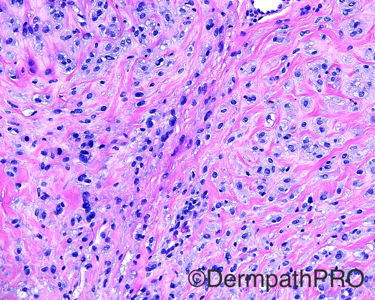
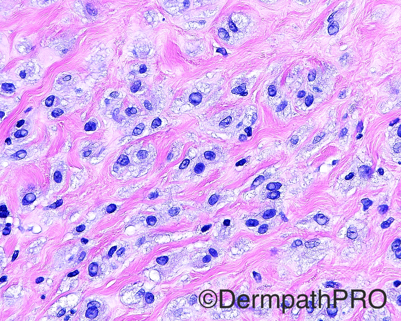
Case History: The patient is a 77 year old man with a shave biopsy of a pearly telangiectatic papule on the left nasal sidewall.

Case posted by Dr Mark Hurt


  Report Record

User Feedback


I think this is some kind of morfologyc variant of intradermal melanocytic nevus with pseudo-physalipherous cells. In a younger patient my first hypothesis would be intradermal Spitz nevus.

Share this comment


Link to comment
share_externally

Raul Perret

Posted

Me I was thinking more of a neuroid variant of fibrous papule. I agree that the nuclei and architecture of cells makes think of nevus. Would consider doing s100, factor XIIIa and cd34 

Share this comment


Link to comment
share_externally

Dr. Mona Abdel-Halim

Posted

I think this is just a fibrous papule of the nose with the rare feature mentioned in Weedon's Skin Pathology in which there are numerous fibroblasts/histiocytes/dendrocytes with clear vacuolated cytoplasm embedded in dense sclerotic and hyalinized stroma. The clinical fits very well.

Share this comment


Link to comment
share_externally

Nitin Khirwadkar

Posted

Would favour a fibrous papule. Would do S-100 to rule out a naevus variant, and a cytokeratin.

Share this comment


Link to comment
share_externally

Guest Romualdo

Posted

Agree with Raul and Mona: clear cell variant of fibrous papule of the nose.

Share this comment


Link to comment
share_externally

Agree with fibrous papule with clear cells.  I think Dr Hurt posted a similar case a few months ago. 

Share this comment


Link to comment
share_externally

Sasi Attili

Posted

My first thought was a cellular variant of a fibrous papule. Obviously would not sign this out without immunos- S100, Fac XIIIa at least....

Share this comment


Link to comment
share_externally

Mark A. Hurt MD

Posted

One can never see too many fibrous papules, which was my diagnosis in this case.  Below are the specifics from my report:


SKIN, LEFT NASAL SIDEWALL , SHAVE BIOPSY :

-- FIBROUS PAPULE

COMMENT:  I think this is an unusual fibrous papule.  It reminded me to some degree of melanocytes or some type of neural-like cells.  I even considered the possibility of cellular neurothekeoma and chordoma.  Another possibility for consideration was PEComa.  In essence, however, all of these conditions in the differential are excluded because of one reason or another, mostly from negative staining with the various markers used to attempt to rule them in.  As a rule fibrous papules are strongly positive with Vimentin, CD63 and Factor XIIIa.  Some of them in the literature have been described as having CD34 positivity, but this one does not.  Thus, I think this is a variant on the theme of fibrous papule.


Micro:


This lesion from the left nasal sidewall, is characterized by two epicenters of a sharply circumscribed lesion containing cells that are outlined as 1-2 cellular units in small nests.  There is fibrous tissue between these cells, and the entire unit is somewhat circumscribed.  There is no nuclear pleomorphism.  The cells also fail to show any mitotic figures.  A few of them have nucleoli that are slightly off center but most of the nucleoli are relatively small.  There are some pseudoinclusions in these cells.  Immunohistochemical stains show that the cells are negative with S100 protein, negative with Melan-A, negative with p40, negative with CAM5.2, negative with EMA, negative with actin and desmin, negative with adipophilin, NSE, D240 and CD34.  The lesion is positive strongly with vimentin, CD63, and has dendritic cell positivity with Factor XIIIa.  Ki-67 shows a very low index of less than 1% positive.  

Share this comment


Link to comment
share_externally


Join the conversation

You can post now and register later. If you have an account, sign in now to post with your account.

Guest
Add a comment...

×   Pasted as rich text.   Paste as plain text instead

  Only 75 emoji are allowed.

×   Your link has been automatically embedded.   Display as a link instead

×   Your previous content has been restored.   Clear editor

×   You cannot paste images directly. Upload or insert images from URL.

Loading...

×
×
  • Create New...