Jump to content
In this section we have spot diagnoses posted on a daily basis since June 2010, now over 4000! You can review the archived cases and read the suggested diagnoses by users and the final comment by the contributors.
Case are uploaded each week day by 10 am UK time with the correct diagnosis will generally be posted at 8 pm UK time. Why not view the most recent spot diagnosis and proffer a diagnosis?

Case Number : Case 1430- 16 December Posted By: Guest

Please read the clinical history and view the images by clicking on them before you proffer your diagnosis.
Submitted Date :
   (0 reviews)

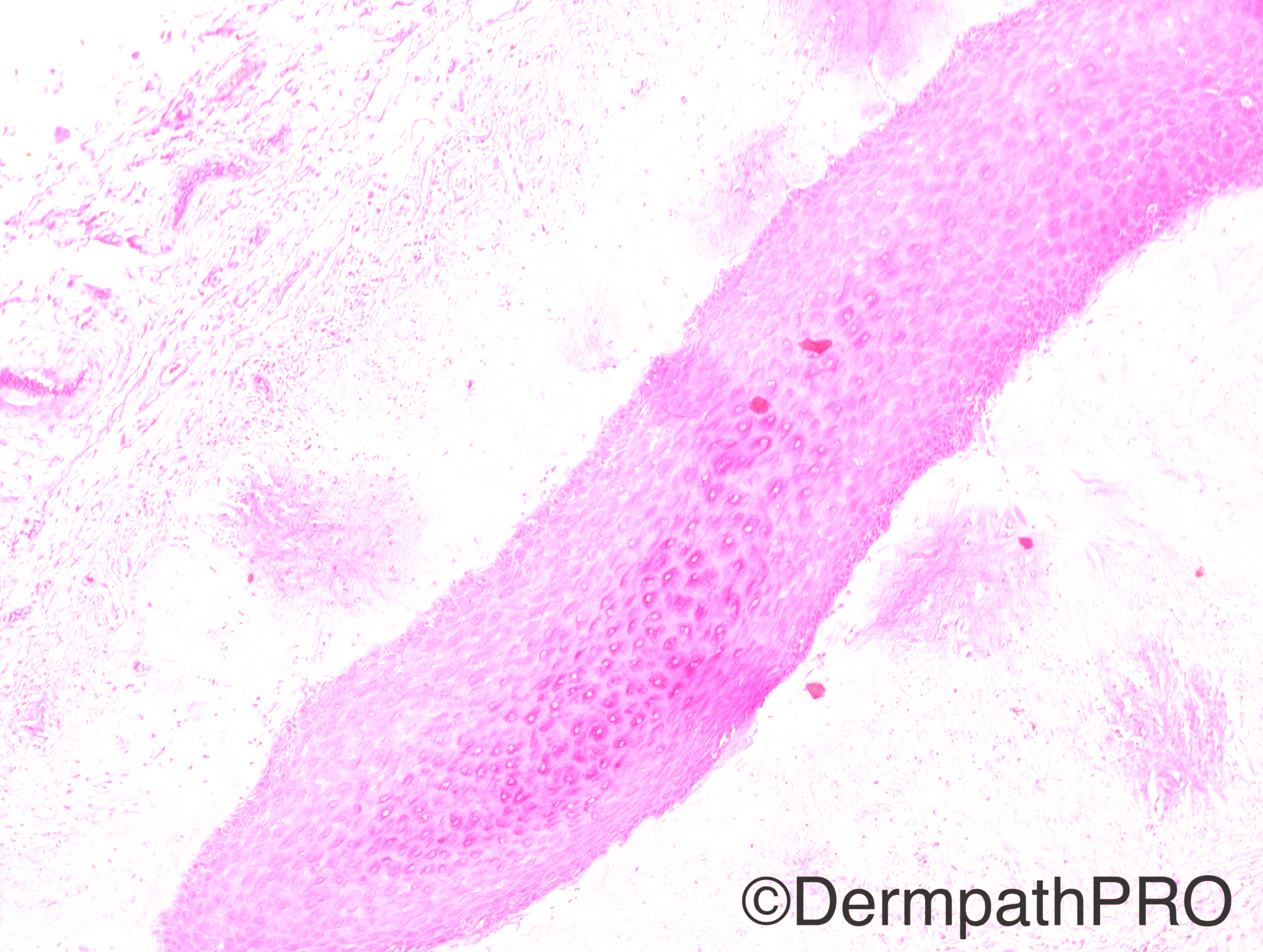
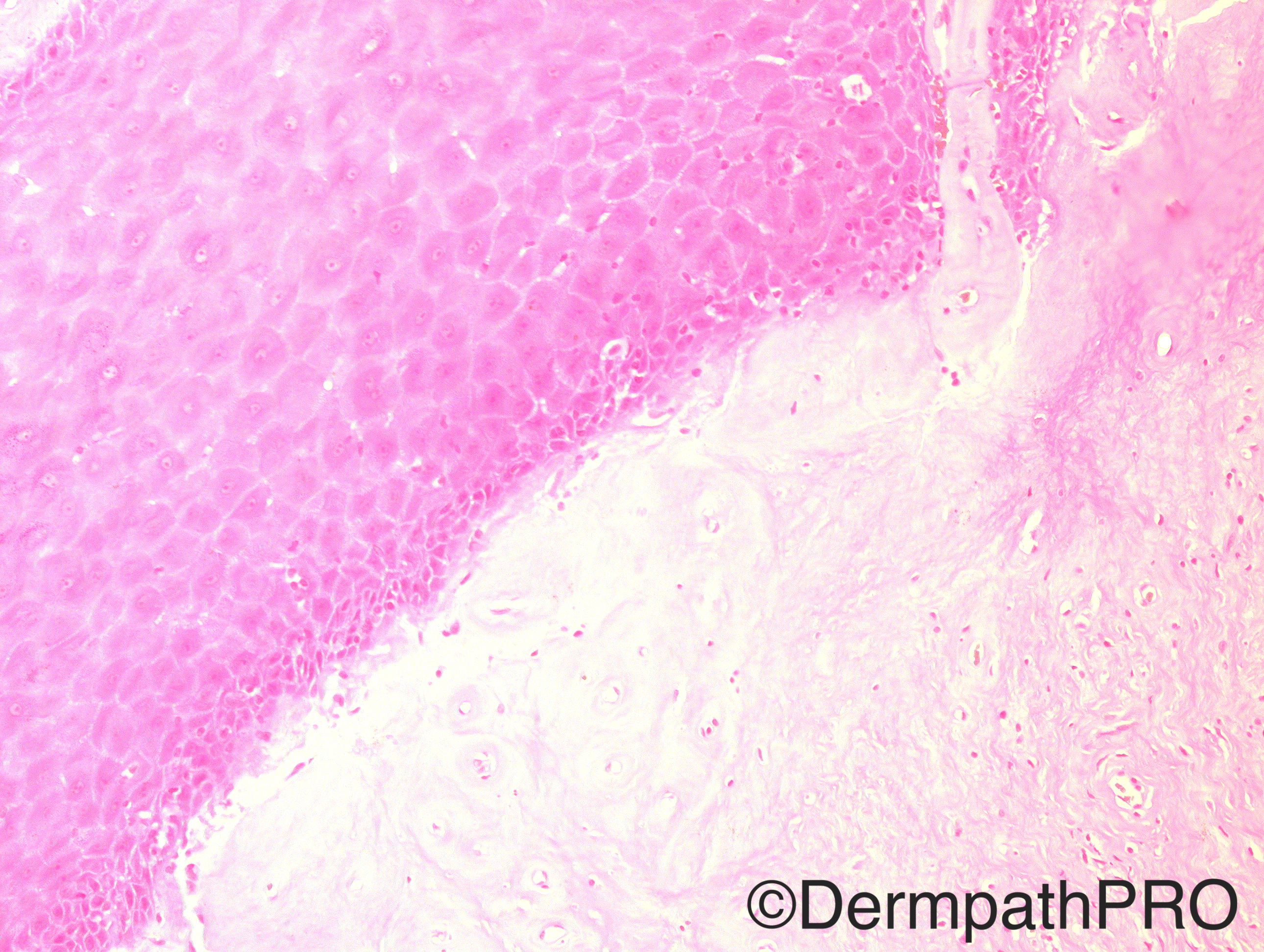
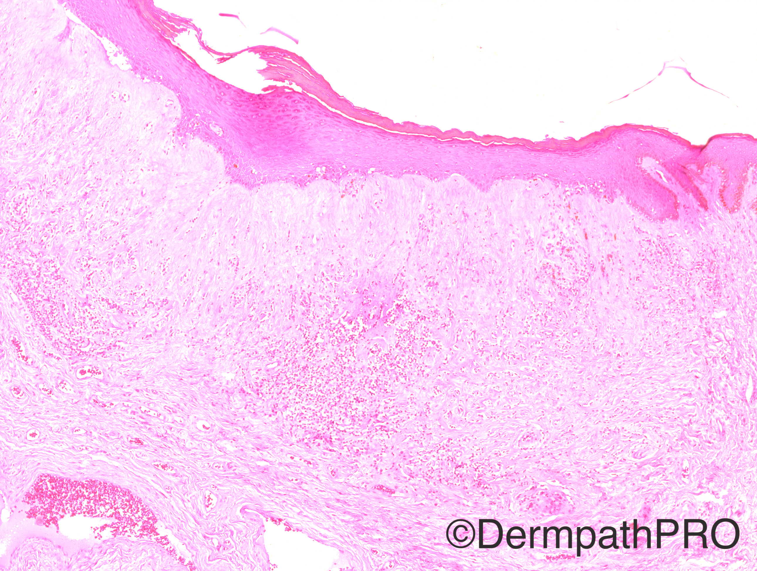
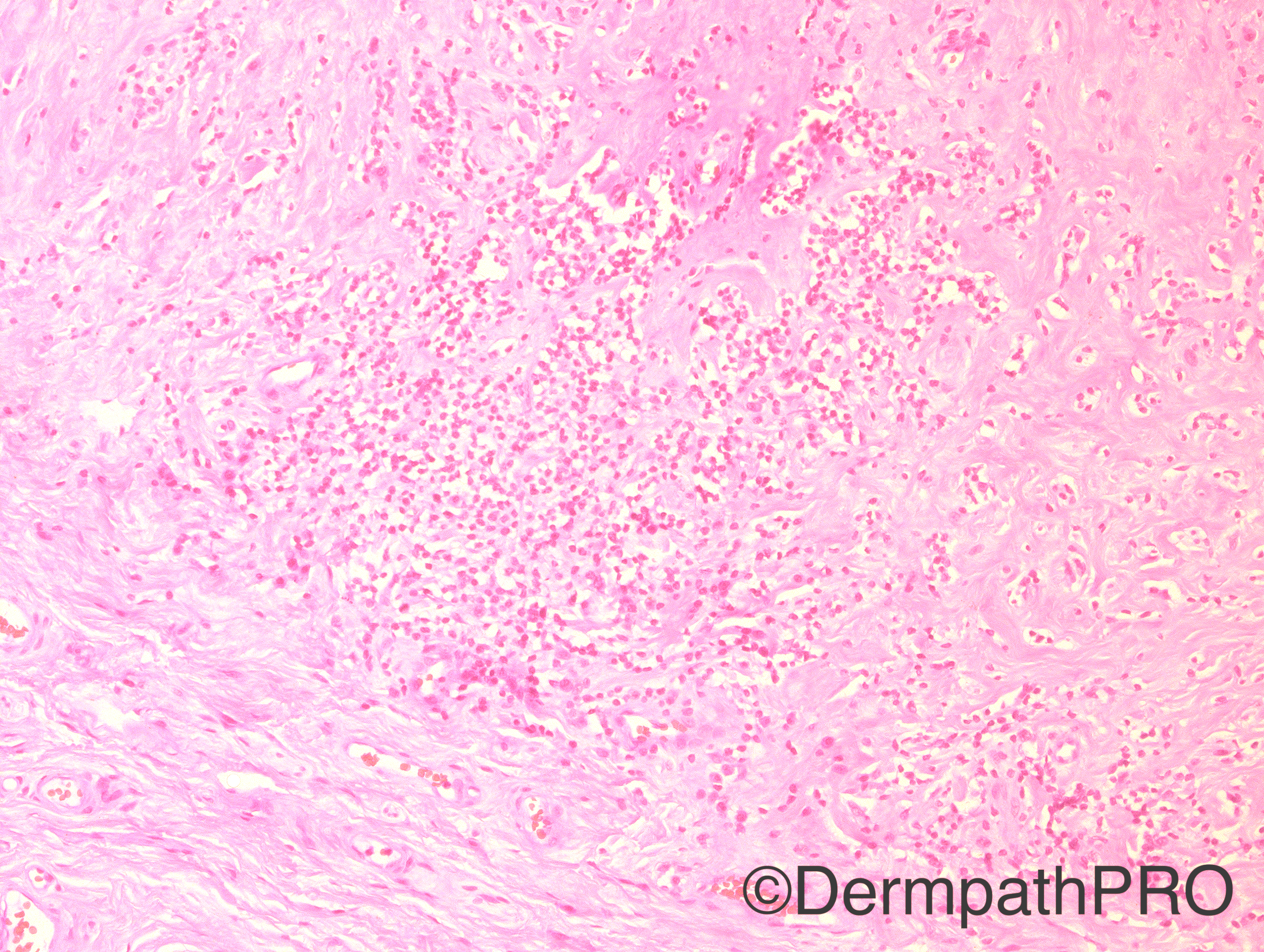
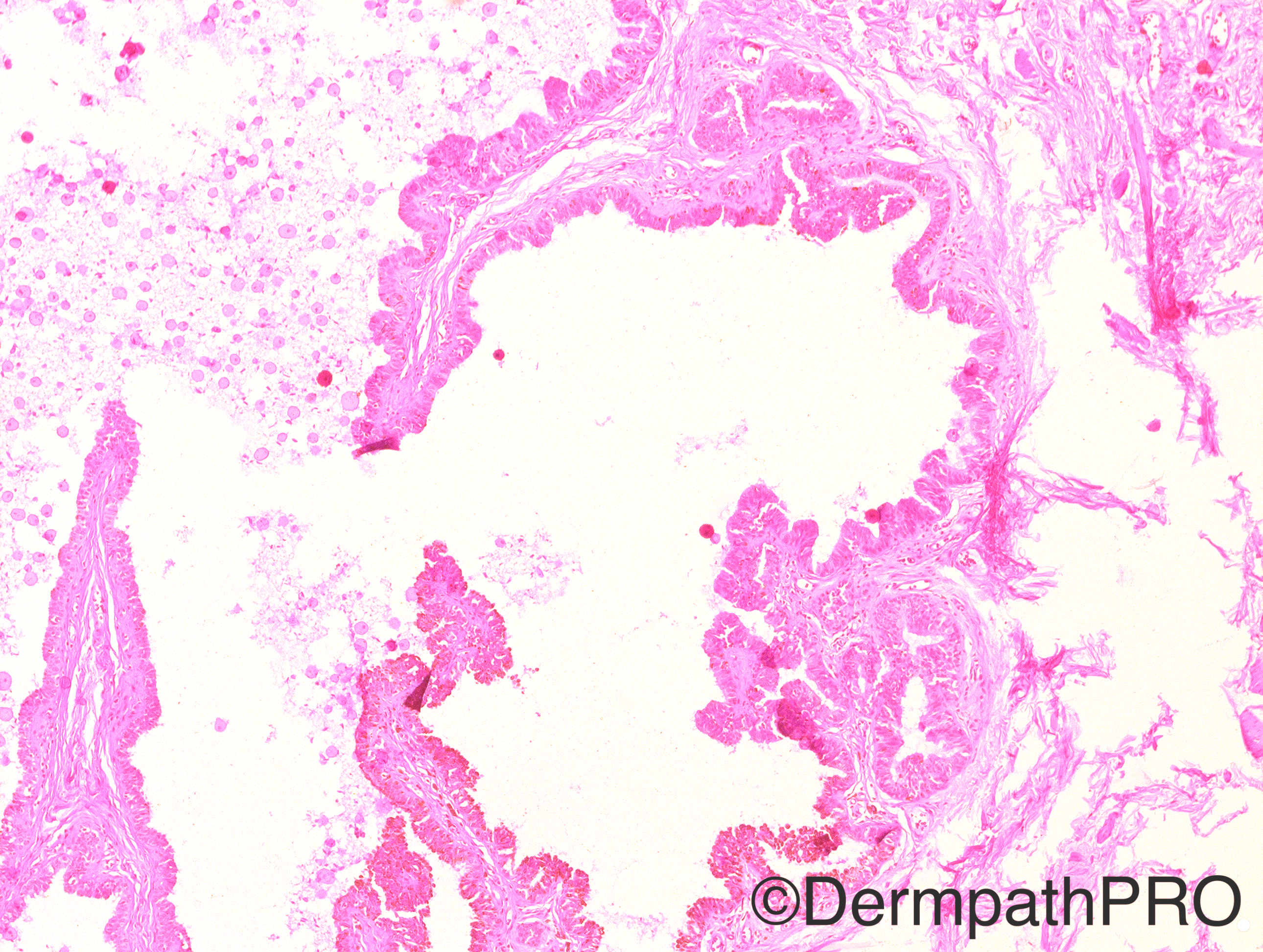
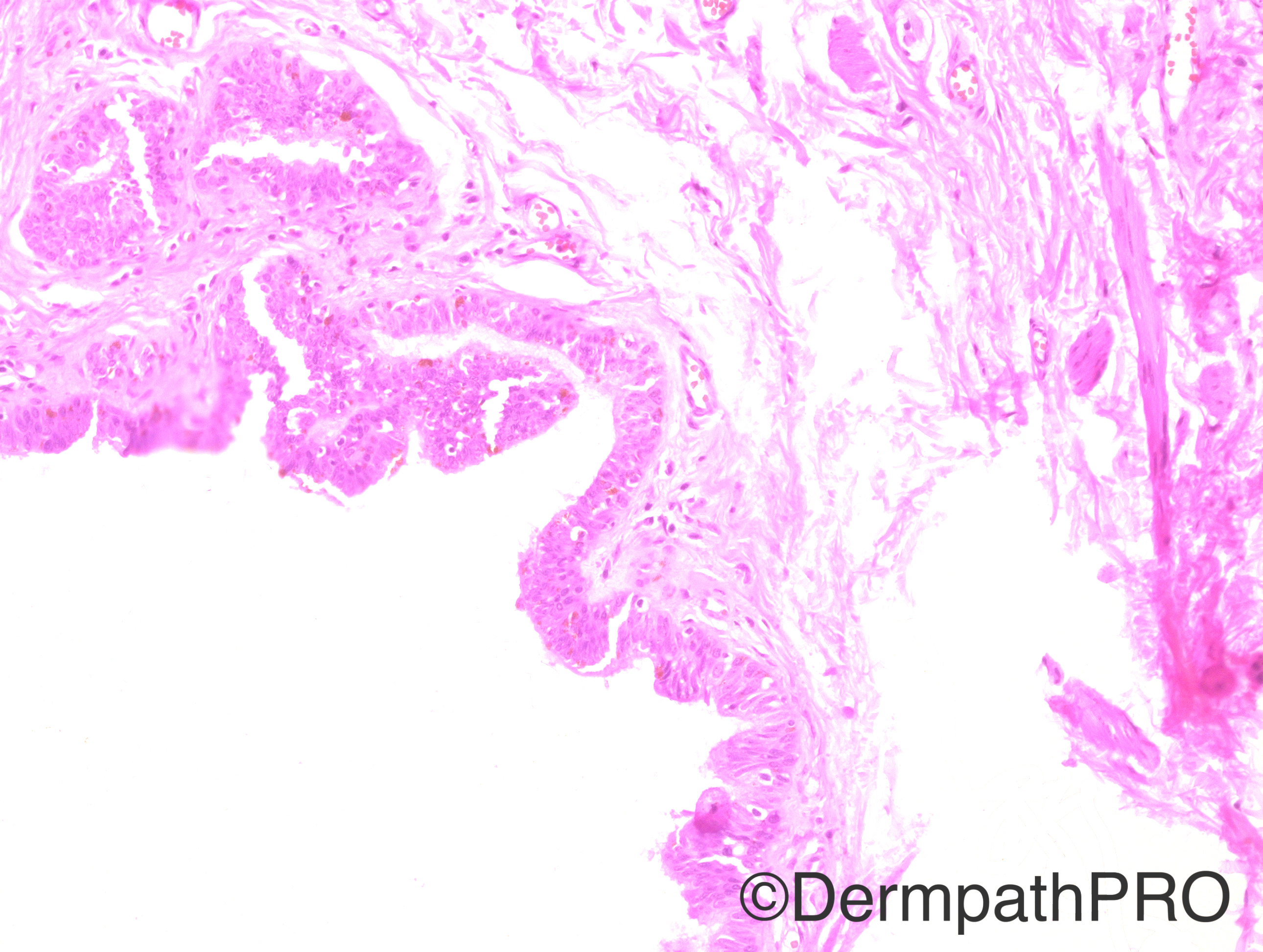
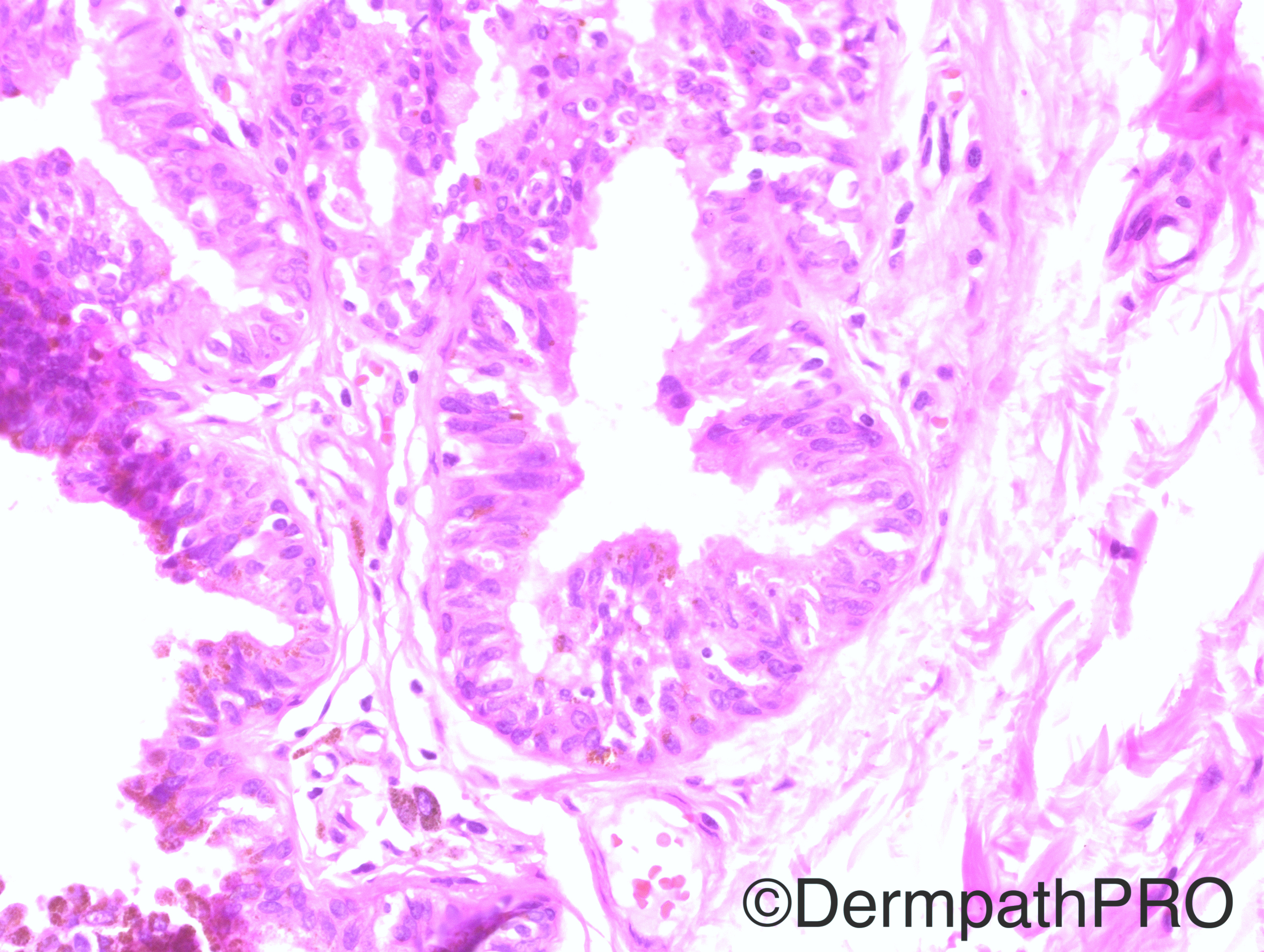
Case History: 67 year-old male who underwent circumcision for severe phimosis.

Case posted by Dr Hafeez Diwan

  Report Record

User Feedback


Raul Perret

Posted

I propose an adnexal neoplasm arising on a contex of balanitis xerotica obliterans. It looks like a papillary eccrine neoplasm with intracelular pigmentation (hemosiderin?). I have read that complex adnexal neoplasms arise on penis but I find hard to perform an specific diagnosis  based only on these images. Makes me think a bit of syringocistadenoma papilliferum

Share this comment


Link to comment
share_externally

Guest Romualdo

Posted

Difficult case. Lichen sclerosus associated with cystic ectopic prostatic tissue? Lipofuscin pigment granules are present in apical cytoplasm of secretory cells.

Share this comment


Link to comment
share_externally

I see two unrelated lesions. There´s a Lichen sclerosus and a lesion that is compoed of a columnar epithelium with some pigments, like Raul said. I believe this is a median raphe cyst. In some of these cysts there´s pigmentation due to the presence of melanocytes proliferating within the cyst.

Share this comment


Link to comment
share_externally

Raul Perret

Posted

another differential a papillary hidrocystoma

Share this comment


Link to comment
share_externally

Raul Perret

Posted

Agree with Igor on the differential of median raphe cyst

Share this comment


Link to comment
share_externally

vincenzo polizzi

Posted

Urethral caruncle on a context of penile amyloidosis and...what are that round bodies? amoebas? penile amoebiasis?

Share this comment


Link to comment
share_externally

Sasi Attili

Posted

Interesting! How can phimotic foreskin hide a cyst this size without the clinician noticing? In some cases of severe phimosis, it is easy for an inexperienced surgeon to incise part of the urethra. But I am not convinced this is urethral tissue. Look forward to the answer..

Share this comment


Link to comment
share_externally

vincenzo polizzi

Posted

I agree with Sasi!

I have never seen a urethral caruncle in a male, but i'm observing something so resembling!

Share this comment


Link to comment
share_externally

Dr. Mona Abdel-Halim

Posted

I am definite about LSEA, not sure about the cyst like structure but thought also of a pigmented median raphe cyst.

Share this comment


Link to comment
share_externally

Nitin Khirwadkar

Posted

Lichen sclerosus+ median raphe cyst. The latter can rarely have melanin. Looks more like lipofuschin here. Some resemblance to hidrocystoma also seen.

Share this comment


Link to comment
share_externally

Arti Bakshi

Posted

Lichen sclerosus clearly explains the phimotic foreskin.

Interesting cyst…agree with Sasi that difficult to reconcile with the fact that this wasn't noted clinically!

There are papillary infolding, intraluminal secretions, pseudo stratified epithelium with snouting  and pigment. The pigment looks more lipofuscin to me than melanin. So, like Romualdo's suggestion of ectopic prostatic tissue …or even ectopic seminal vesicle!!

Share this comment


Link to comment
share_externally

Dr. Richard Carr

Posted

Always consider the possibility of an error.  The cyst is not in the low powers after all.  Also like Romualdo's suggestion although I was thinking of a apocrine cystadenoma (can have lipofuscin++).


Aside: I once saw a case of lentigo maligna in one whole cross-section of a case of a lesion removed from the leg of a relatively young person and the pathologist reported it as melanoma in situ despite there being lot's of solar elastosis and sebaceous units confined the the "floating" section.  I also remember the same happening in a referral case on a foot lesion one piece with a carcinoma clearly did not look like foot skin.

Share this comment


Link to comment
share_externally

I agree with Dr Carr.  We need a low power view to see if and how the two pathologies are related. 

Share this comment


Link to comment
share_externally

Dr. Hafeez Diwan

Posted

I should have included a low power shot.  The cyst was in the same piece of tissue as the rest.  It is lined by pseudostratified epithelium, and so I called this lichen sclerosus with an associated median raphe cyst.

Share this comment


Link to comment
share_externally

Guest Romualdo

Posted

Dr. Hafeez Diwan, what is the nature of the pigment, melanin or lipofuscin?

Share this comment


Link to comment
share_externally


Join the conversation

You can post now and register later. If you have an account, sign in now to post with your account.

Guest
Add a comment...

×   Pasted as rich text.   Paste as plain text instead

  Only 75 emoji are allowed.

×   Your link has been automatically embedded.   Display as a link instead

×   Your previous content has been restored.   Clear editor

×   You cannot paste images directly. Upload or insert images from URL.

Loading...

×
×
  • Create New...