Jump to content
In this section we have spot diagnoses posted on a daily basis since June 2010, now over 4000! You can review the archived cases and read the suggested diagnoses by users and the final comment by the contributors.
Case are uploaded each week day by 10 am UK time with the correct diagnosis will generally be posted at 8 pm UK time. Why not view the most recent spot diagnosis and proffer a diagnosis?

Case Number : Case 1431- 17 December Posted By: Guest

Please read the clinical history and view the images by clicking on them before you proffer your diagnosis.
Submitted Date :
   (0 reviews)

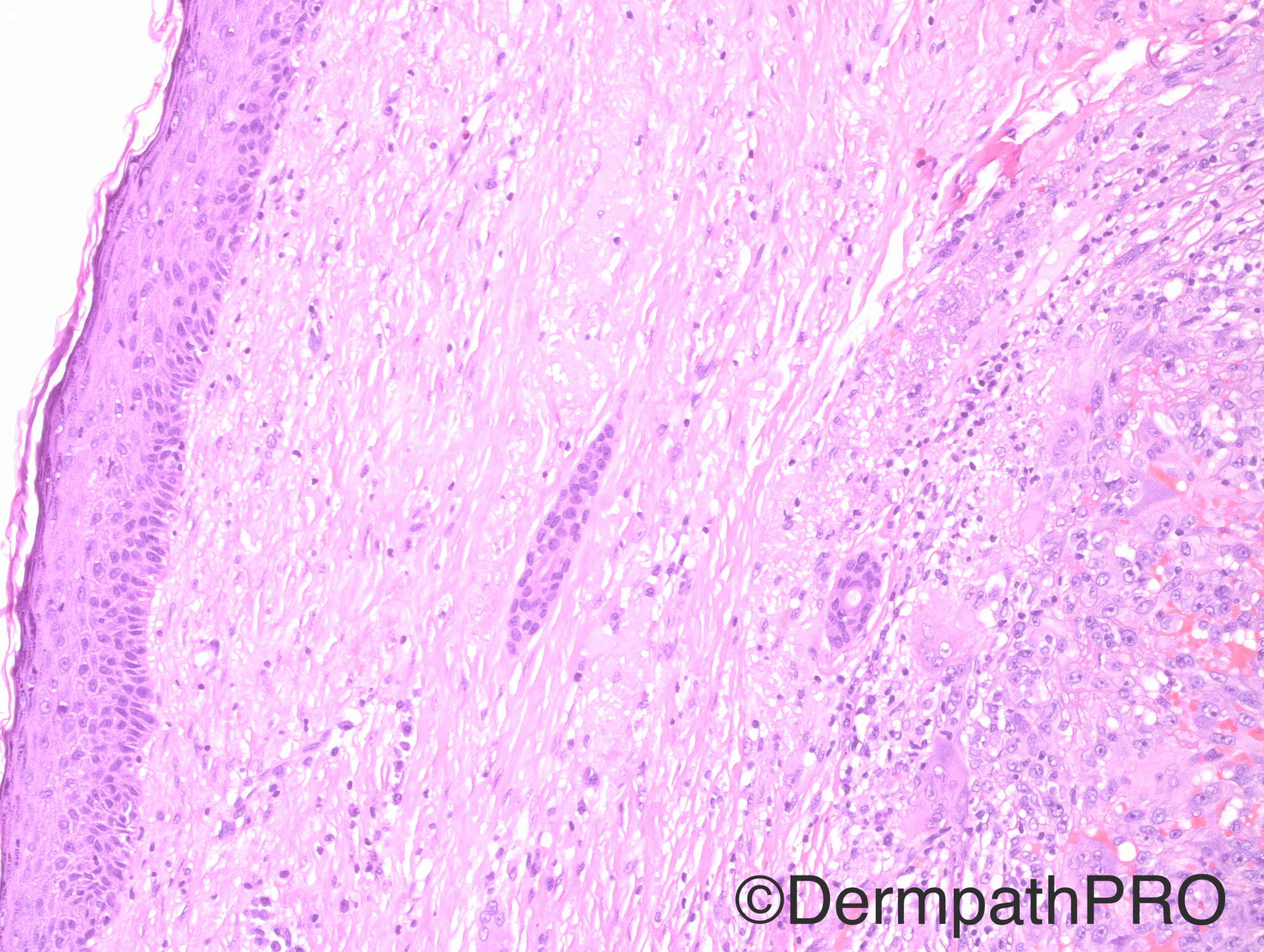
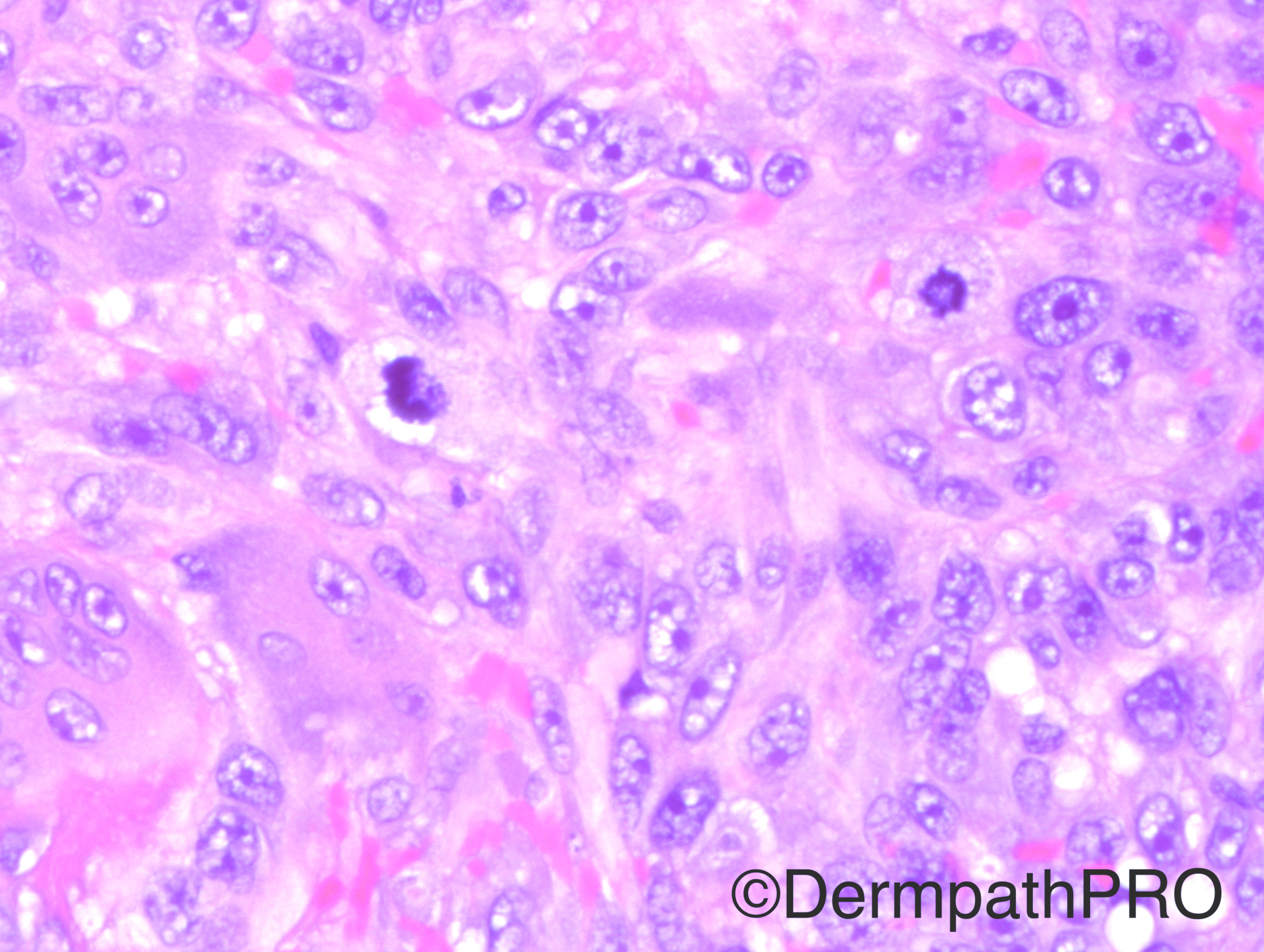
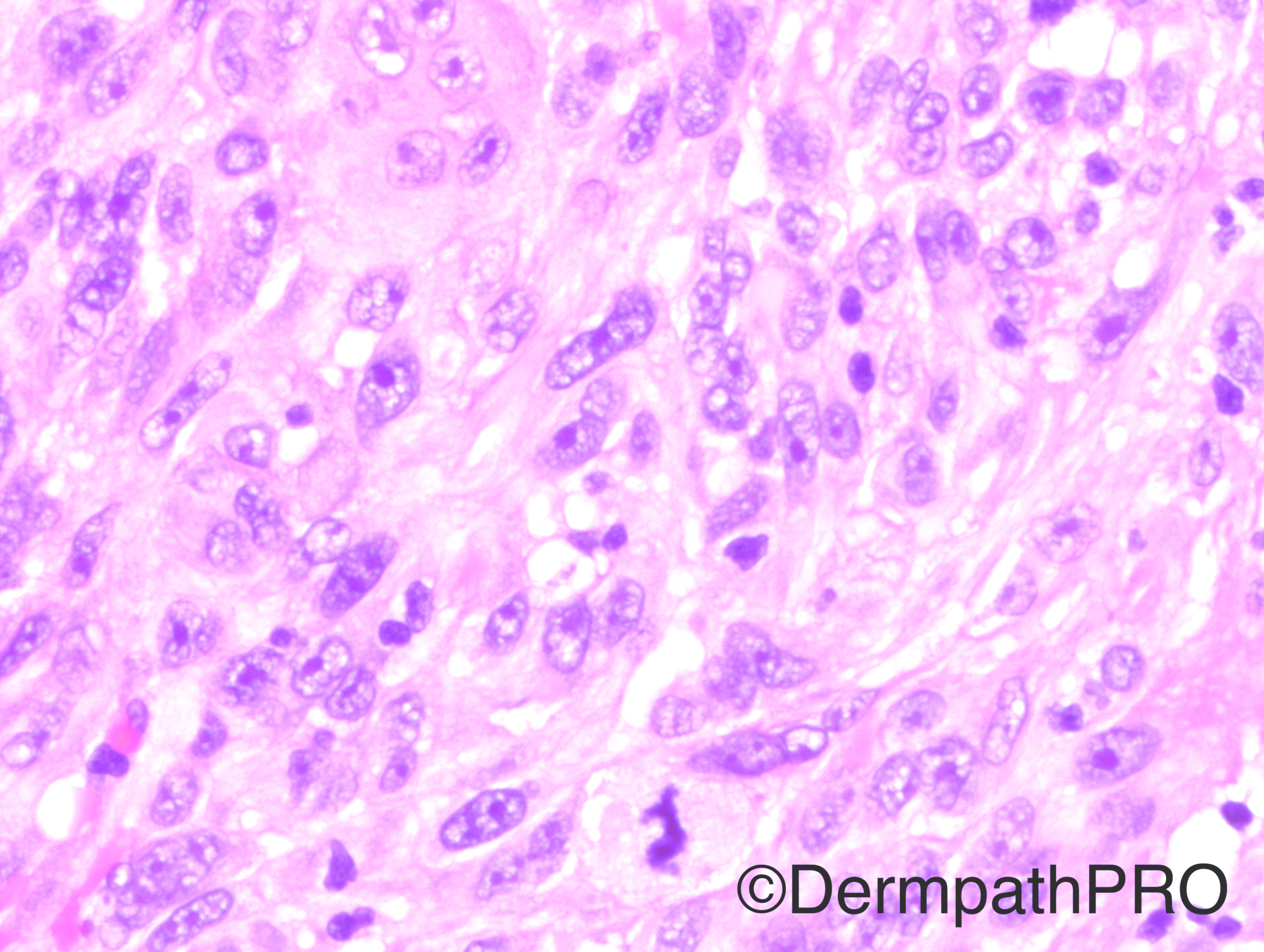
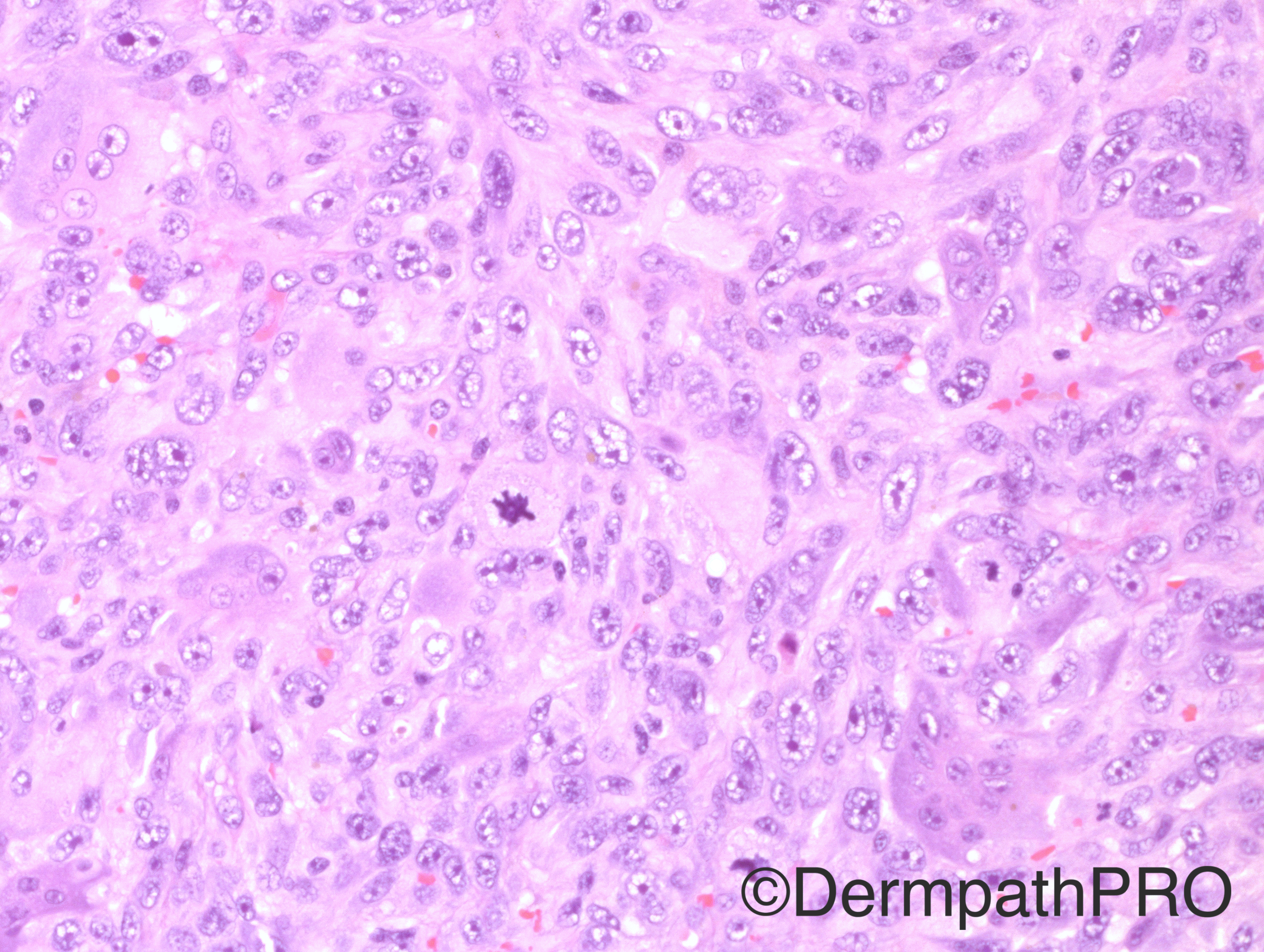
Case History: 93 year-old male with right face lesion.

Case posted by Dr Hafeez Diwan


  Report Record

User Feedback


Nitin Khirwadkar

Posted

Looks like an angiosarcoma. A small IHC panel to rule out usual mimics like pseudovascular SCC etc.

Share this comment


Link to comment
share_externally

Dr. Mona Abdel-Halim

Posted

The face affection and the presence of large multinucleated giant cells made me think of AFX/pleomorphic dermal (undifferentiated) sarcoma spectrum. Looks deep not contiguous to the epidermis and separated from it by a wide zone of collagen favouring pleomorphic sarcoma over AFX. IHC is definitely needed before finalizing the diagnosis. Would love to see the scanning power to assess exact depth and SC affection.

Share this comment


Link to comment
share_externally

Raul Perret

Posted

I favor atypical fibroxanthoma with osteoclast giant cells.  Epithelioid angiosarcoma is my second differential. But of course that a wide panel of immunos should be performed. The differential of AFX vs pleomorphic undifferentiated sarcoma as far as I have read is usually based on: size of the tumor >2cm, Deep infiltration (subcutis, fascia, etc.), presence of necrosis and vascular invasión, all these characteristics favour PUS.

Share this comment


Link to comment
share_externally

Guest Romualdo

Posted

Dr. Hafeez, in relation to yesterday's case, what was the nature of the pigment, lipofuscin or melanin?

Share this comment


Link to comment
share_externally

vincenzo polizzi

Posted

what about metastasis of undifferentiated carcinoma ( lung? ), beacuase of round-pushing margins?

Share this comment


Link to comment
share_externally

Sasi Attili

Posted

Difficult without seeing silhouette. Thinking metastasis as looks to have no epidermal connection....need immunos

Share this comment


Link to comment
share_externally

Dr. Richard Carr

Posted

I think there is a recent report of SCC with malignant giant cell tumour areas (these here looked more like malignant GCs to me but any high grade tumour with haemorrhage can attract reactive osteoclastic GC - IHC will sort).

Share this comment


Link to comment
share_externally

Dr. Hafeez Diwan

Posted

Atypical fibroxanthoma.  Multiple keratins were negative, as were CD31, CD34, and melanocytic markers.

Share this comment


Link to comment
share_externally

Dr. Hafeez Diwan

Posted

In response to Dr. Romualdo's question about the pigment in yesterday's case, I am guessing melanin. 

Share this comment


Link to comment
share_externally


Join the conversation

You can post now and register later. If you have an account, sign in now to post with your account.

Guest
Add a comment...

×   Pasted as rich text.   Paste as plain text instead

  Only 75 emoji are allowed.

×   Your link has been automatically embedded.   Display as a link instead

×   Your previous content has been restored.   Clear editor

×   You cannot paste images directly. Upload or insert images from URL.

Loading...

×
×
  • Create New...