Jump to content
In this section we have spot diagnoses posted on a daily basis since June 2010, now over 4000! You can review the archived cases and read the suggested diagnoses by users and the final comment by the contributors.
Case are uploaded each week day by 10 am UK time with the correct diagnosis will generally be posted at 8 pm UK time. Why not view the most recent spot diagnosis and proffer a diagnosis?

Case Number : Case 1436- 24 December Posted By: Guest

Please read the clinical history and view the images by clicking on them before you proffer your diagnosis.
Submitted Date :
   (0 reviews)

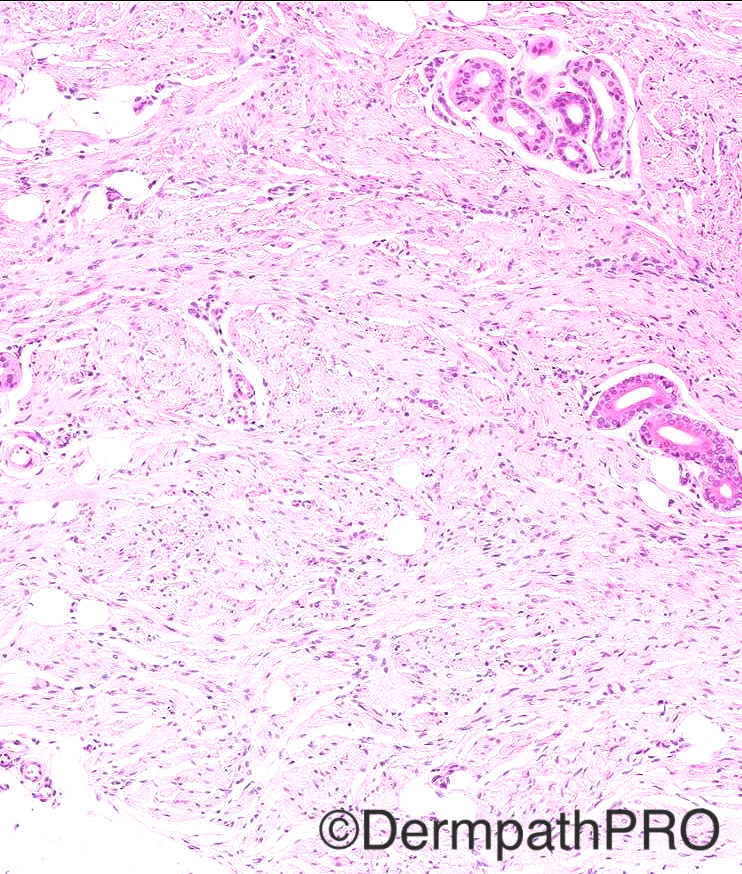
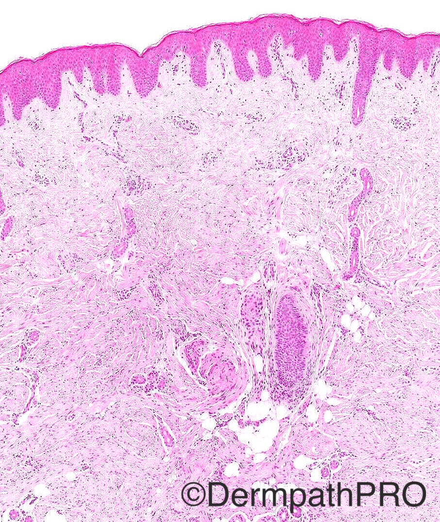
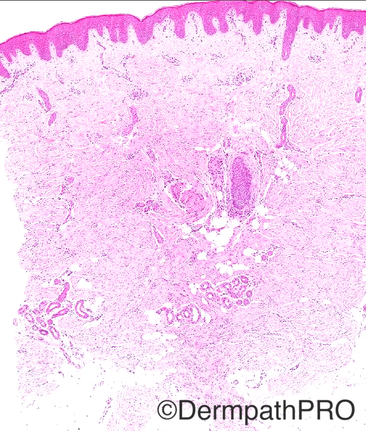
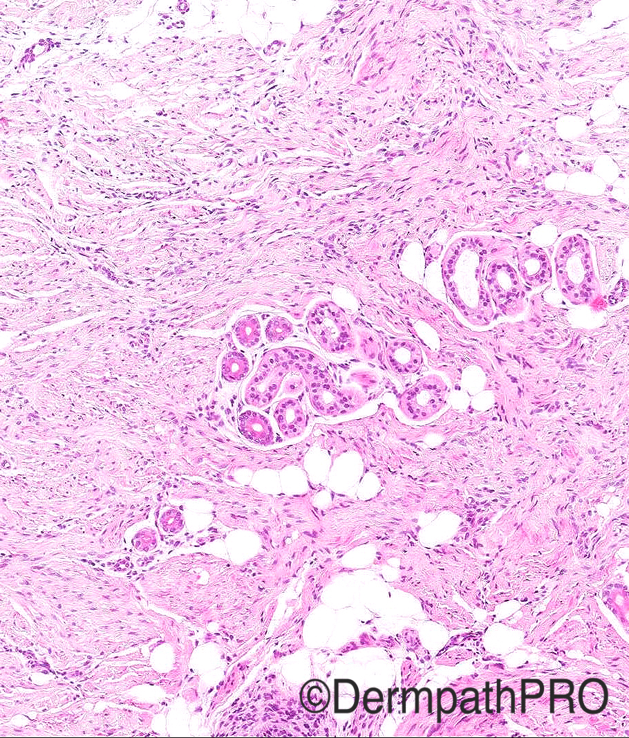
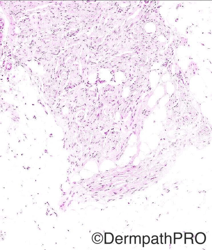
Case History: 3yr/boy with slowly growing painless area of skin thickening in paraumbilical region since 1 year. ?morphoea

Case posted by Dr Arti Bakshi


  Report Record

User Feedback


Raul Perret

Posted

I would include a panel of markers including desmin, s-100, CD34, cyclin d-1 and beta catenin. I think that fibroblastic connective tissue nevus is an excellent suggestion (I did not know this entity) but i would include also in the differential Gardners fibroma 

Share this comment


Link to comment
share_externally

Agree with Fibroblastic connective tissue nevus, but my first impression (without clinical history) was Dermatomyofibroma.

Share this comment


Link to comment
share_externally

vincenzo polizzi

Posted

Avere With fibroblastic connective nevus.

Share this comment


Link to comment
share_externally

vincenzo polizzi

Posted

Sorry agree With fibroblastic connective nevus

Share this comment


Link to comment
share_externally

vincenzo polizzi

Posted

Now i'm thinking of fibrous hamarthoma of the infancy. I agree with Raul about panel of markers. Merry Christmas.

Share this comment


Link to comment
share_externally

Guest Romualdo

Posted

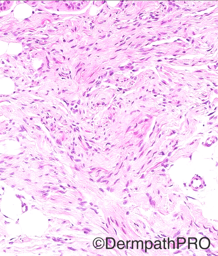
I think there are pseudovascular spaces. I am thinking of giant cell fibroblastoma.

Share this comment


Link to comment
share_externally

Dr. Mona Abdel-Halim

Posted

Fibroblastic connective tissue nevus is a good suggestion but would have expected it at an earlier onset. I think also as Romualdo said that there are slits lined by tumor cells (pseudovascular spaces), so I am thinking also of giant cell fibroblastoma however there is no enough floret giant cells.

Share this comment


Link to comment
share_externally

Arti Bakshi

Posted

  Merry Christmas to all !!

I called this a Fibroblastic Connective Tissue Naevus. (well done to Eman for getting the diagnosis straight away and making light of a difficult case!)

All of the excellent d/d considered above, went through my mind too.  Immunos were done: S100, SMA, Desmin, beta- catenin were completely negative.  CD34 was very focally positive in lesional cells, and showed mainly backgpound stromal positivity. (thus excluding DFSP/giant cell fibroblastoma)

Although these lesions are described as CD34+ in the  original series by Fletcher (http://www.ncbi.nlm.nih.gov/pubmed/22892597 ), in the full text, they state and I quote '' In most cases, CD34 positivity was weak and multifocal in a very variable proportion of lesional cells (Fig. 2). In only 2 cases was there focally strong positivity in the majority of cells''.

Interestingly, in another review (published at around the same time from France ( (http://www.ncbi.nlm.nih.gov/pubmed/22014540 ), the same entity was described as 'Cellular Connective Tissue Naevus' and all their 4 cases were CD34 negative.

Share this comment


Link to comment
share_externally

Arti Bakshi

Posted

In response to Mona's comment, I think the age of presentation can be quite variable as per literature.


Also found another more recent reference:

 
J Cutan Pathol. 2015 Aug 13. doi: 10.1111/cup.12605. [Epub ahead of print]
Fibroblastic connective tissue nevus.

Fibroblastic connective tissue nevus (FCTN) is a newly recognized, benign cutaneous mesenchymal lesion of fibroblasts/myofibroblastic lineage, which expands the classification of connective tissue nevi. We present three cases of FCTN nd discuss significant clinical, morphologic and immunophenotypic overlap with dermatomyofibroma. Our cases were from young women, aged 32, 24 and 10, and presented as 1.2 and 1 cm nodules on the posterior neck and right upper flank, respectively while presenting as a linear plaque of the right posterior thigh in the latter case. The lesions showed a poorly circumscribed proliferation of hypercellular spindle cells arranged in short to longer intersecting fascicles entrapping adnexal structures. Superficial adipose tissue was also entrapped in one case. The spindle cells had fibroblastic features with pale eosinophilic cytoplasmic extensions and inconspicuous nucleoli. The spindle cells were positive for CD34 in two cases. One case was negative for CD34, smooth muscle actin (SMA), desmin and S100. The overall features were consistent with a diagnosis of FCTN. In two cases, we further elucidated the fibroblastic differentiation of the spindle cells in FCTN with electron microscopy, which has not been previously described.

Share this comment


Link to comment
share_externally


Join the conversation

You can post now and register later. If you have an account, sign in now to post with your account.

Guest
Add a comment...

×   Pasted as rich text.   Paste as plain text instead

  Only 75 emoji are allowed.

×   Your link has been automatically embedded.   Display as a link instead

×   Your previous content has been restored.   Clear editor

×   You cannot paste images directly. Upload or insert images from URL.

Loading...

×
×
  • Create New...