Jump to content
In this section we have spot diagnoses posted on a daily basis since June 2010, now over 4000! You can review the archived cases and read the suggested diagnoses by users and the final comment by the contributors.
Case are uploaded each week day by 10 am UK time with the correct diagnosis will generally be posted at 8 pm UK time. Why not view the most recent spot diagnosis and proffer a diagnosis?

Case Number : Case 1437- 25 December Posted By: Guest

Please read the clinical history and view the images by clicking on them before you proffer your diagnosis.
Submitted Date :
   (0 reviews)

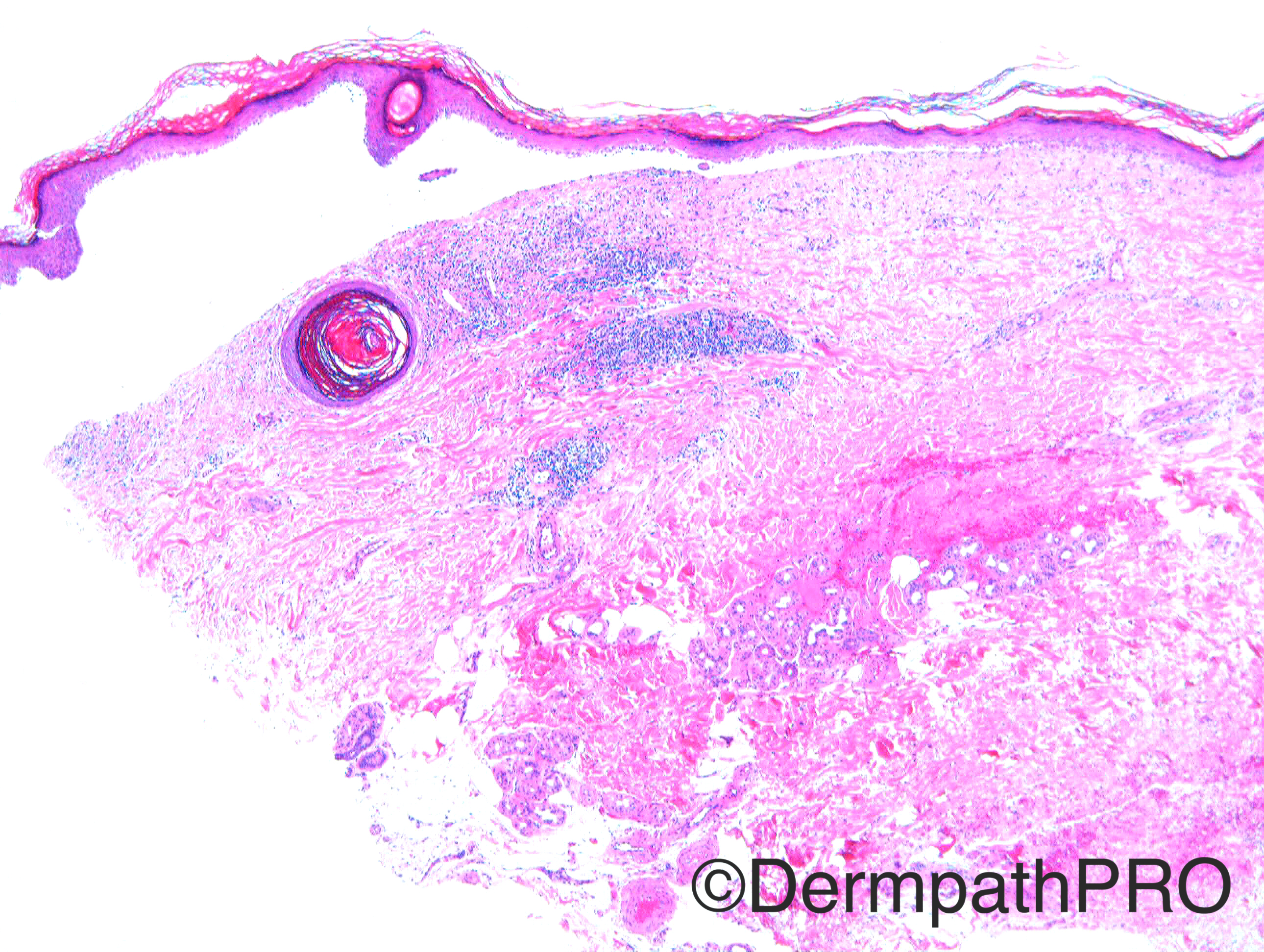
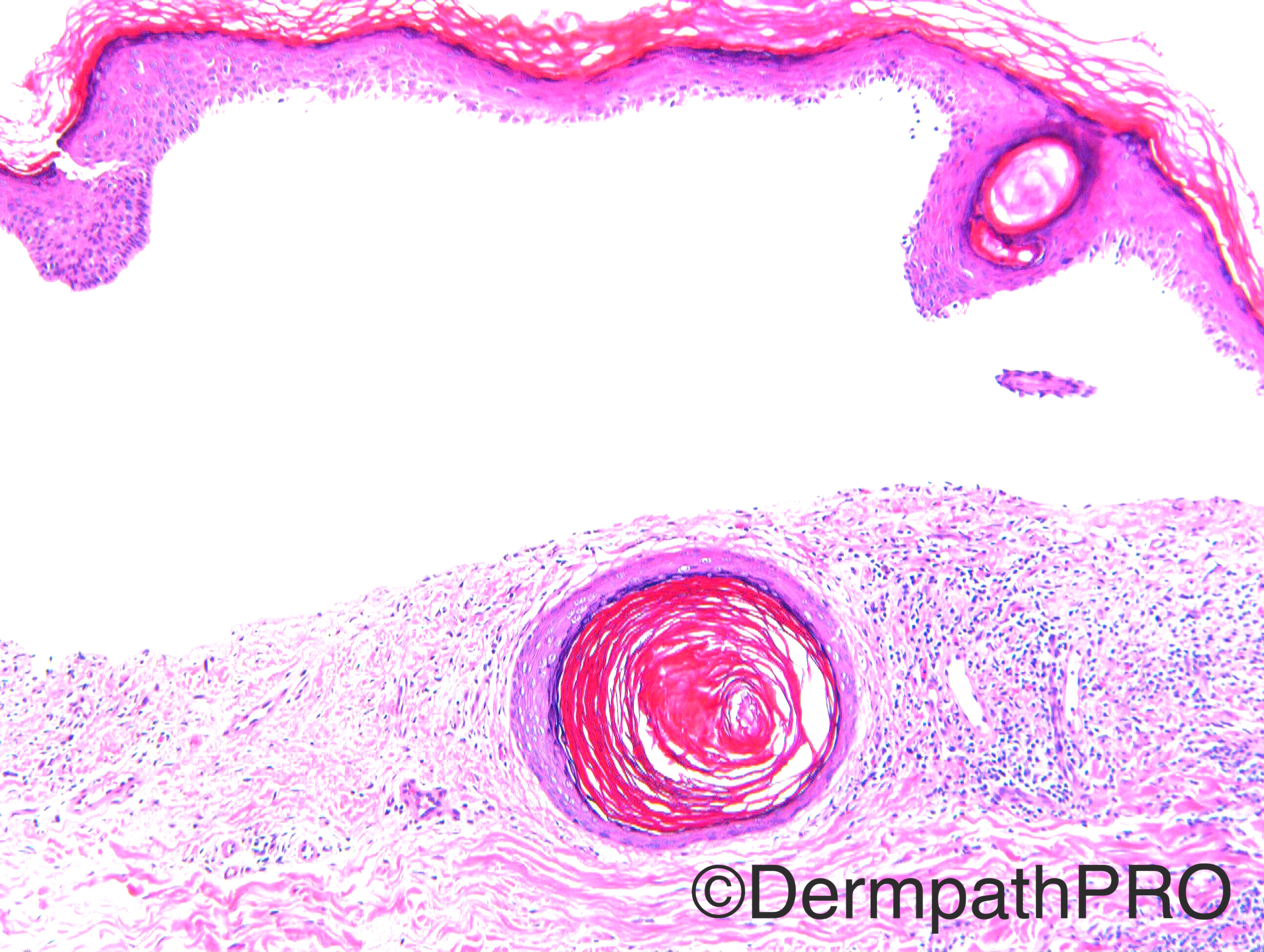
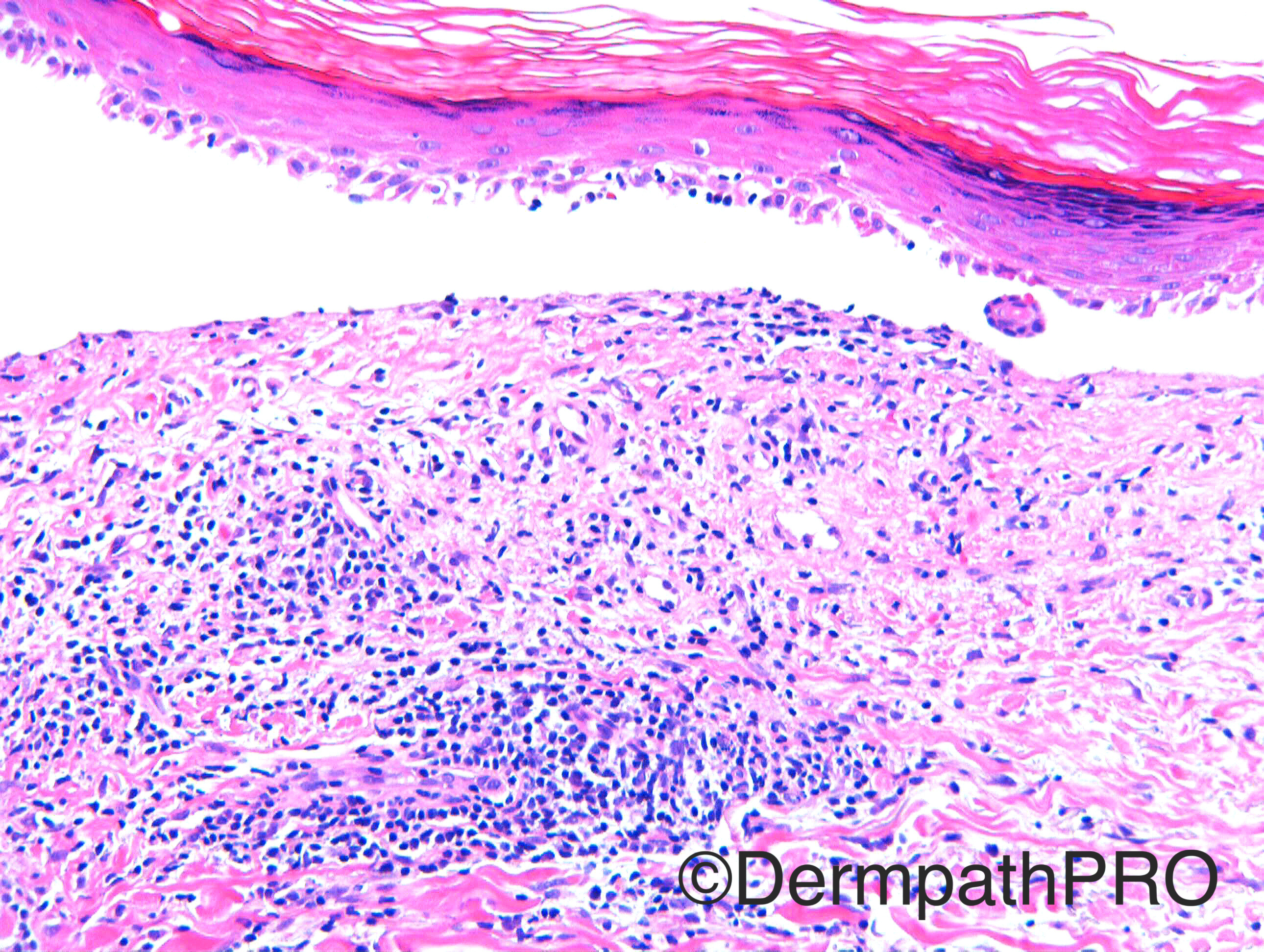
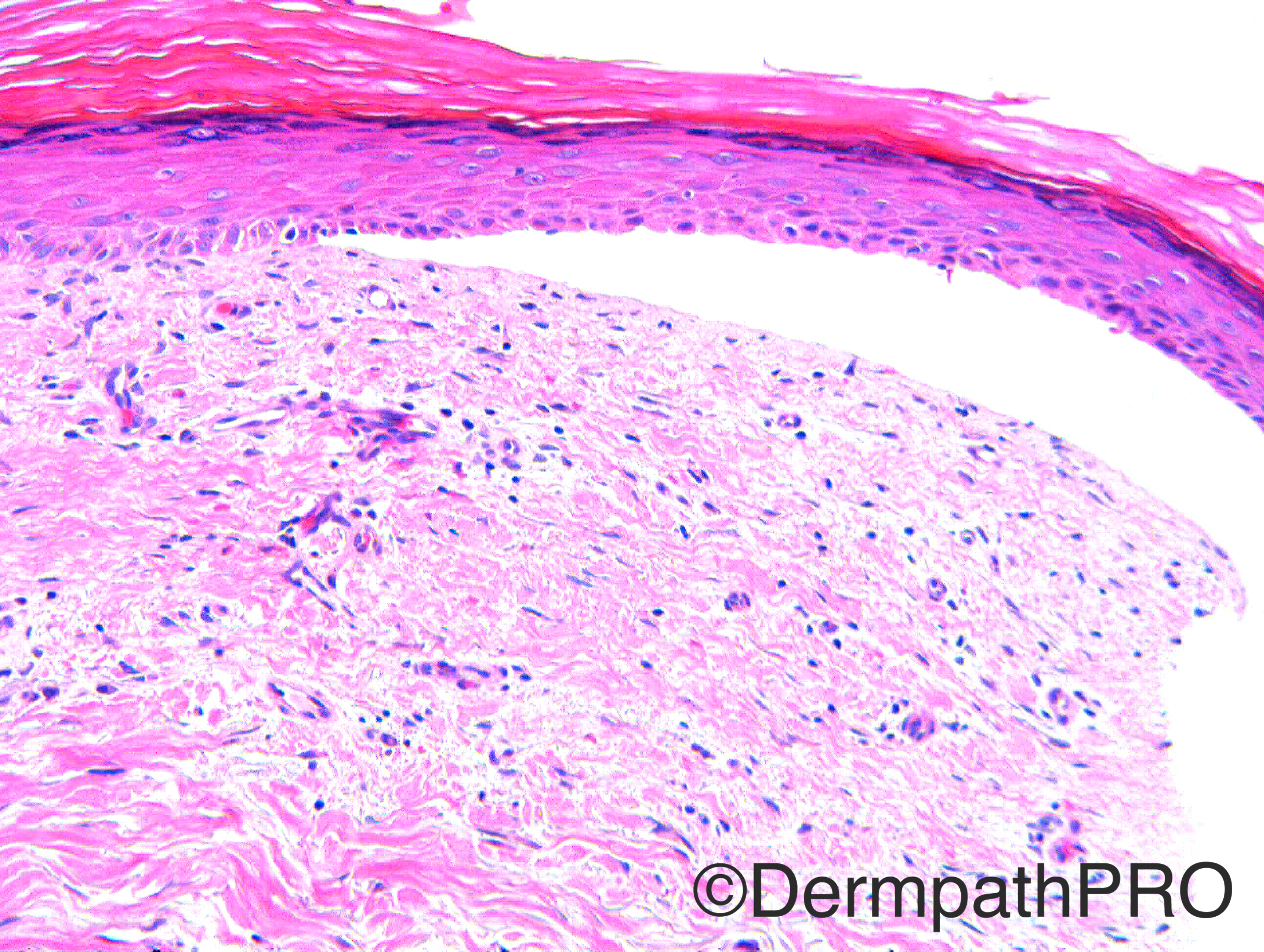
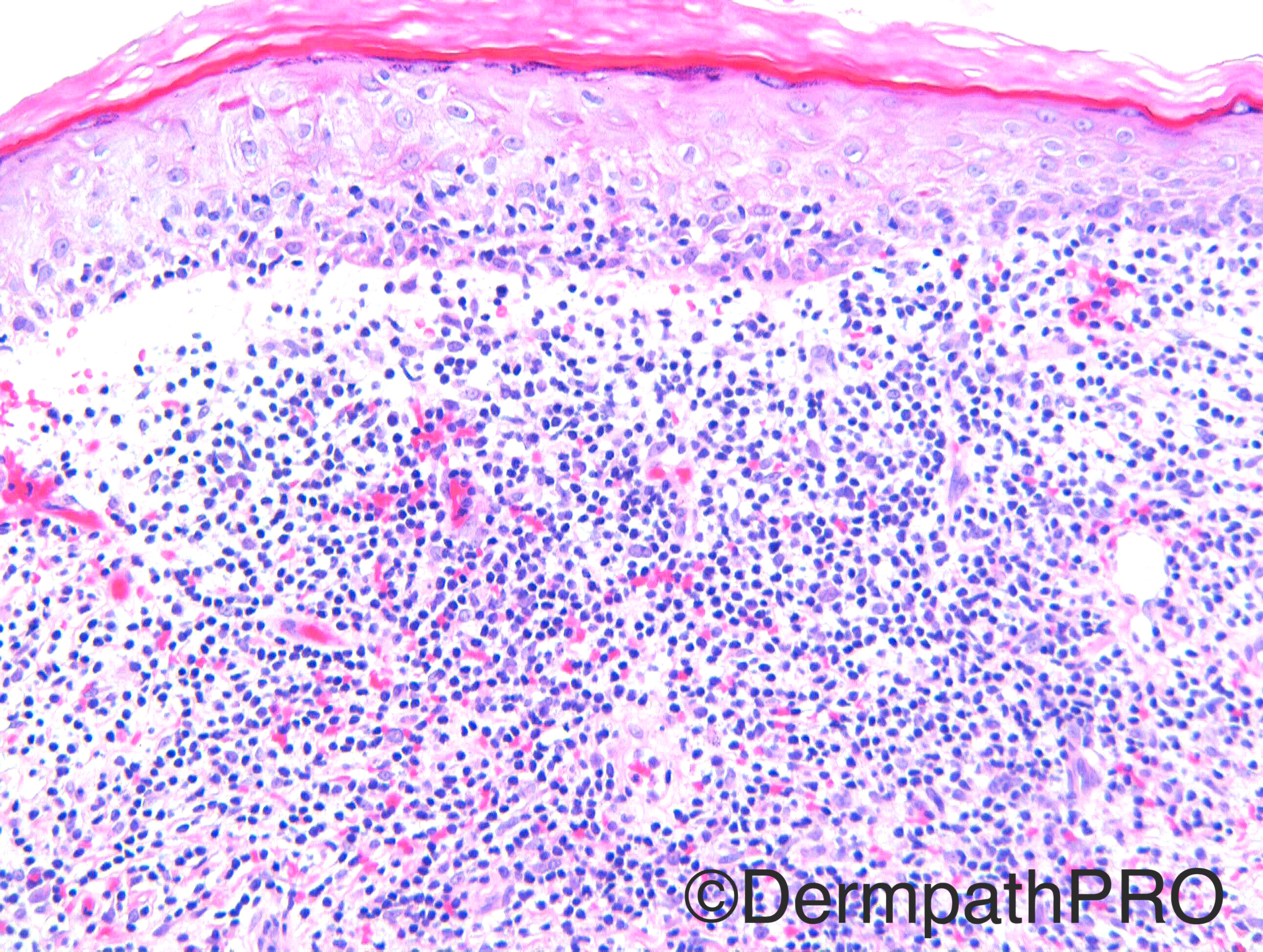
Case History: M50. Blistering eruption arm and chest. ?Bullous lichen planus, ?Pemphigus. Case c/o Dr Nitin Khirwadkar

Case posted by Dr Richard Carr


  Report Record

User Feedback


vincenzo polizzi

Posted

Follicular plugs, interface lichenoid cell-poor dermatitis, epidermal atrophy, mucin(?)...what about bulls subacute lupus?

Share this comment


Link to comment
share_externally

Dr. Mona Abdel-Halim

Posted

As Eman said, I am thinking in lichen planus pemphigoides. In lichen planus pemphigoides, blisters may develop away from lichen papules on normal or erythematous skin or may develop in lichen papules. If developing in lichen papules, they will show esinophils in the infiltrate which are not seen in bullous lichen planus. The presence of hypergranulosis, squamatized basal cell layer (fig4) and definite interface change (fig6) may favour blisters of lichen planus pemphigoides developing within lichen papules. Bullous SLE would have shown a neutrophil rich blister with neutrophilic microabscesses.

Share this comment


Link to comment
share_externally

Raul Perret

Posted

Agree with lichen planus pemphigoides, hypergranulosis and interface change as well of eosinophils seen on picture 4 made me favor it. Nevertheless, the presence of extravasated red blood cells, interstitial eosinophils and a lymphocytic infiltrate that surrounds in some areas the blood vessels makes me put in the differential a bullous drug reaction. I would like to ask you what do you think is the reason of follicular  plugging in this case.  Greetings!

Share this comment


Link to comment
share_externally

I agree with Lichen planus pemphigoides, but clinical picture is essential in this case. As I can see many lymphocytes within the epidermis without proportionate interface changes, I would perform immunostains for MF (bullous MF) even with popsitive DIF, because bullae in MF can be triggered by autoimmune bullous disorders.

Share this comment


Link to comment
share_externally

Dr. Richard Carr

Posted

Apologies for the delay - I was on hols but I am pleased to see you all pretty much nailed it. 


This was a case of lichen planus pemphigoides.


The follicular hyperkeratosis my reflect the element of lichen planus / lichen planopilaris in this case as some of you thought there was also evidence of LP in the images in addition to the immunobullous process with eosinophils - I would not dissagree.

Share this comment


Link to comment
share_externally


Join the conversation

You can post now and register later. If you have an account, sign in now to post with your account.

Guest
Add a comment...

×   Pasted as rich text.   Paste as plain text instead

  Only 75 emoji are allowed.

×   Your link has been automatically embedded.   Display as a link instead

×   Your previous content has been restored.   Clear editor

×   You cannot paste images directly. Upload or insert images from URL.

Loading...

×
×
  • Create New...