Jump to content
In this section we have spot diagnoses posted on a daily basis since June 2010, now over 4000! You can review the archived cases and read the suggested diagnoses by users and the final comment by the contributors.
Case are uploaded each week day by 10 am UK time with the correct diagnosis will generally be posted at 8 pm UK time. Why not view the most recent spot diagnosis and proffer a diagnosis?

Case Number : Case 1439- 29 December Posted By: Guest

Please read the clinical history and view the images by clicking on them before you proffer your diagnosis.
Submitted Date :
   (0 reviews)

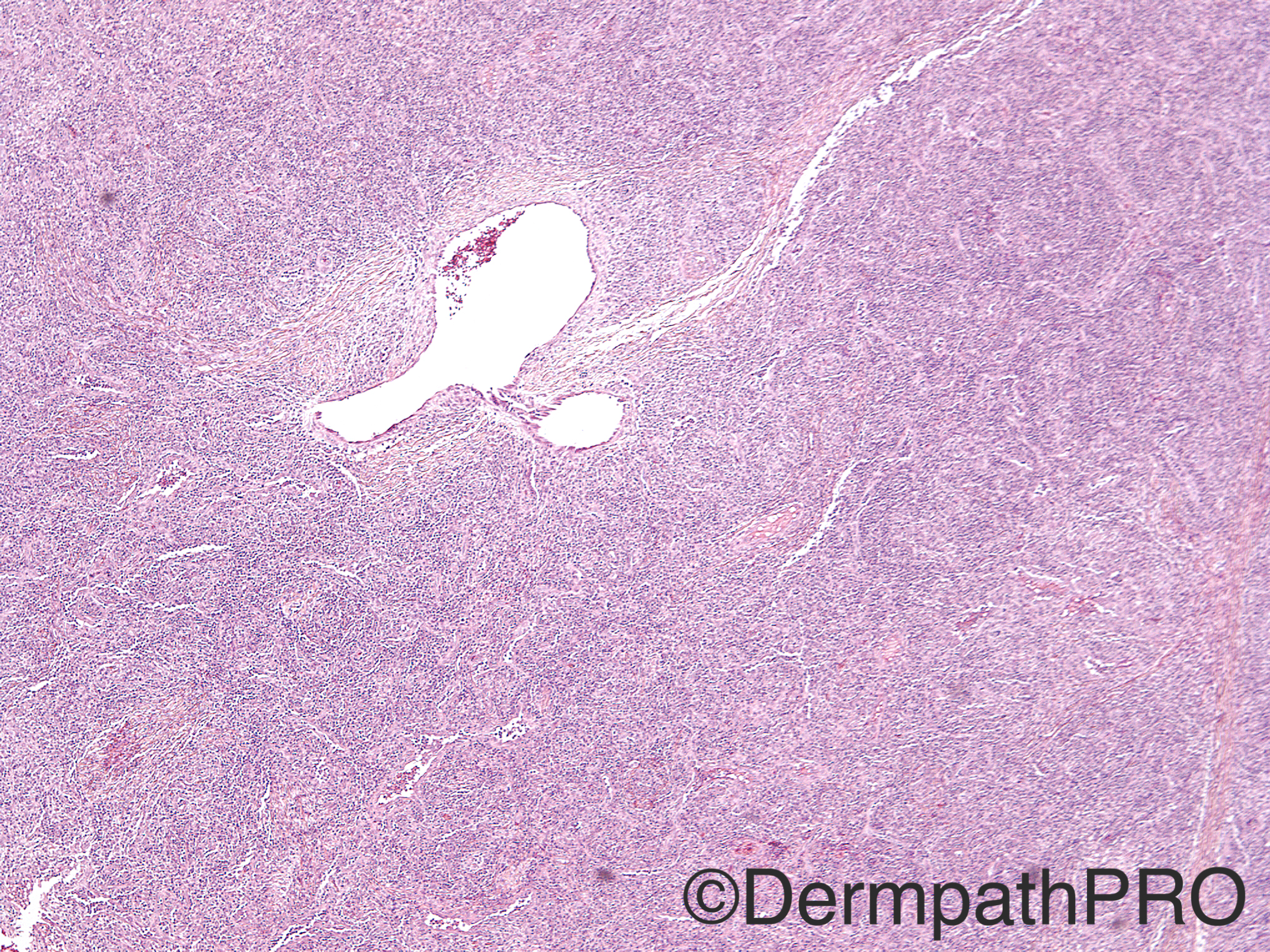
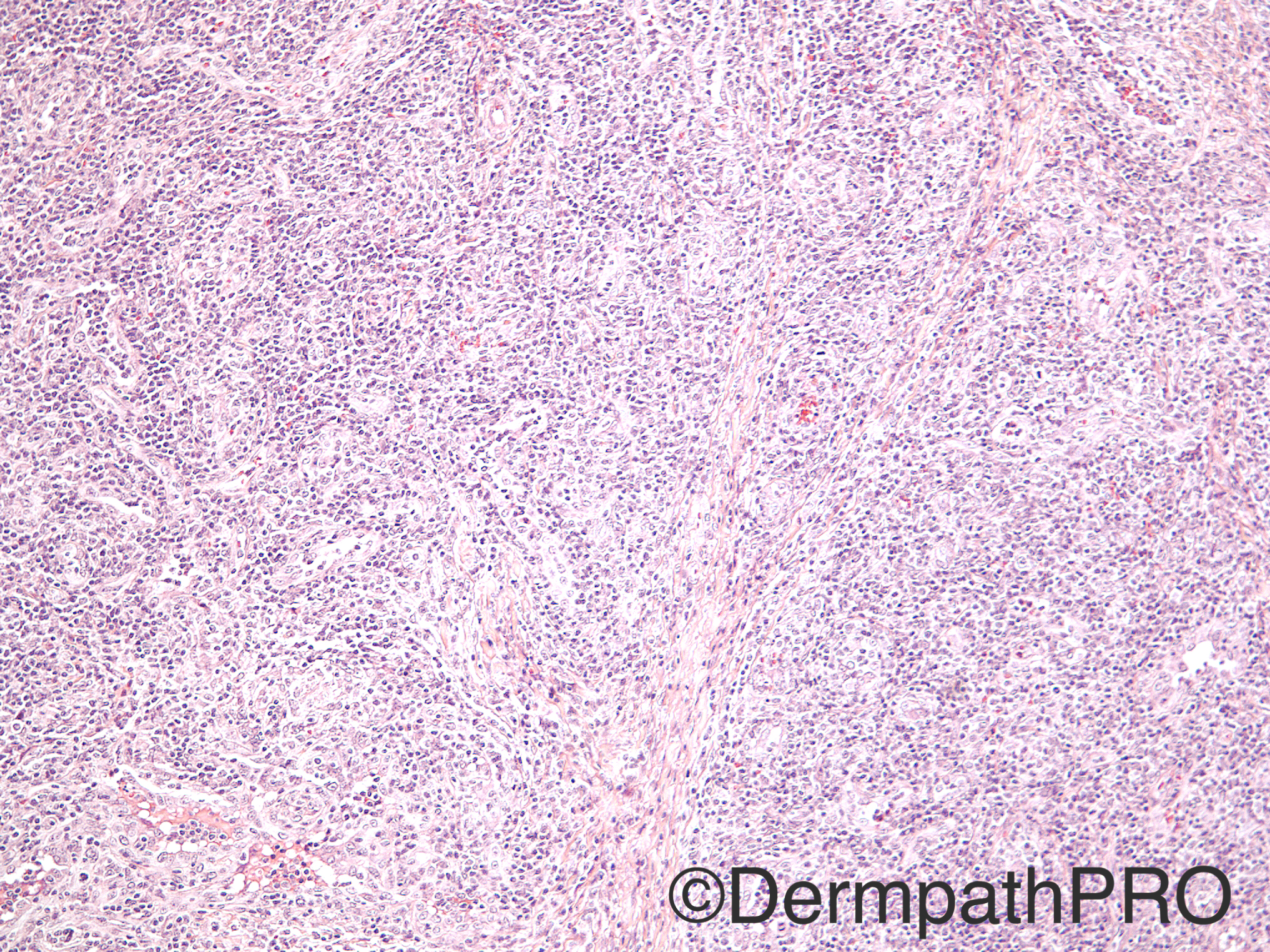
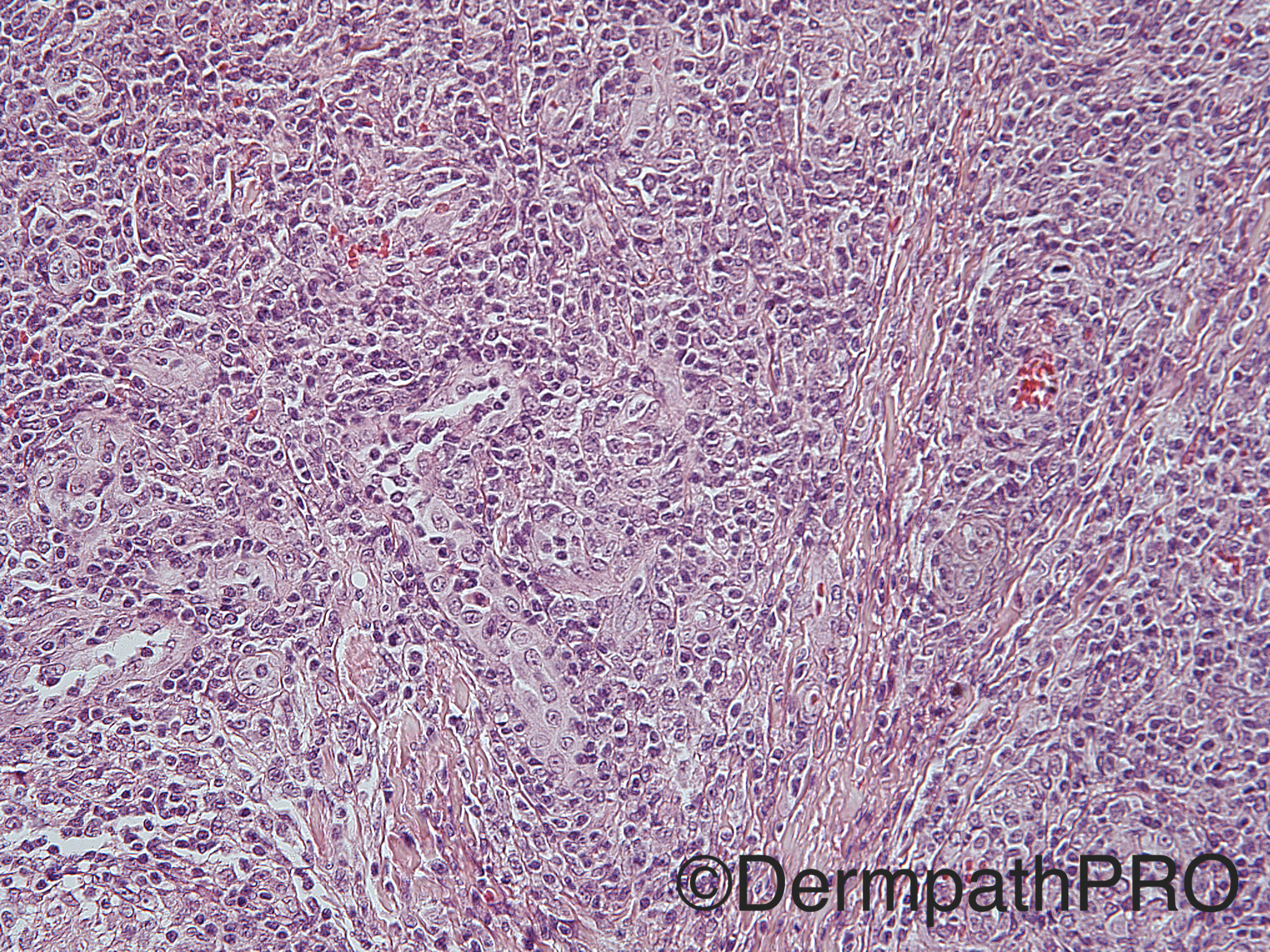
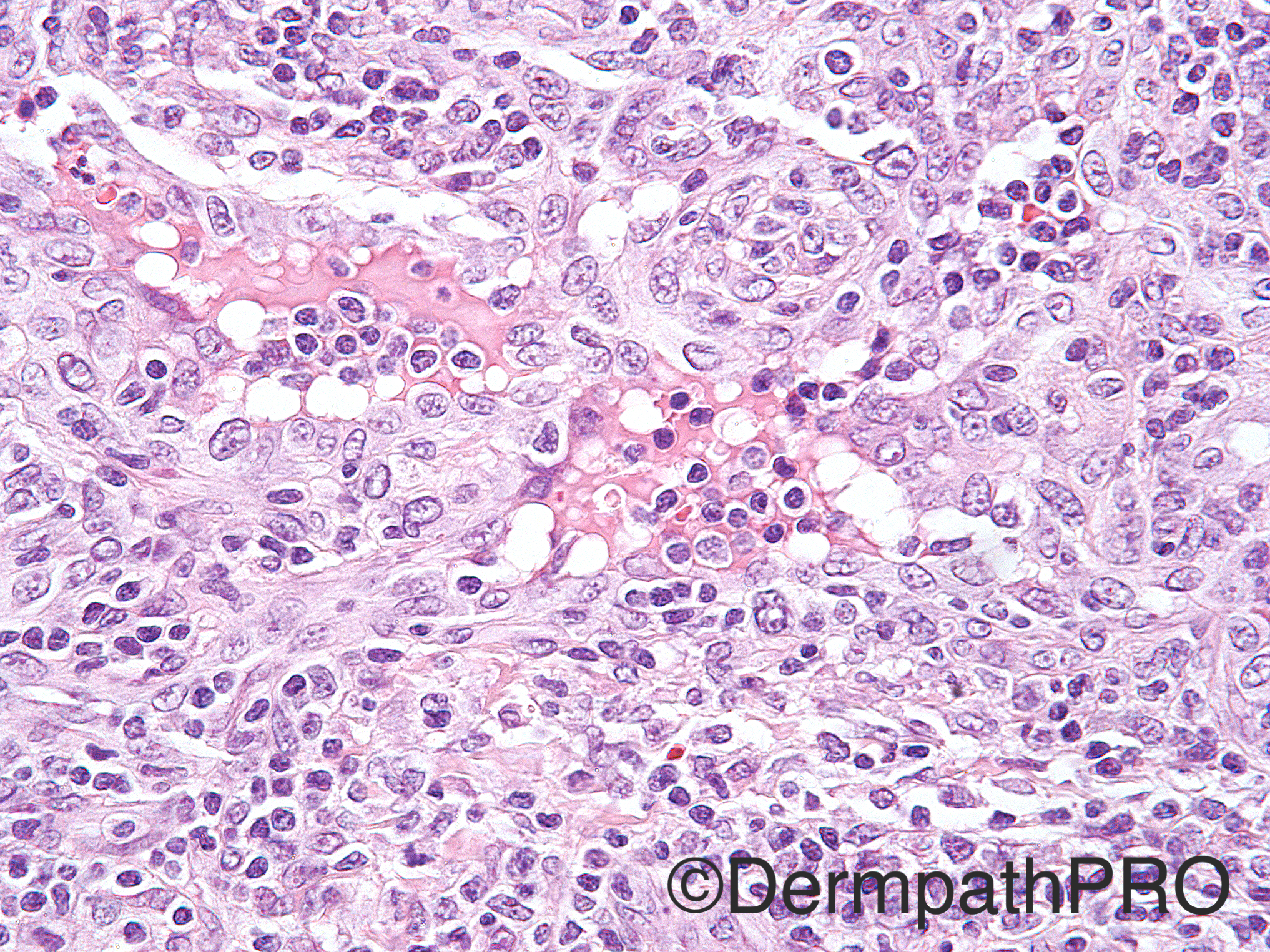
Case History: 12-year-old girl with subcutaneous nodule of left hand.

Case posted by Dr Uma Sundram


  Report Record

User Feedback


vincenzo polizzi

Posted

Agree completely with Romualdo, although the unusual site: ALHE.

Share this comment


Link to comment
share_externally

Arti Bakshi

Posted

What about cutaneous epithelioid angiomatous nodule?

Share this comment


Link to comment
share_externally

Nitin Khirwadkar

Posted

Dermal based vascular proliferation. Vessels lined by plump epithelioid cells. Abundant lymphoid tissue, along with scattered eosinophils- angiolymphoid hyperplasia with eosinophilia.

Cutaneous epithelioid angiomatous nodule did cross my mind, but the vessels in this condition are usually not well formed.

Share this comment


Link to comment
share_externally

Sasi Attili

Posted

Given the well circumscribed nature of the lesion, would prefer EAN

Share this comment


Link to comment
share_externally

Raul Perret

Posted

Agree that Epithelioid hemangioma comes in the differential. I would still perform CD34, CD31, vimentin, CK and EMA. I think that epithelioid sarcoma comes to the differential also  because it can have highly inflammed áreas.

Share this comment


Link to comment
share_externally

Guest Romualdo

Posted

Cutaneous epithelioid angiomatous nodule is a nice suggestion, but this lesion (today's case) was described as a subcutaneous nodule. CEAN are typically located at superficial dermis and deep dermis and subcutaneous tissue are generally not involved.

Share this comment


Link to comment
share_externally

Dr. Mona Abdel-Halim

Posted

I am thinking of epithelioid hemangioma as Romualdo said, possibly intravascular hence the very circumscribed and the sub cutaneous localization. CEAN would have been a superficial dermal lesion.

Share this comment


Link to comment
share_externally

Uma Sundram

Posted

Cutaneous epithelioid angiomatous nodule. This was not an intravascular lesion and was very well circumscribed. The lesion was located in the superficial subcutis. Vascular markers were positive but keratin markers were negative. Unusual site for EH and usually this entity has more eosinophils.  Importantly, this is not hemangioendothelioma or angiosarcoma.

Share this comment


Link to comment
share_externally


Join the conversation

You can post now and register later. If you have an account, sign in now to post with your account.

Guest
Add a comment...

×   Pasted as rich text.   Paste as plain text instead

  Only 75 emoji are allowed.

×   Your link has been automatically embedded.   Display as a link instead

×   Your previous content has been restored.   Clear editor

×   You cannot paste images directly. Upload or insert images from URL.

Loading...

×
×
  • Create New...