Jump to content
In this section we have spot diagnoses posted on a daily basis since June 2010, now over 4000! You can review the archived cases and read the suggested diagnoses by users and the final comment by the contributors.
Case are uploaded each week day by 10 am UK time with the correct diagnosis will generally be posted at 8 pm UK time. Why not view the most recent spot diagnosis and proffer a diagnosis?

Case Number : Case 1207 - 06 February Posted By: Guest

Please read the clinical history and view the images by clicking on them before you proffer your diagnosis.
Submitted Date :
   (0 reviews)

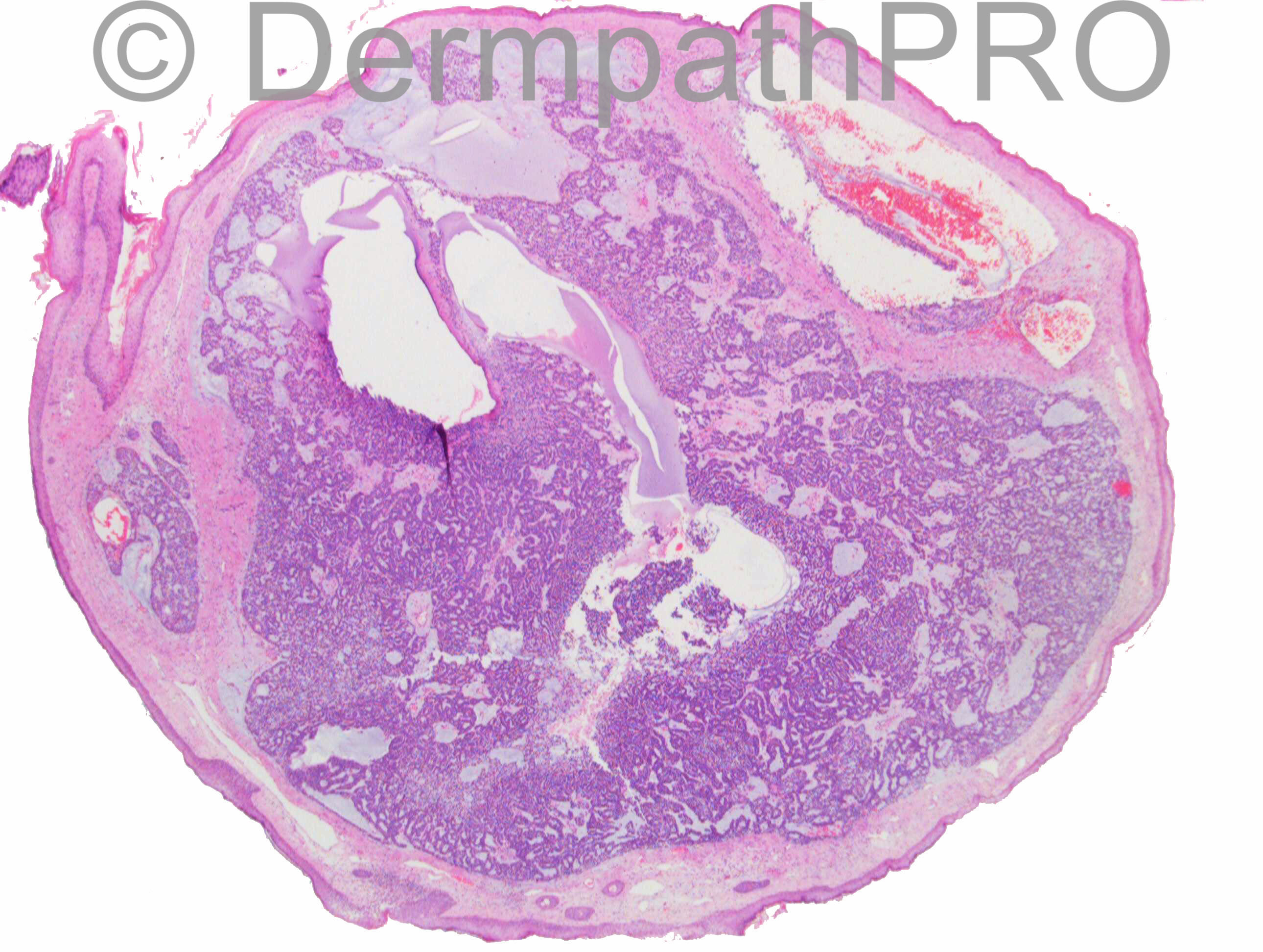
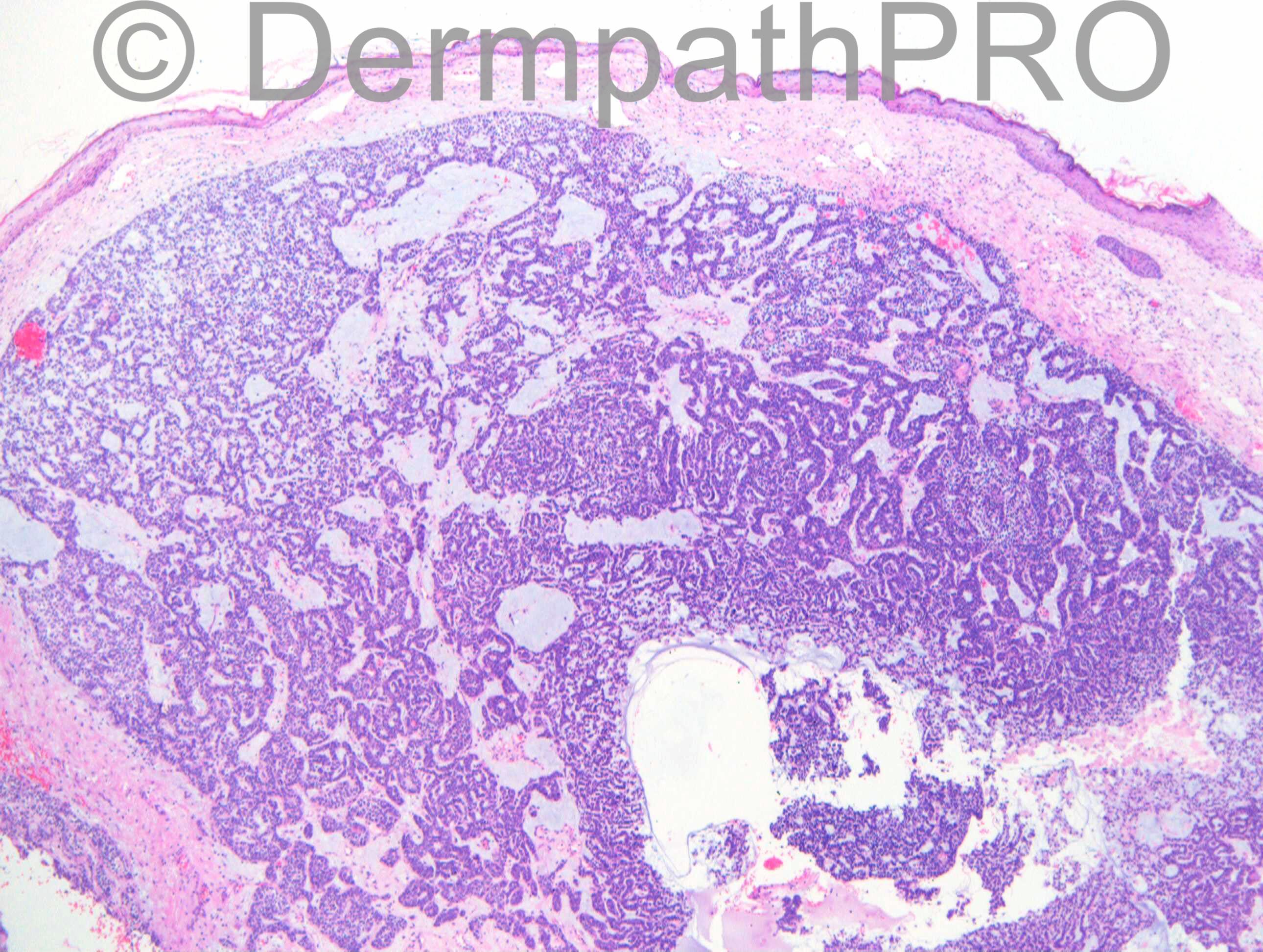
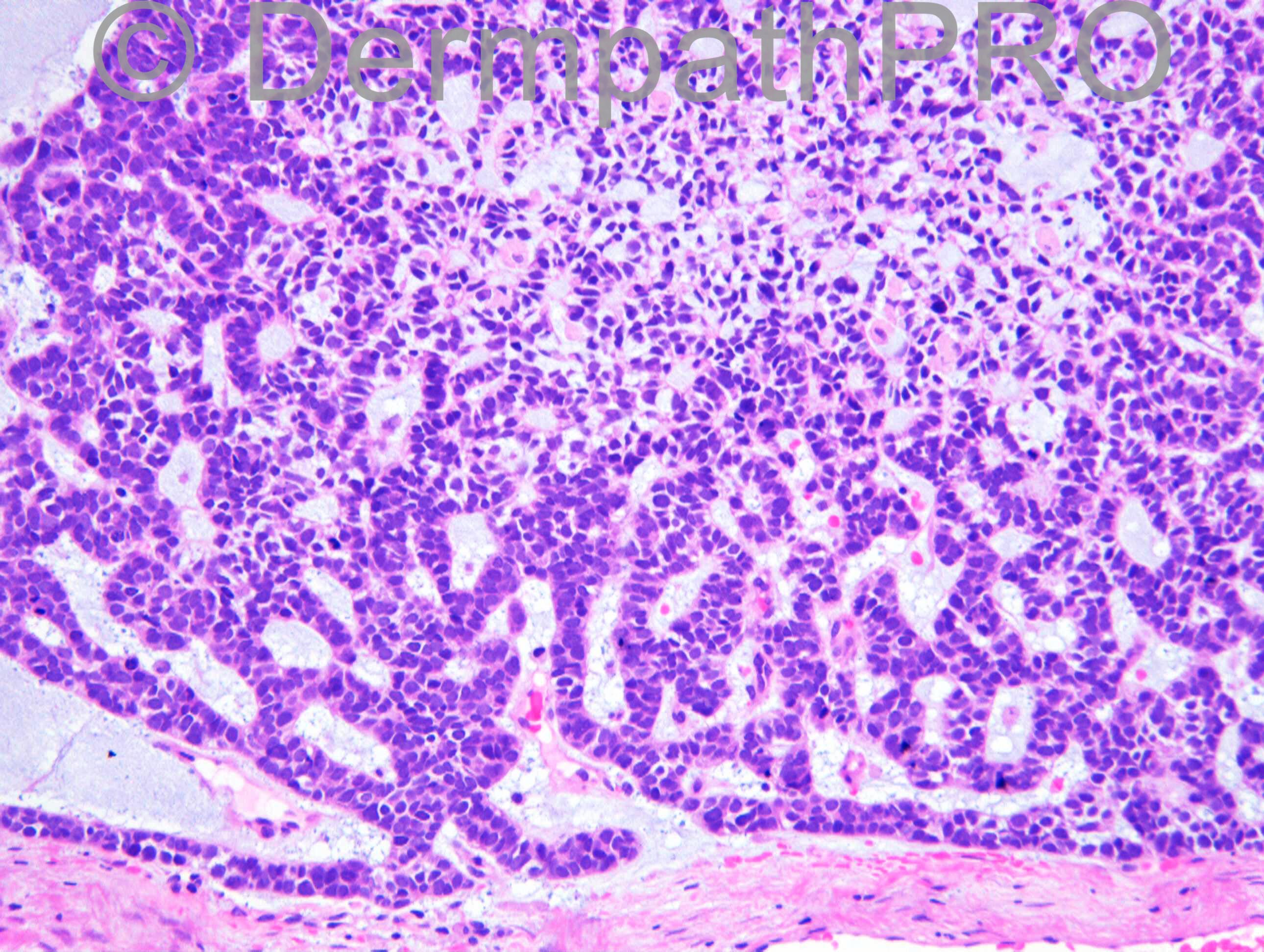
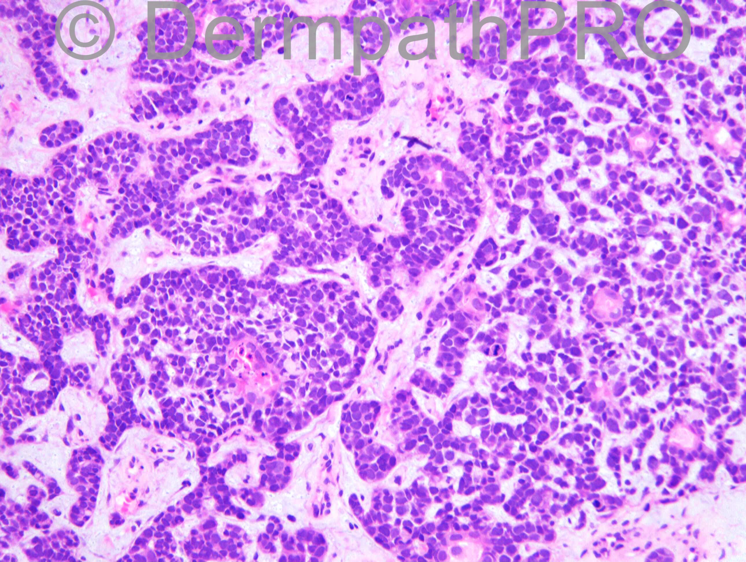
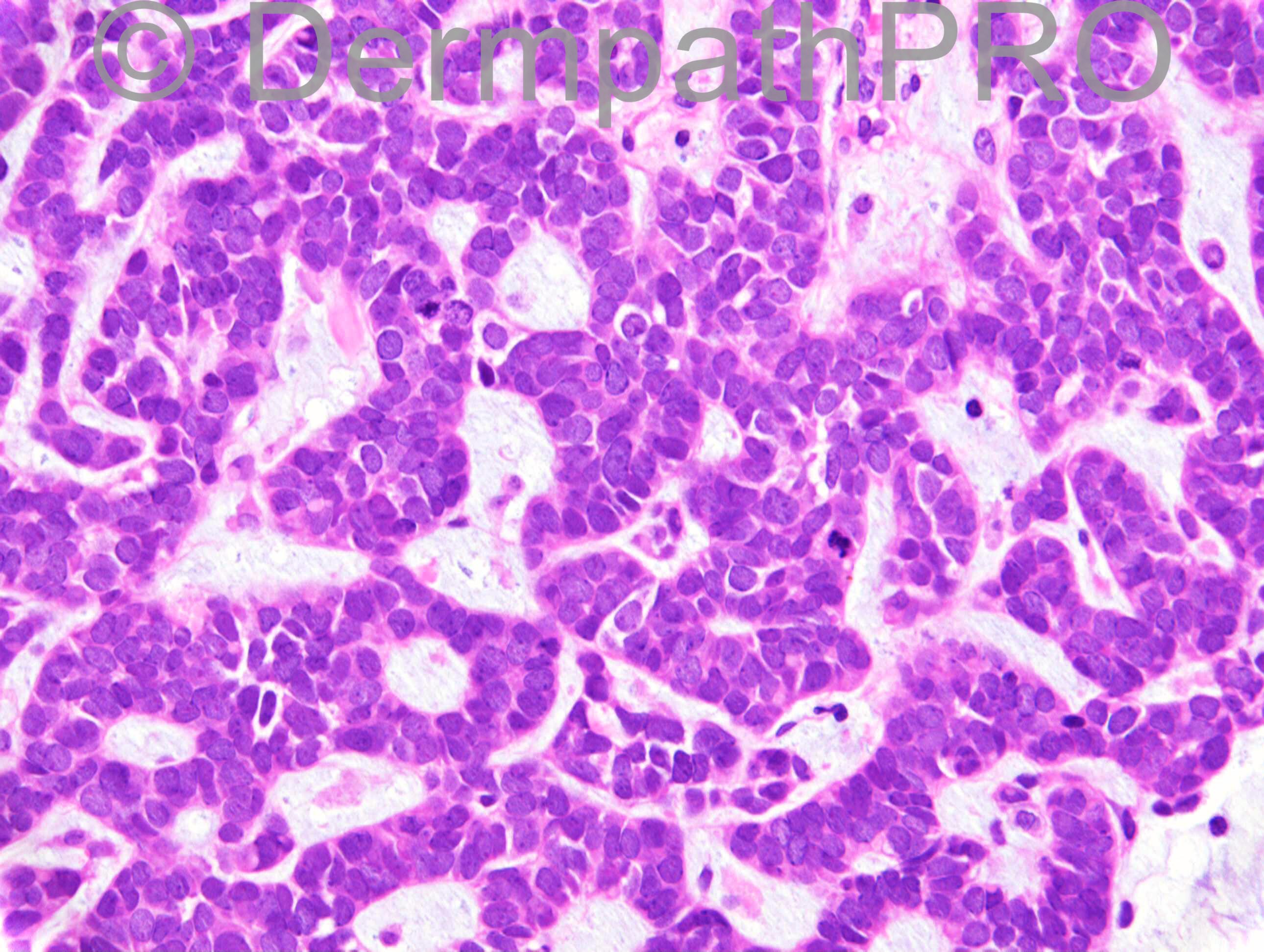
F86. Shiny nodule ?BCC right side of nose.

Case posted by Dr Richard Carr


  Report Record

User Feedback


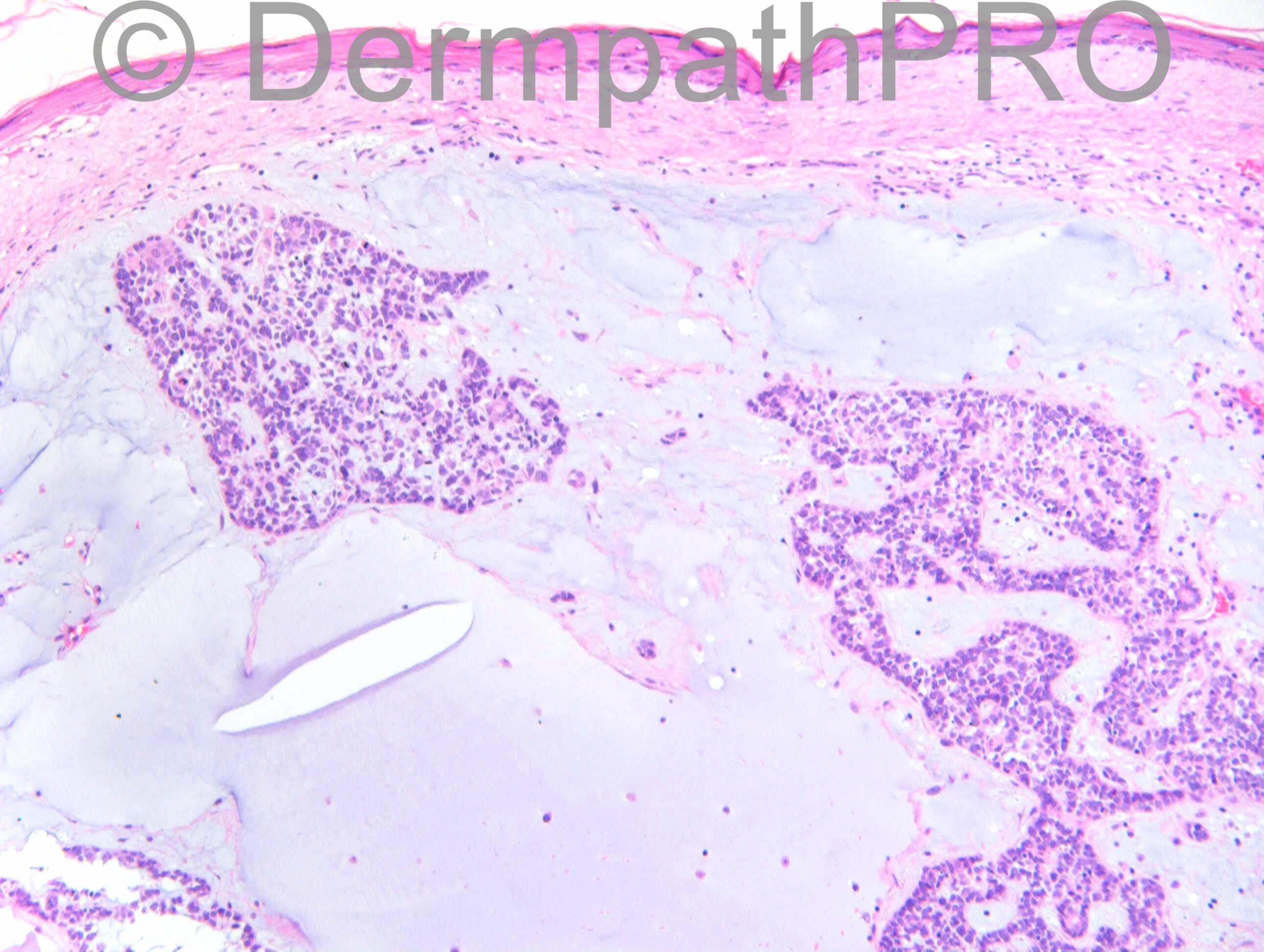
Mucin producing carcinoma with neuroendocrine cytology. Need to exclude metastasis.

Share this comment


Link to comment
share_externally

Guest Romualdo

Posted

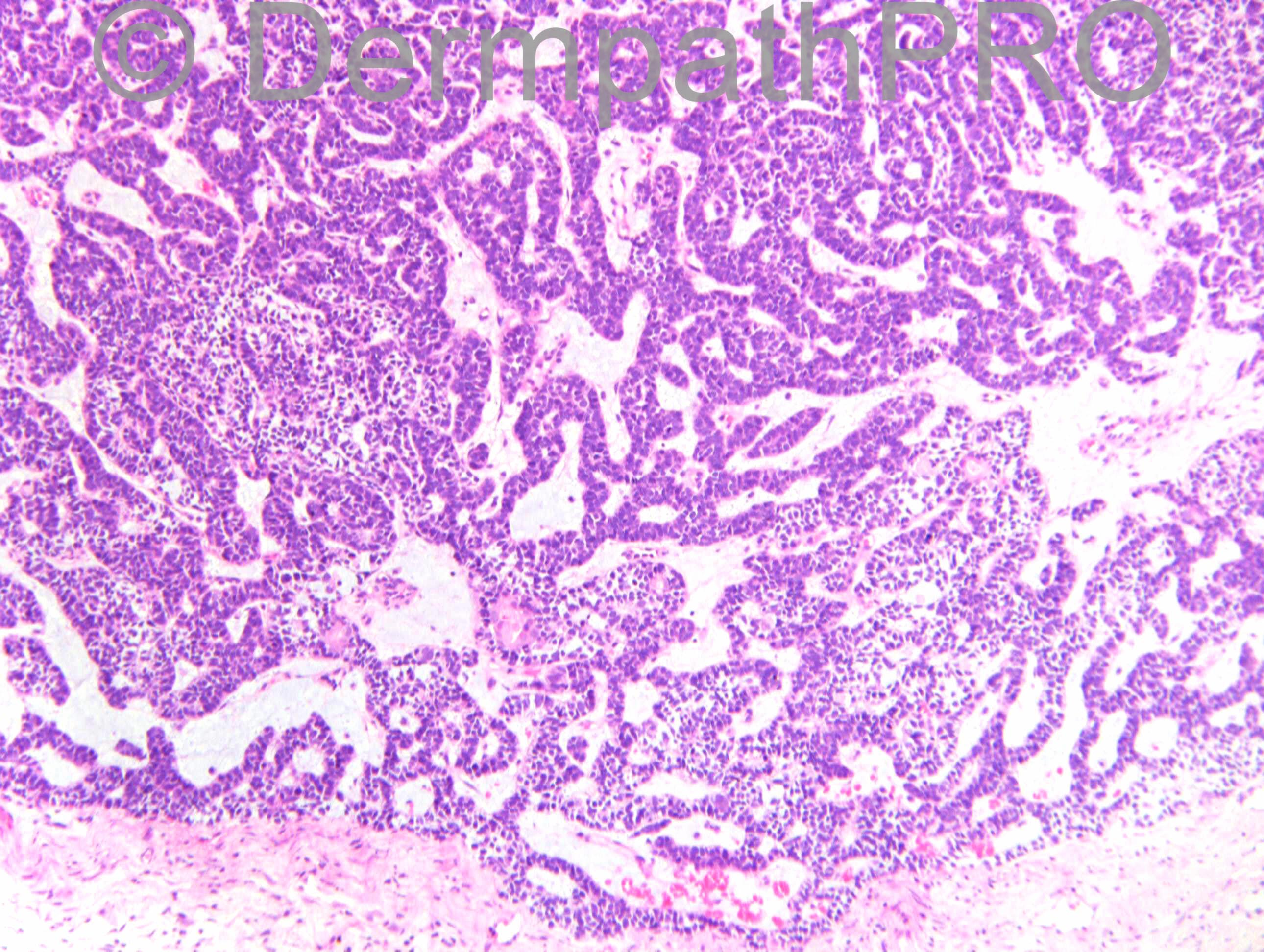
Merkel cell carcinoma with eccrine differentiation. Classical cytology of Merkel carcinoma cells is present besides small eccrine ducts. See the following reference: [url="http://www.ncbi.nlm.nih.gov/pubmed/18995205"]http://www.ncbi.nlm.nih.gov/pubmed/18995205[/url]

Share this comment


Link to comment
share_externally

Dr. Mona Abdel-Halim

Posted

I think it is mucinous carcinoma of the skin, exclusion of metastasis is needed before considering it primary. As Abdul Kadir said, neuroendocrine differentiation may be present, for confirmation with IHC. I am not aware of extensive mucin production by MCC. Lovely case as usual. Hope to see some immunos.

Share this comment


Link to comment
share_externally

Guest Giovanni Falconieri

Posted

Fully agree with the broad differential and the assessment above. If primary, I would vote for papillary/solid neuroendocrine carcinoma of skin, a rare analogue to breast mostly seen in women especially in lids. Of course confirmatory IHC is needed
[url="http://www.ncbi.nlm.nih.gov/pubmed/16160476"]http://www.ncbi.nlm....pubmed/16160476[/url]

Share this comment


Link to comment
share_externally

Dr. Richard Carr

Posted

Forgot to thank Dr Miroslav Radojkovic for kindly sending me this case.

Share this comment


Link to comment
share_externally

Guest Jim Davie MD

Posted

Agree with the above excellent differential. Resembles a Type B mucinous carcinoma of the breast with neuroendocrine differentiation, which can also can also show up in the skin as a primary with similar IHC staining profile.

[color=#000080]Primary mucinous carcinomas of the skin express TFF1, TFF3, estrogen receptor, and progesterone receptors. Hanby AM1, [b]McKee P[/b], Jeffery M, Grayson W, Dublin E, Poulsom R, Maguire B. Am J Surg Pathol. 1998 Sep;22(9):1125-31. [/color][url="http://www.ncbi.nlm.nih.gov/pubmed/9737246"][color=#000080]http://www.ncbi.nlm.nih.gov/pubmed/9737246[/color][/url]

Share this comment


Link to comment
share_externally

Dr. Richard Carr

Posted

[img]https://dermpathpro.com/uploads/spot_diagnosis_comment_img/Case_1207_RAC7027x20_p53_4pm.jpg[/img]

[img]https://dermpathpro.com/uploads/spot_diagnosis_comment_img/Case_1207_RAC7027x20_p63_4pm.jpg[/img]

[img]https://dermpathpro.com/uploads/spot_diagnosis_comment_img/Case_1207_RAC7027x20_Ki67_4pm.jpg[/img]

[img]https://dermpathpro.com/uploads/spot_diagnosis_comment_img/Case_1207_RAC7027x20_BerEP4_4pm.jpg[/img]

Share this comment


Link to comment
share_externally

Dr. Richard Carr

Posted

I did not think of a mucinous or neuroendocrine carcinoma and have asked Dr Radojkovic to undertake neuroendocrine markers. Any other thoughts?

Share this comment


Link to comment
share_externally

Dr. Mona Abdel-Halim

Posted

With the BerEP4 positivity, it might be just a BCC with sweat gland differentiation (evident duct formations).

Share this comment


Link to comment
share_externally

Guest Romualdo

Posted

After a rapid search on Google I found various articles discussing p53, BerEP4 and p63 positivity in Merkel cell carcinomas. p63 positivity is said to imply more agressiviness in localized tumors. One article says BerEP4 is positive in all Merkel cell carcinomas. I think CK20 and neuroendocrine markers should be included in the IHQ panel.

Share this comment


Link to comment
share_externally

Guest Jim Davie MD

Posted

[size=4]Interesting stain pattern that would (in combination with squamous/eccrine differentiation) favor primary cutaneous mucinous CA[/size]

[size=4]-- Low Ki67 index argues against MCC (usually have high >60% positive).[/size]
[size=4]-- The P63 (+) supports a primary cutaneous mucinous CA over metastatic mucinous CA
[url="http://www.ncbi.nlm.nih.gov/pubmed/19788442"] J Cutan Pathol. 2010 Apr;37(4):411-5.[/url]

Would suggest adding: CAM5.2 (LMW cytokeratin), CK15, and EMA.

CK15: may be useful stain to exclude metastatic CA (only 40% sensitive, but 98% specific for primary adnexal CA vs.metastatic adenoCA). Reference: [url="http://www.ncbi.nlm.nih.gov/pubmed/20190734?dopt=Abstract"]Mod Pathol. 2010 May;23(5):713-9. 2010 Feb 26:[/url]

CAM5.2: I have seen Merkel cell carcinoma be particularly treacherous as they sometimes decide "not to read the textbooks" and stain negatively for CK20 in 10-20% of cases. This can cause a fatal exclusion of MCC from a small blue cell tumor workup.
For that reason I routinely add low molecular weight CK (CAM5.2) as it is more sensitive than CK20 in showing perinuclear dots. Reference: [url="http://www.ncbi.nlm.nih.gov/pubmed/16164706"]J Eur Acad Dermatol Venereol. 2005 Sep;19(5):546-51.[/url]

EMA: Positive staining for both EMA and BER-EP4 would support a diagnosis of MCC. Sometimes EMA has characteristic perinuclear dots in MCC...similar to either CK20 or LMW cytokeratin. (In contrast, BCC is usually only positive for BER-EP4, whereas sebaceous CA/squamous CA are only positive for EMA.)[/size]

Share this comment


Link to comment
share_externally

Guest Tiberiu Tebeica

Posted

I see no evidence against an adenoid BCC so far...

Share this comment


Link to comment
share_externally

Dr. Richard Carr

Posted

Dear All. Thank-you for the opinions.
My report as follows:
I agree with you (Dr Radojkovic), this looks like the malignant counterpart of spiradenoma. I could not identify a pre-existing benign spiradenoma however. The lesion is low grade and well circumscribed, and resembles the salivary basal cell adenocarcinoma. It would be prudent just to rule out a primary elsewhere. However, I suspect this is most likely a cutaneous primary. The lesion abuts the base of the specimen and there is a possibility for local recurrence. With the nice circumscription and relatively low grade, I would expect metastasis to be unlikely from this lesion.

Notes: p53 diffuse strong (supports malignant); p63 diffuse sparing central ducts (supports myoepithelial element), Ki67 ~ 10% (supports low-grade and not MCC); BerEP4 +/++50 weak to moderate with accentuation of duct lumina (the lack of peripheral palisading and lack of strong peripheral accentiation of BerEP4 argue against BCC in my opinion) In my opinion there is a clear myoepithelial look with stromal type mucin - a little mixed tumour like).

I will report back on the neuroendocrine markers in due course.

Share this comment


Link to comment
share_externally

Dr. Richard Carr

Posted

Hi Richard,
Here are the results of immunohistochemical investigation that I performed on that tumour that we thought to be probably malignant eccrine spiradenoma and some people suggested neuroendocrine differentiation.
p63 - Diffuse nuclear positivity in all cells (spared in central glands)
p53 - Diffuse, mostly strong positivity in over 75% of cells,
CK5/6 - Strong membranous positivity in some, predominantly 'luminal' cells,
CK7 - Similar to CK5/6, membranous positivity in some 'luminal' cells,
BerEP4 - Weak positivity in a few, predominantly 'luminal' cells,
EMA - Strong membranous or intracytoplasmic positivity in a few, predominantly 'luminal' cells,
CD117 - Weak, predominantly membranous positivity in quite a few cells;
S-100 protein - Weak to moderate intracytoplasmic positivity in occasional cells and strong positivity in numerous dendritic dermal cells;
Cam 5.2, CK20, CEA, Chromogranin, Synaptophysin, CD56 and Oestrogen receptors - All negative.
Proliferative activity of this tumour is relatively high with 30 - 40% of cells showing strong nuclear positivity with the antibody against Ki67 antigen.
[b][u]CONCLUSION[/u][/b]
There are [b]no features of neuroendocrine differentiation[/b] and the tumour's immunophenotype, although not quite typical, is supporting the original diagnosis of an [b]adnexal skin tumour, apparently of the eccrine gland origin, consistent with eccrine spiradenoma and features indicating malignant transformation[/b] (diffuse positivity with p53 and relatively high proliferative activity).
With kind regards.
Miro Radojkovic

Share this comment


Link to comment
share_externally


Join the conversation

You can post now and register later. If you have an account, sign in now to post with your account.

Guest
Add a comment...

×   Pasted as rich text.   Paste as plain text instead

  Only 75 emoji are allowed.

×   Your link has been automatically embedded.   Display as a link instead

×   Your previous content has been restored.   Clear editor

×   You cannot paste images directly. Upload or insert images from URL.

Loading...

×
×
  • Create New...