Jump to content
In this section we have spot diagnoses posted on a daily basis since June 2010, now over 4000! You can review the archived cases and read the suggested diagnoses by users and the final comment by the contributors.
Case are uploaded each week day by 10 am UK time with the correct diagnosis will generally be posted at 8 pm UK time. Why not view the most recent spot diagnosis and proffer a diagnosis?

Case Number : Case 1212 - 13 February Posted By: Guest

Please read the clinical history and view the images by clicking on them before you proffer your diagnosis.
Submitted Date :
   (0 reviews)

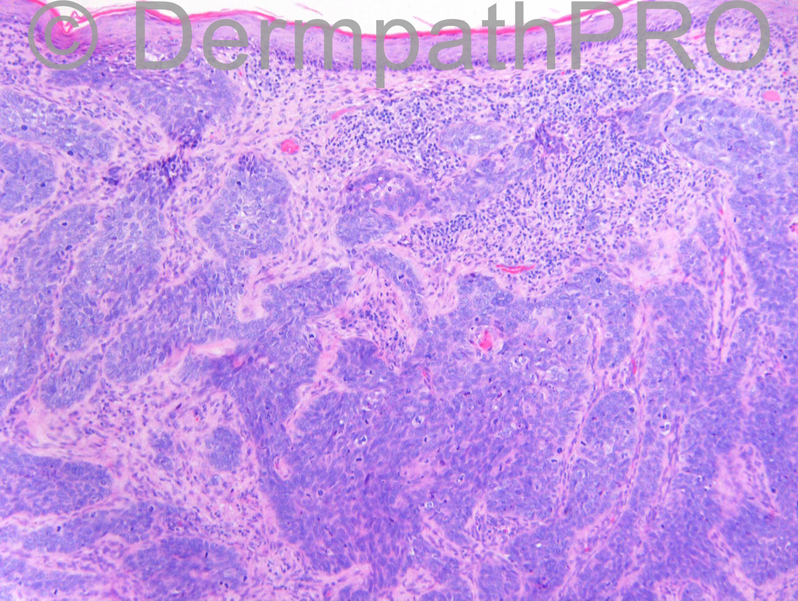
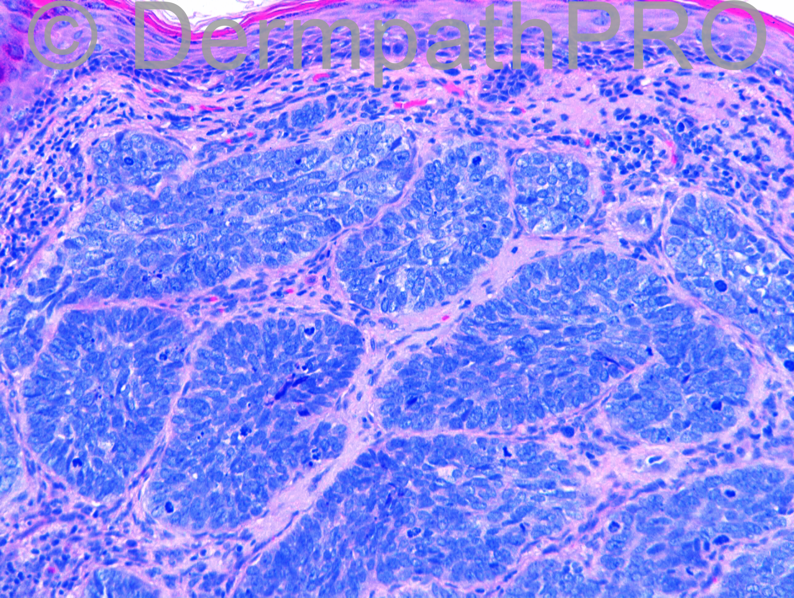
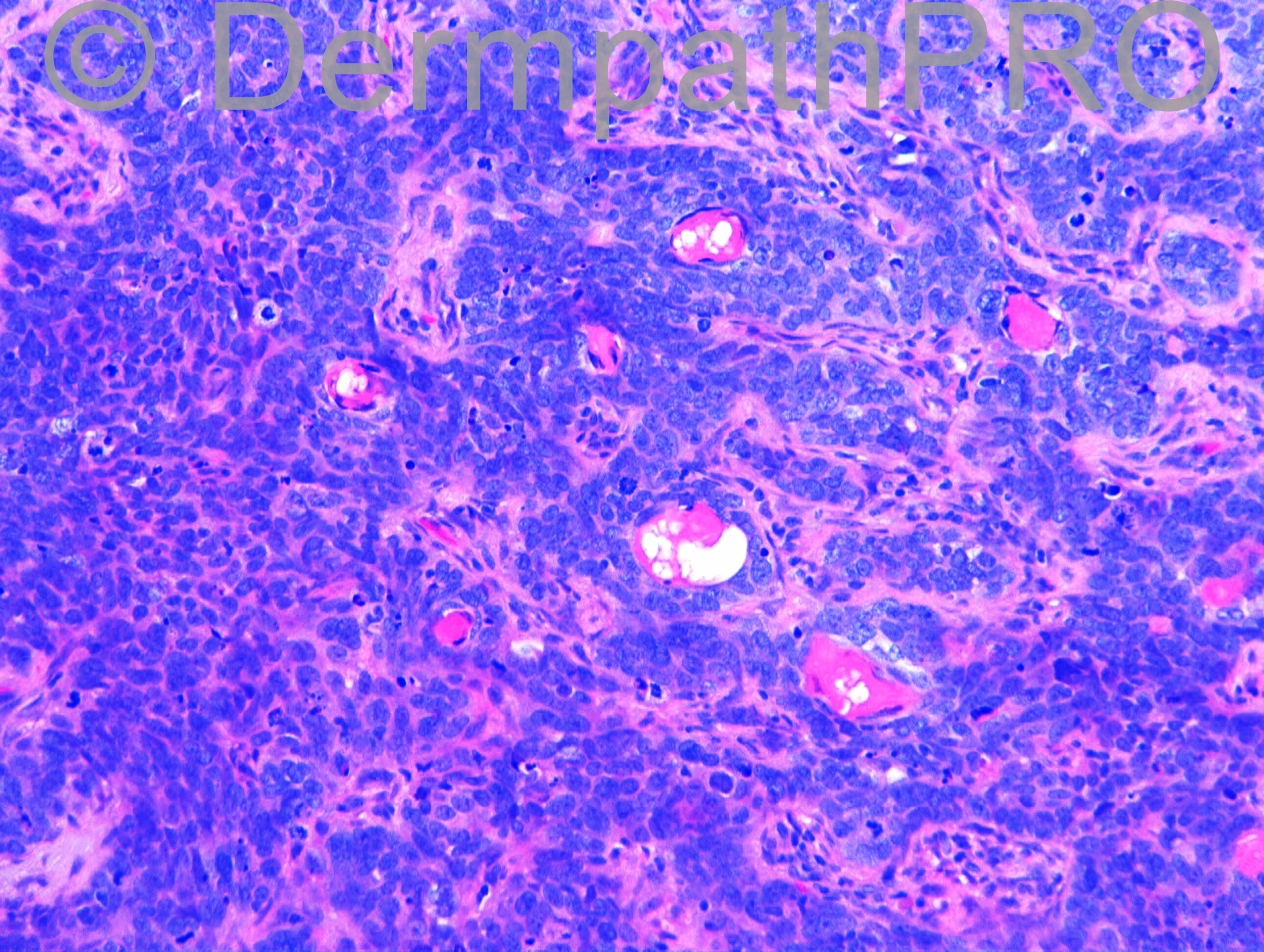
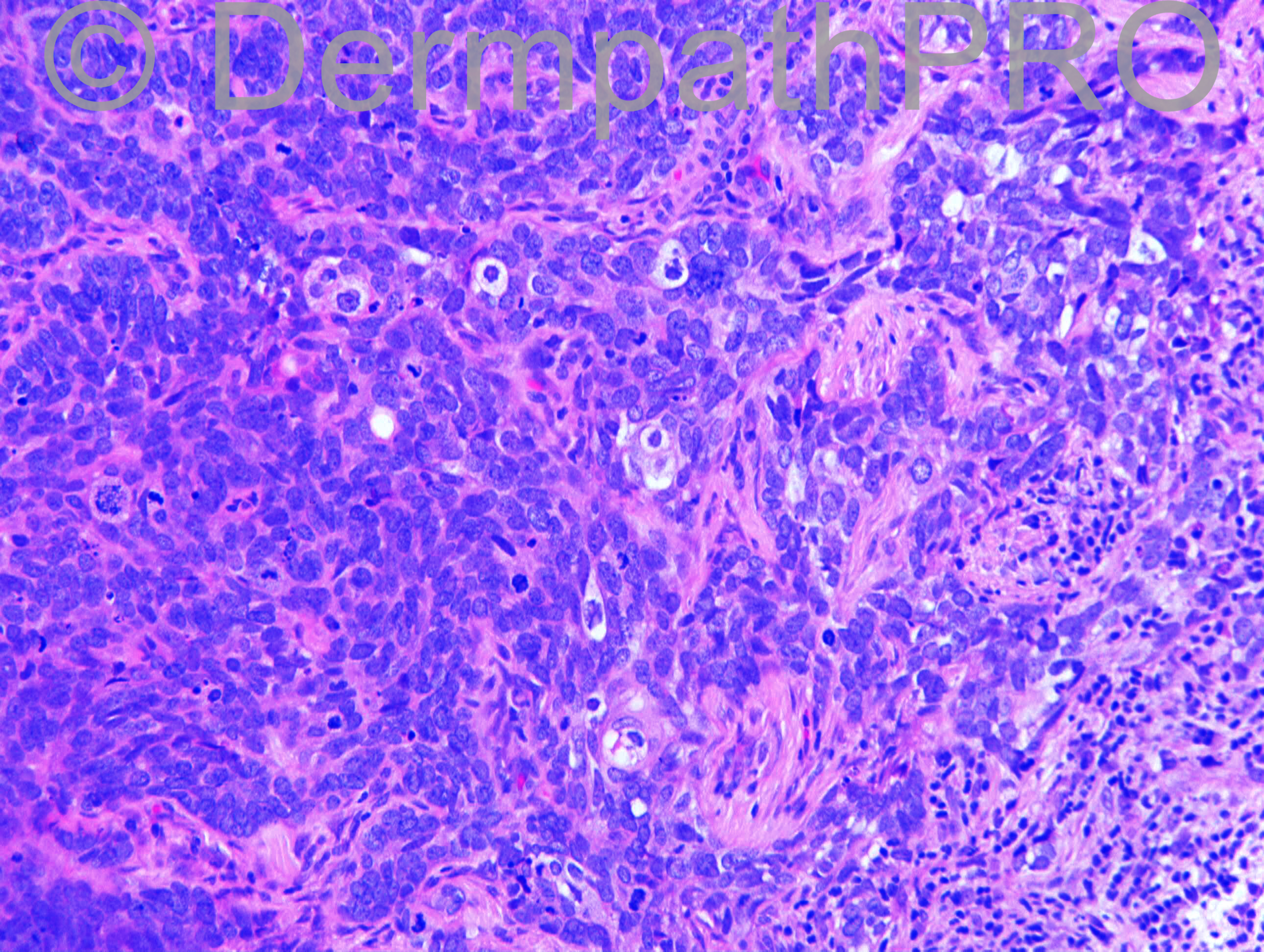
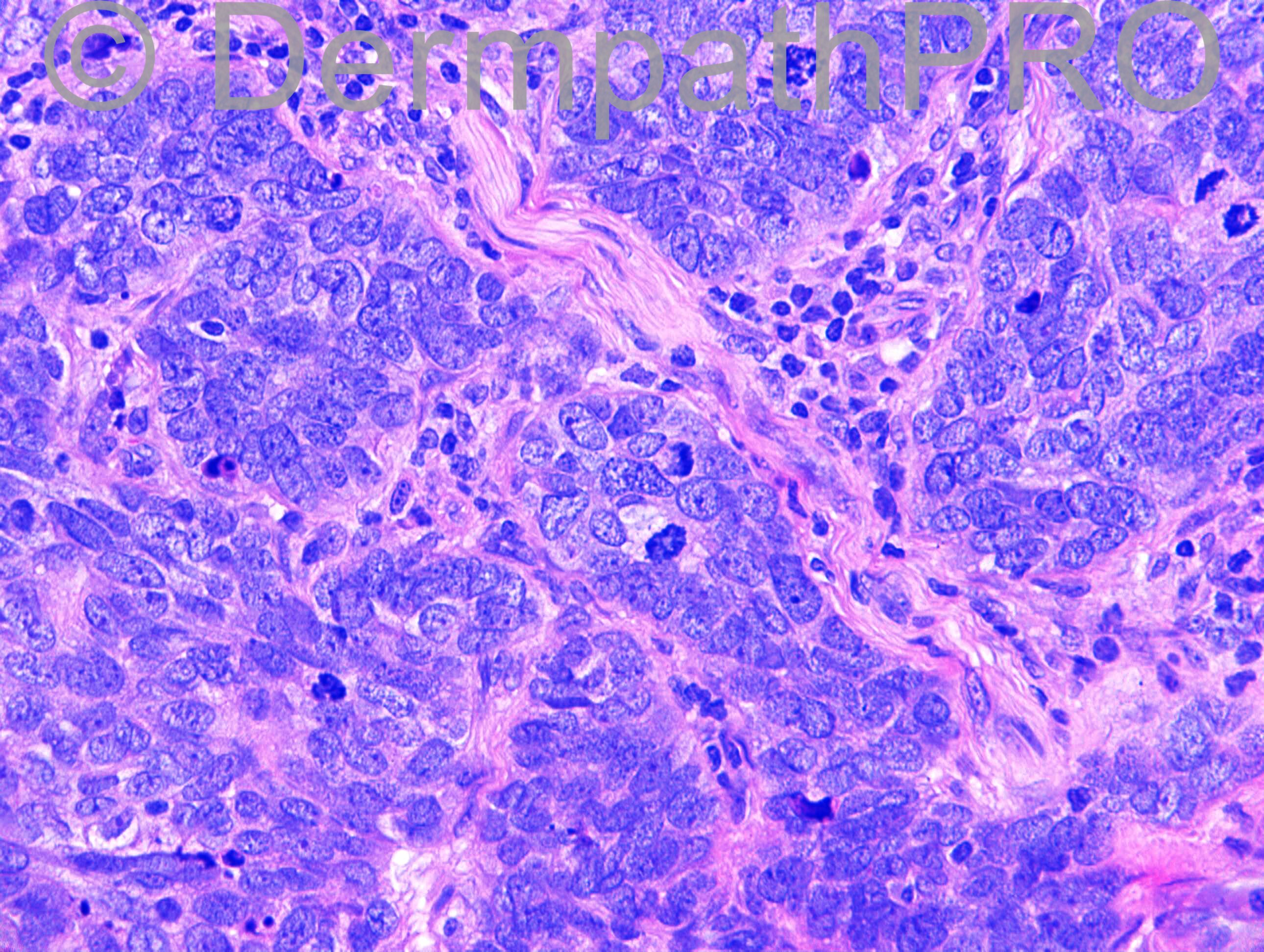
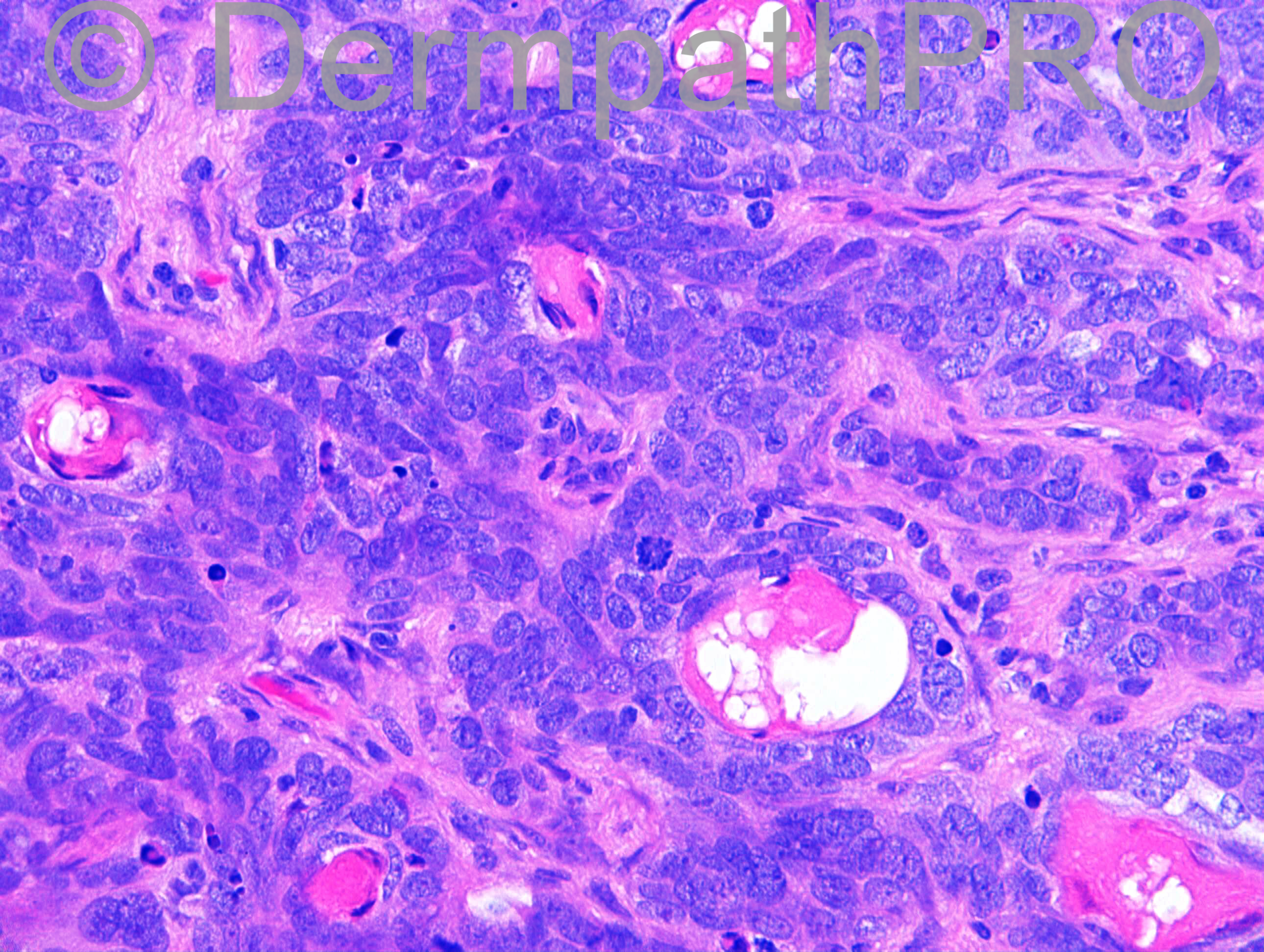
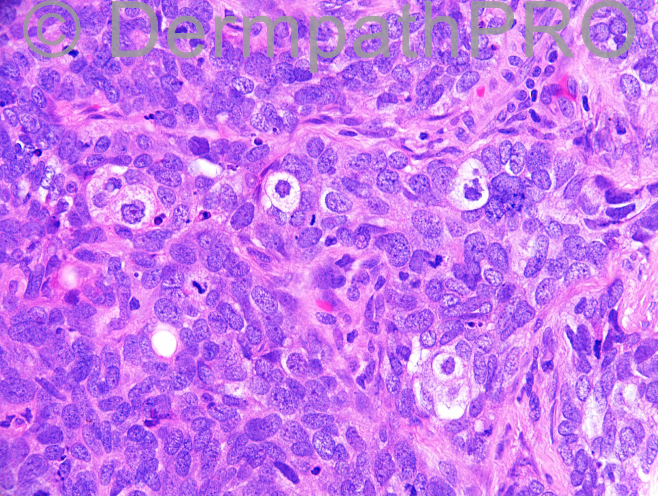
F87. Lesion on nose. ?BCC

Case posted by Dr Richard Carr

  Report Record

User Feedback


Dr. Mona Abdel-Halim

Posted

From sebaceous tumors, I worked out in mind: Well differentiated seb carcinoma vs. Sebaceoma.
I think the architecture (not that well circumscribed and the slightly infiltrative lower border) and the significant atypical mitoses will favour well differentiated seb carcinoma.
The rare BCC with seb differentiation should have shown typical peripheral palisading and peritumoral mucinous artefacts which is not the case (only minimal focal palisading)
Would love to see EMA and BerEP4

Share this comment


Link to comment
share_externally

Guest Romualdo

Posted

I think the presence of numerous mitotic figures, some of them apparently atypical, and the extremely limited foci of sebaceous differentiation favor a poorly differentiated sebaceous carcinoma.

Share this comment


Link to comment
share_externally

Eman El-Nabarawy

Posted

Favor BCC with sebaceous differentiation.

Share this comment


Link to comment
share_externally

Dr. Richard Carr

Posted

[img]https://dermpathpro.com/uploads/spot_diagnosis_comment_img/Case_1212_RAC6949x40b_EMA_4pm.jpg[/img]

Share this comment


Link to comment
share_externally

Dr. Richard Carr

Posted

[img]https://dermpathpro.com/uploads/spot_diagnosis_comment_img/Case_1212_RAC6949x10_BerEP4_4pm.jpg[/img]

Share this comment


Link to comment
share_externally

Dr. Mona Abdel-Halim

Posted

EMA is highlighting ducts and focally staining some cells (mature sebocytes) , immature cells/ basaloid cells are negative. This is seen in Sebaceoma and Seb Carc.
BerEP4 is negative apart from a tiny focus. This speaks against BCC.
Seb Carc is usually BerEP4 positive while Sebaceoma is BerEP4 negative.
So this case on IHC is following Sebaceoma pattern.
Although, after some reading, I found that sebaceomas can have conspicuous mitotic activity, still I am not comfortable with the architecture of this lesion being a benign architecture and I am still favoring Seb Carc and I need help !!
Also, I think that exclusion of Muir Torre syndrome is important.

Share this comment


Link to comment
share_externally

nick turnbull

Posted

sebaceoma with prominent mitotic activity? I'm anxious about sebaceous carcinoma. , ki67, p53, p21? Looking forward to the answer when I get up in the morning. Goodnight all from NZ

Share this comment


Link to comment
share_externally

Dr. Richard Carr

Posted

This was a difficult case. I reported the prior punch biopsy as a poorly differentiated sebaceous carcinoma then reviewed it at an MDM and in view of close resemblance to BCC watered down my report initial report as follows:
SUPPLEMENTARY REPORT: DR R A CARR
Immunostain shows moderate focal BerEP4 staining. EMA is limited to sebaceous duct differentiation. I cannot entirely rule out BCC with sebaceous differentiation.

The excision (illustrated) was reported at a sister institution as a nodular BCC with no mention of the sebaceous differentiation (which was pretty focal and these images are representative).

I reviewed the case and issued a supplementary as follows:
Sections reviewed for Skin MDM. Malignant basaloid tumour with distinctive amphophilic rounded nuclei. There is no significant palisading, retraction or interstitial mucin. BerEP4 is negative. EMA confirms morphological impression of focal sebaceous duct differentiation. A difficult case, but in combination with the prior biopsy, the weight of evidence favours poorly differentiated sebaceous carcinoma rather than basal cell carcinoma. The deep margin is very close.

Learning points: As Mona says be careful to make sure you seen good palisading with retraction artefact and interstial mucin before excluding mimics of BCC like basaloid SCC and sebaceous carcinoma. I suspect a good proportion (though certainly not all) metastasising BCC are other tumours. Like Romualdo & Mark I called it poorly differentiated (basaloid predominant and highly mitotic). This case had particularly well developed focal sebaceous ductal differentiation (distincitve abrupt compact keratin with bubbly effect). Solitary sebaceous carcinomas on the H&N of the elderly have a low association with Muir Torre syndrome.

Share this comment


Link to comment
share_externally


Join the conversation

You can post now and register later. If you have an account, sign in now to post with your account.

Guest
Add a comment...

×   Pasted as rich text.   Paste as plain text instead

  Only 75 emoji are allowed.

×   Your link has been automatically embedded.   Display as a link instead

×   Your previous content has been restored.   Clear editor

×   You cannot paste images directly. Upload or insert images from URL.

Loading...

×
×
  • Create New...