Jump to content
In this section we have spot diagnoses posted on a daily basis since June 2010, now over 4000! You can review the archived cases and read the suggested diagnoses by users and the final comment by the contributors.
Case are uploaded each week day by 10 am UK time with the correct diagnosis will generally be posted at 8 pm UK time. Why not view the most recent spot diagnosis and proffer a diagnosis?

Case Number : Case 1214 - 17 February Posted By: Guest

Please read the clinical history and view the images by clicking on them before you proffer your diagnosis.
Submitted Date :
   (0 reviews)

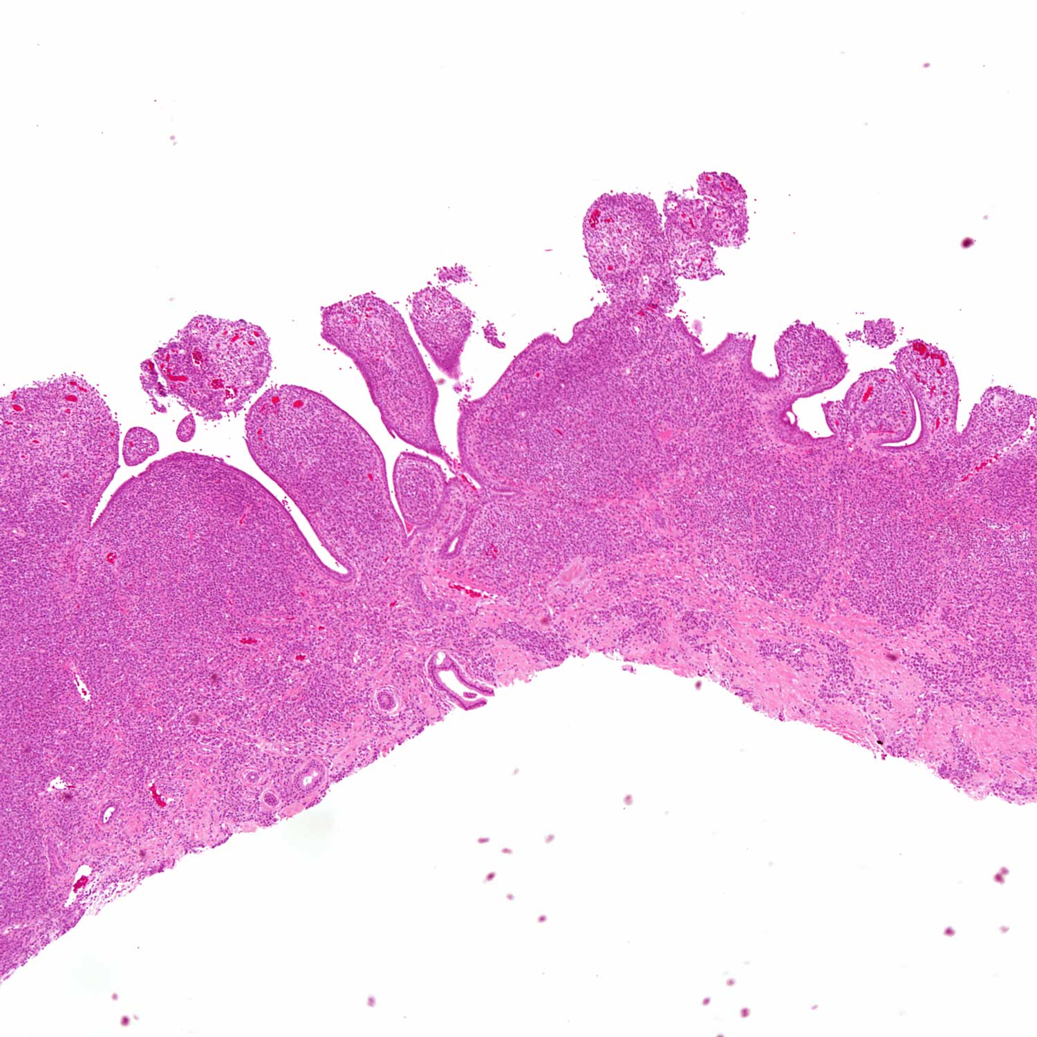
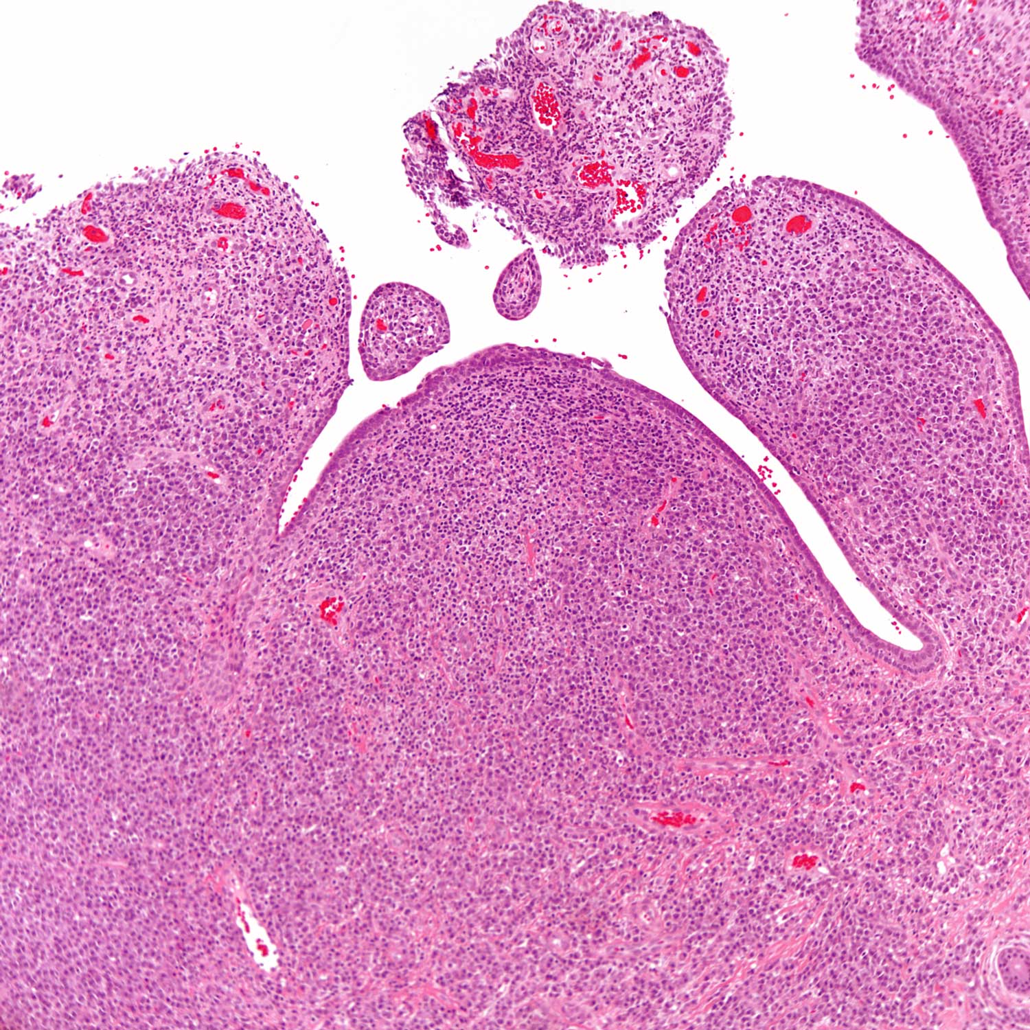
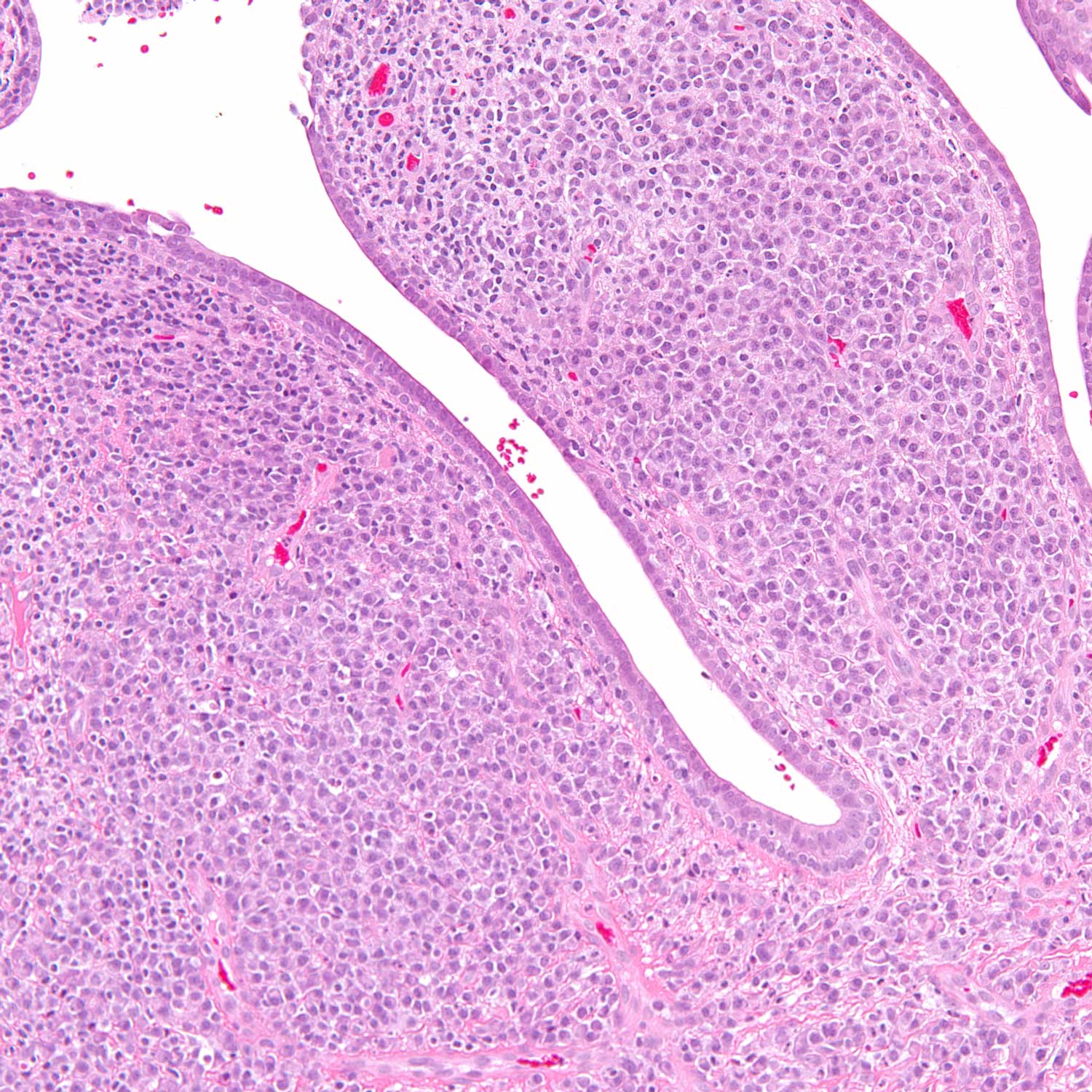
65 year old woman with a lesion on the mid nose.

Case posted by Dr Uma Sundram


  Report Record

User Feedback


Guest Giovanni Falconieri

Posted

Heavy plasma cell infiltrate. If polytypic, I suspect chronic infection (rhinoscleroma?). PAS stain may be revealing

Share this comment


Link to comment
share_externally

Guest Romualdo

Posted

Except for some neutrophils near the surface I see a pure plasma cell population. I think this is plasmacytoma.

Share this comment


Link to comment
share_externally

Eman El-Nabarawy

Posted

Lower part of syringocystadenoma papilliferum.

Share this comment


Link to comment
share_externally

Dr. Mona Abdel-Halim

Posted

As my colleague Eman said, this is nothing but Syringocystadenoma Papilliferum... The plasma cells are a clue as well as the tall columner epithelial lining of the papillary projections with a hint of decapitation secretion in some areas...

Share this comment


Link to comment
share_externally

Guest Jim Davie MD

Posted

Agree, syringocystadenoma papilliferum.
Nodular plasma cell-rich infiltrate is supportive for the dx, when it is present. I find the dual-layer of surface epithelial cells with basal layer showing cuboidal features, and decapitation secretion/overall papillary architecture as Mona pointed out, most helpful for making the diagnosis in a head/neck cutaneous lesion. (Very rarely these degenerate into syringocystadenocarcinoma papilliferum, but there is no atypia in the surface bilayer, and no invasive component in these sections.)

Share this comment


Link to comment
share_externally

Uma Sundram

Posted

I also wanted to add that the plasma cell population was polytypic. No infectious agent was found via special stains.

Share this comment


Link to comment
share_externally


Join the conversation

You can post now and register later. If you have an account, sign in now to post with your account.

Guest
Add a comment...

×   Pasted as rich text.   Paste as plain text instead

  Only 75 emoji are allowed.

×   Your link has been automatically embedded.   Display as a link instead

×   Your previous content has been restored.   Clear editor

×   You cannot paste images directly. Upload or insert images from URL.

Loading...

×
×
  • Create New...