Jump to content
In this section we have spot diagnoses posted on a daily basis since June 2010, now over 4000! You can review the archived cases and read the suggested diagnoses by users and the final comment by the contributors.
Case are uploaded each week day by 10 am UK time with the correct diagnosis will generally be posted at 8 pm UK time. Why not view the most recent spot diagnosis and proffer a diagnosis?

Case Number : Case 1218 - 23 February Posted By: Guest

Please read the clinical history and view the images by clicking on them before you proffer your diagnosis.
Submitted Date :
   (0 reviews)

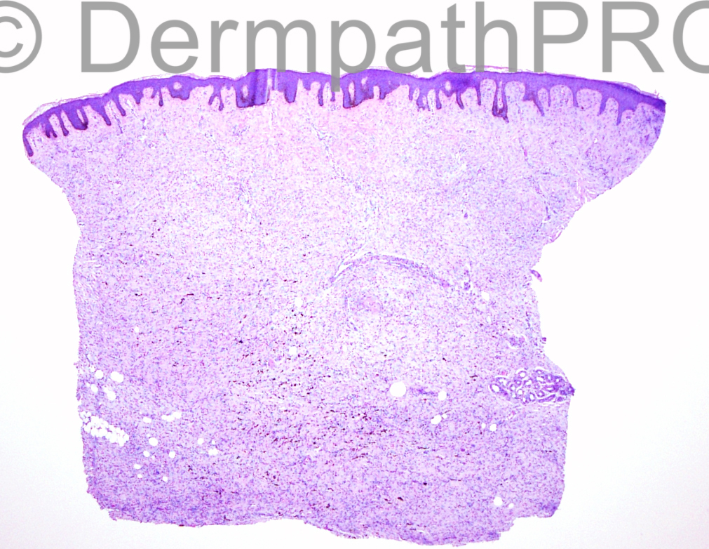
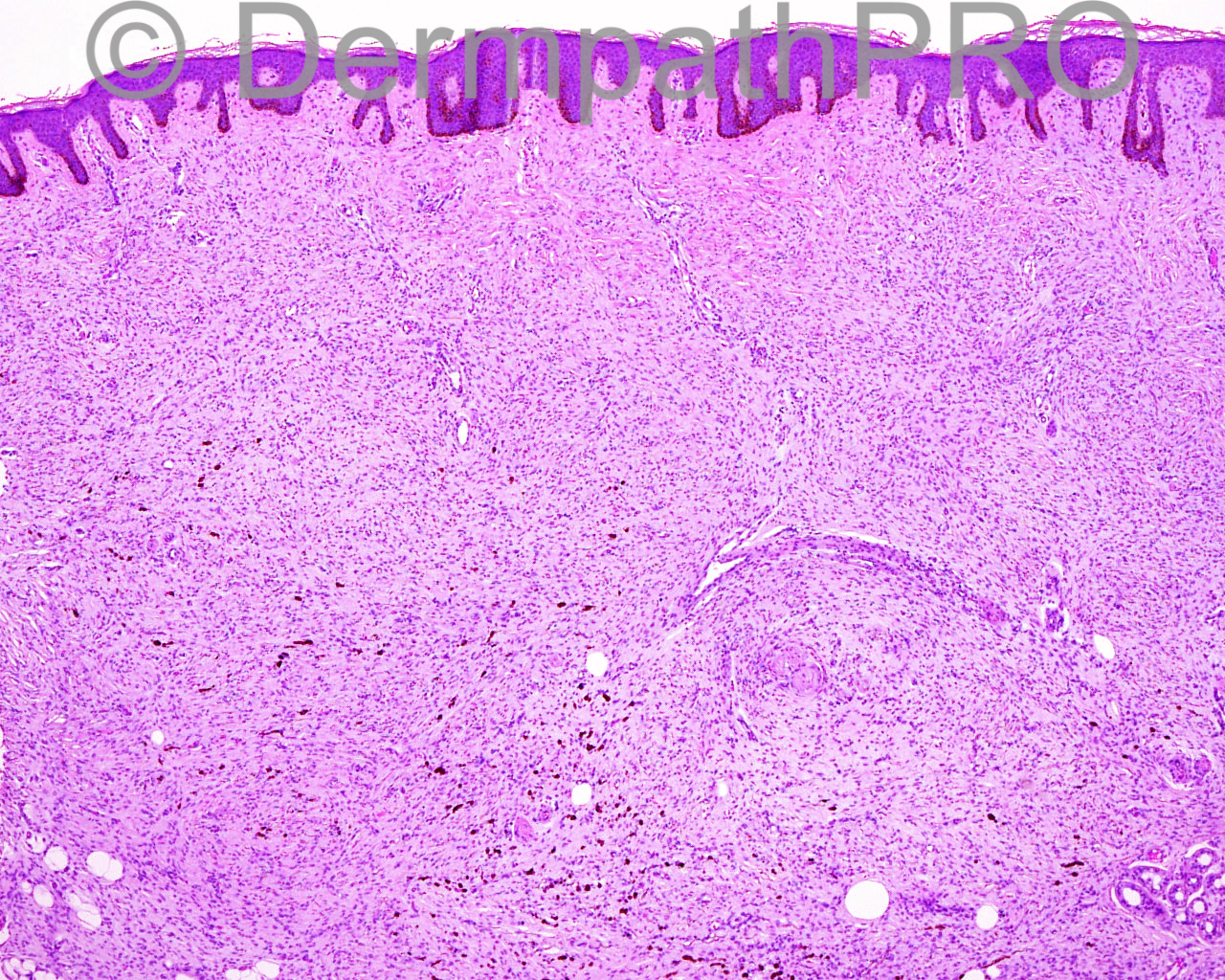
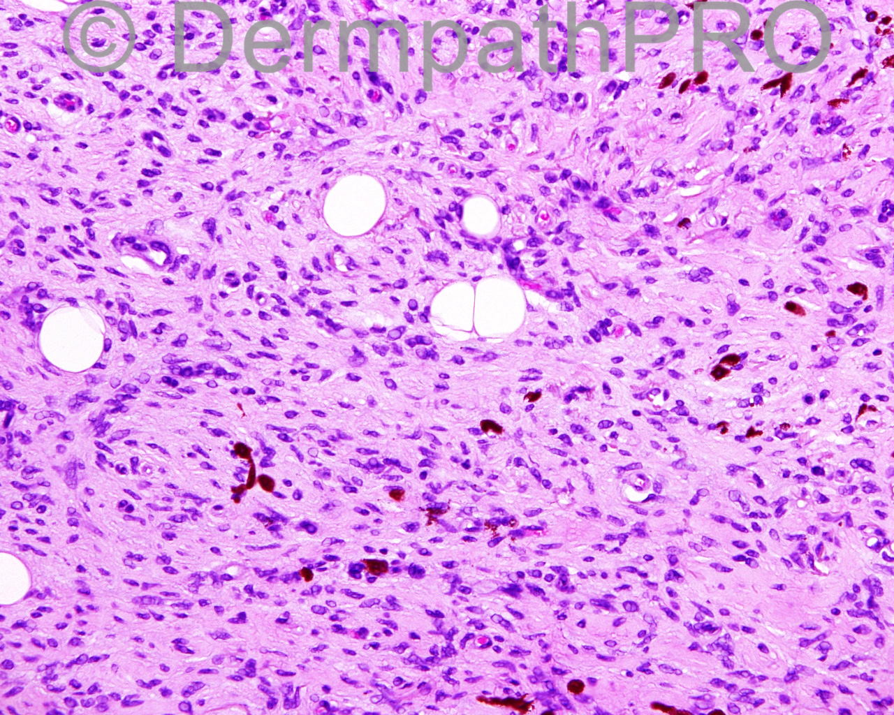
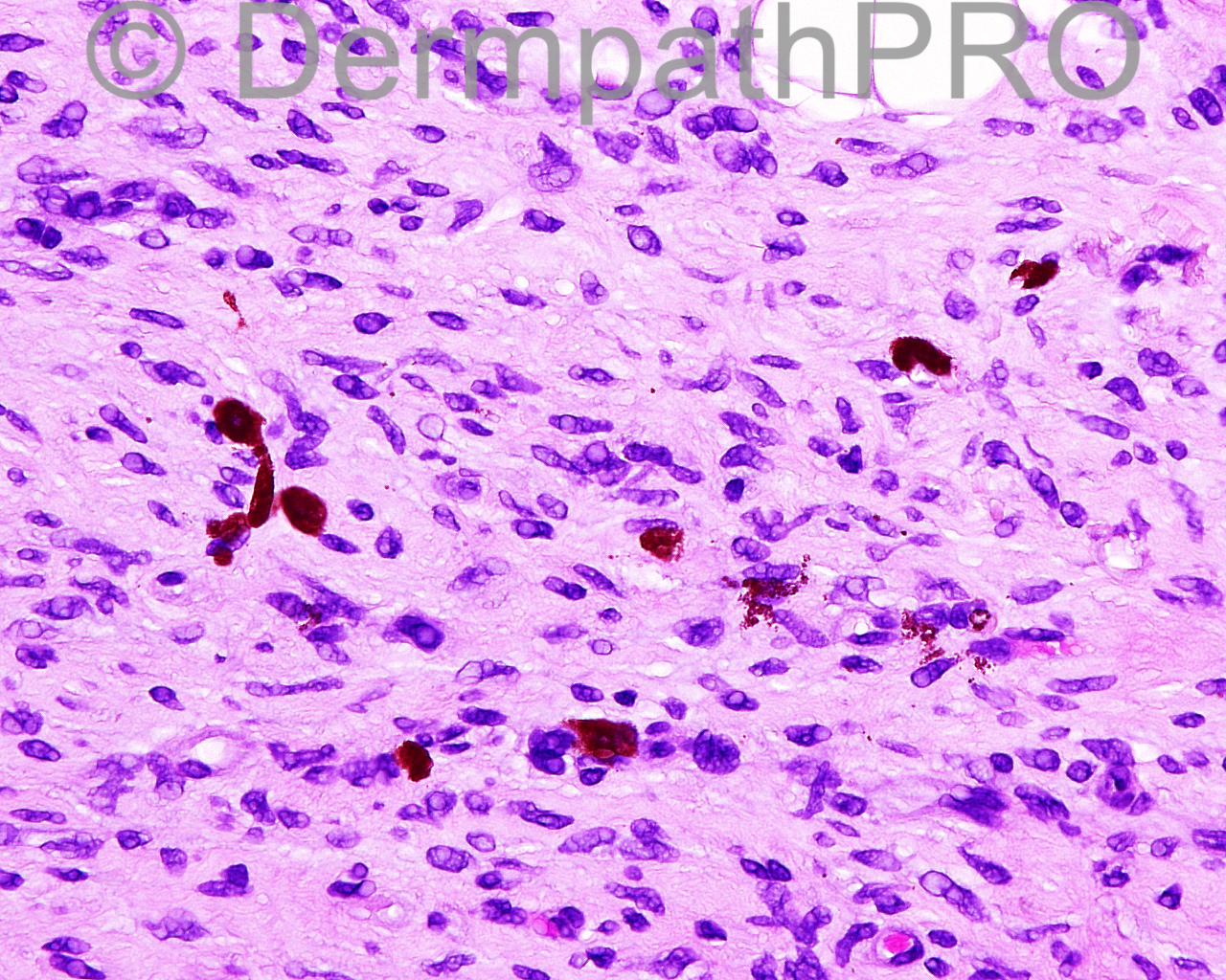
The patient is a 52-year-old man with a punch biopsy of dome-shaped, pink-tan nodule with positive dimple sign on the left buttock.

Case posted by Dr Mark Hurt


  Report Record

User Feedback


Dr. Mona Abdel-Halim

Posted

Agree.. Pigmented DFSP (Bednar) tumor..

Share this comment


Link to comment
share_externally

Dr. Richard Carr

Posted

Myxoid DFSP certainly must but high on the list but something is holding me back (obviously CD34 could help). DFSP usually have a storiform pattern and clearly distinguished collagen and elastic fibres. I thought it had a diffuse neurofibroma-like look as well (would do S100 and because of site MelanA as well although it does not look like blue naevus). A low-grade MPNST and desmoplastic melanoma are other considerations.

Share this comment


Link to comment
share_externally

Mark A. Hurt MD

Posted

Here is my diagnosis:

SKIN, LEFT BUTTOCK , PUNCH BIOPSY :
[b]-- DERMATOFIBROSARCOMA PROTUBERANS, PIGMENTED (BED[/b][b]N[/b][b]ÁR[/b][b] TUMOR)[/b]
[b] WITH MELANOCYTIC COLONIZATION[/b]


[center]Reference[/center]


Goncharuk V, Mulvaney M, Carlson JA. Bednár tumor associated with dermal melanocytosis: melanocytic colonization or neuroectodermal multidirectional differentiation? J Cutan Pathol. 2003 Feb;30(2):147-51. PubMed PMID: 12641795.


Micro: This lesion consists of a relatively diffuse neoplasm of relatively small and uniform cells that produce a sheet-like pattern of infiltration through the dermis. Some areas have a storiform pattern but most areas are diffuse and encase adnexal structures as well as some adipose tissue. A curious finding is the presence of pigmented cells in this lesion, many of them somewhat dendritic and a few epithelioid. This lesion abuts the surface or at least comes very close to the surface. Most of the lesional cells are epithelioid and slightly tapered. The pigmented cells within it are positive with Melan-A in the brown and red chromogen forms. CD68 is negative in those same cells. S100 protein appears to be positive within them. SOX10 with a red chromogen also appears to stain the nuclei of these cells marking them as melanocytes. These cells, however, are a very small part of the entire neoplasm. CD34, for instance, is diffusely positive throughout the lesion. Ki-67 shows an increased signal of about 5 % to 10% in the lower portion of the lesion and less staining in the superficial aspects. Factor XIIIa shows dendritic staining throughout much of the field. The elastic tissue staining shows a paucity of elastic fibers deep and an increase in elastic fibers in the mid portion of the lesion. Smooth muscle actin and desmin are both negative with the exception of blood vessels in the field.

Share this comment


Link to comment
share_externally


Join the conversation

You can post now and register later. If you have an account, sign in now to post with your account.

Guest
Add a comment...

×   Pasted as rich text.   Paste as plain text instead

  Only 75 emoji are allowed.

×   Your link has been automatically embedded.   Display as a link instead

×   Your previous content has been restored.   Clear editor

×   You cannot paste images directly. Upload or insert images from URL.

Loading...

×
×
  • Create New...