Jump to content
In this section we have spot diagnoses posted on a daily basis since June 2010, now over 4000! You can review the archived cases and read the suggested diagnoses by users and the final comment by the contributors.
Case are uploaded each week day by 10 am UK time with the correct diagnosis will generally be posted at 8 pm UK time. Why not view the most recent spot diagnosis and proffer a diagnosis?

Case Number : Case 1222 - 27 February Posted By: Guest

Please read the clinical history and view the images by clicking on them before you proffer your diagnosis.
Submitted Date :
   (0 reviews)

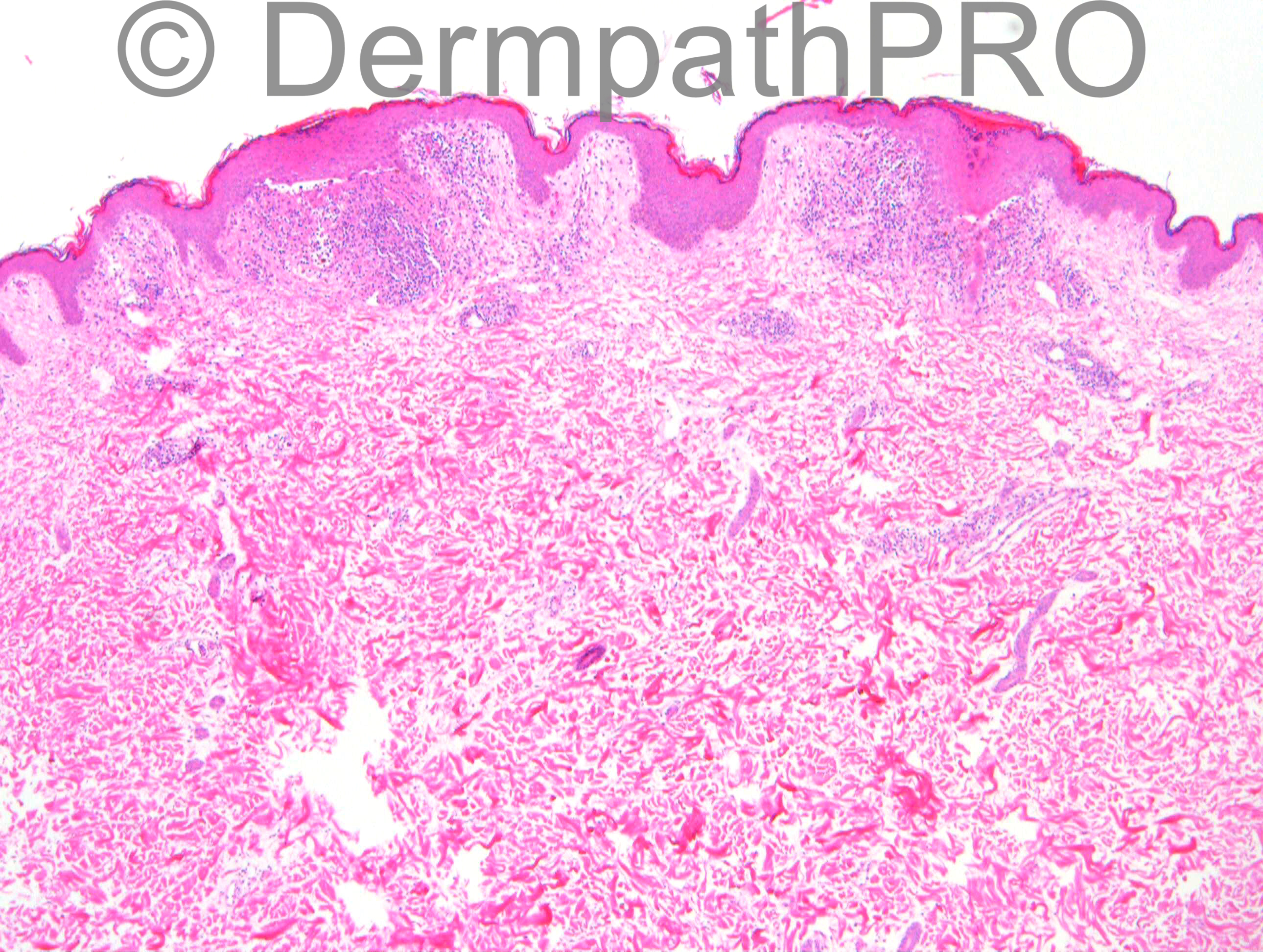
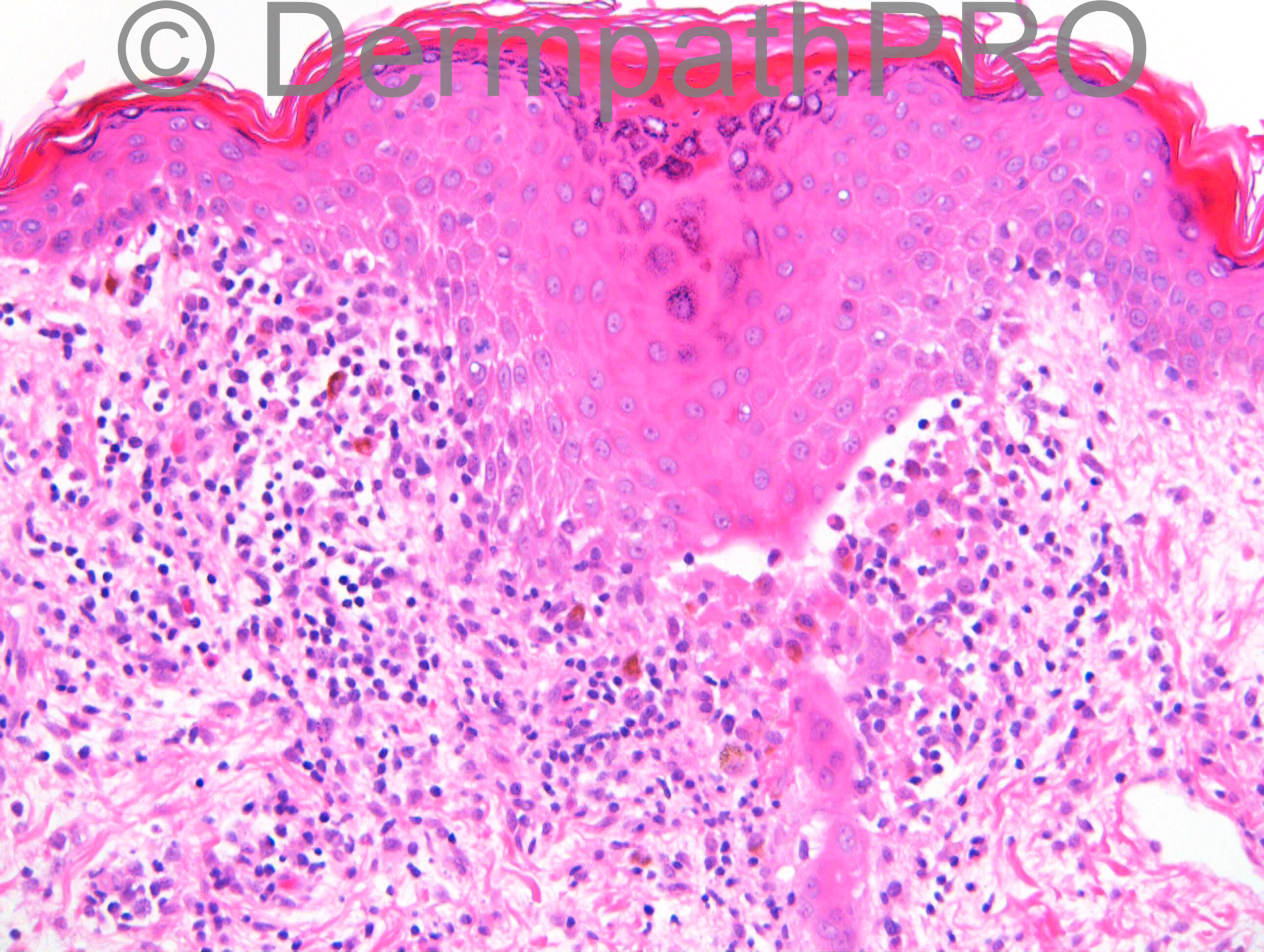
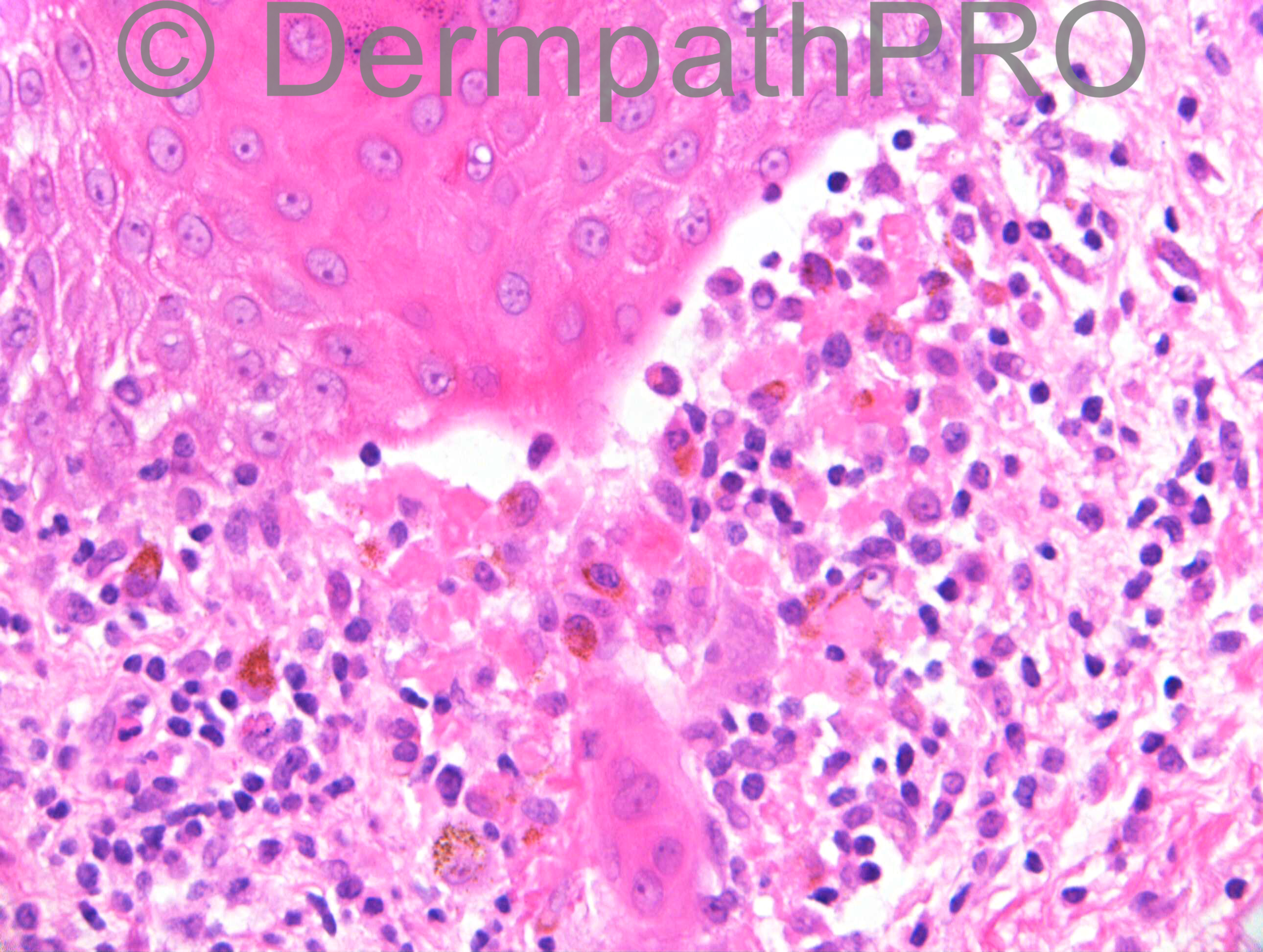
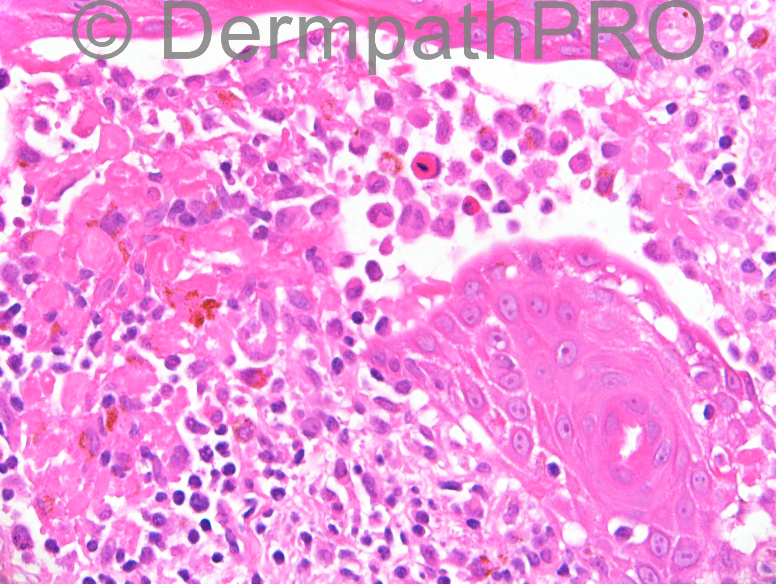
F41. Small asymptomatic erythematous papules both axillae with marked post inflammatory hyperpigmentation. ?Dowling Degos, ?Pityriasis lichenoides – but localised, ??Darier’s.

Case posted by Dr Richard Carr

  Report Record

User Feedback


Dr. Mona Abdel-Halim

Posted

I think we r having a follicle (and apocrine duct) related interface changes, spongiosis, exocytosis, secondary acantholysis, dyskeratotic cells, vesiculation and related dermal inflammatory cell infiltrate with overlying hyperkeratosis and parakeratosis. This together with the clinical presentation, I am thinking of fox fordyce disease (apocrine miliaria), but the strange thing is that fox fordyce is usually pruritic?

Share this comment


Link to comment
share_externally

Guest Romualdo

Posted

In my oppinion the most striking feature is the presence of numerous Langerhans cells. Although the first idea is Langerhans cell histiocytosis, the first images suggest lichen planus. There are foci of epidermal hyperplasia, hyperkeratosis and hypergranulosis associated with band-like lymphohistiocytic infiltrates and vacuolar alteration of dermo-epidermal junction. I think this could be lichen planus pigmentosus inversus with associated Langerhans cell hyperplasia.

Share this comment


Link to comment
share_externally

Eman El-Nabarawy

Posted

Folliculocenteric Langerhans cell histiocytosis mimicking lichen planus.

Share this comment


Link to comment
share_externally

Dr. Richard Carr

Posted

This was a case from far afield but I noted that the patient was taking citalopram.

Share this comment


Link to comment
share_externally

Miliarial spongiosis and acantholysis. ?Acrosyringeal Grover's disease vs. drug reaction

Share this comment


Link to comment
share_externally

Dr. Mona Abdel-Halim

Posted

Citalopram is a SSRI anti- depressant. These drugs are effective in controlling severe and chronic pruritus. Looking at the images again, I still think this is apocrine miliaria and may be the lesions are asymptomatic because of the citalopram... May be..

Share this comment


Link to comment
share_externally

Dr. Richard Carr

Posted

I was struck by the acrosyringeal centric location of this flexural lichen planus-like reaction and raised the posibility of a a drug (citalopram) induced lichen planus. The marked individual cell necrosis in addition to numerous Langerhan's cells and spongiosis to me together with the odd clinical features are good for a drug reaction. I was able to ascertain some evidence of reports of Citalopram induced lichen planus. Whether the drug is somehow being concentrated by the sweat ducts at the acrosyringium is obviously speculative but an interesting idea. I do not think this is Langerhan's cell histiocytosis because of the mixed infiltrate and clear cut lichenoid reaction pattern.

Share this comment


Link to comment
share_externally


Join the conversation

You can post now and register later. If you have an account, sign in now to post with your account.

Guest
Add a comment...

×   Pasted as rich text.   Paste as plain text instead

  Only 75 emoji are allowed.

×   Your link has been automatically embedded.   Display as a link instead

×   Your previous content has been restored.   Clear editor

×   You cannot paste images directly. Upload or insert images from URL.

Loading...

×
×
  • Create New...