Jump to content
In this section we have spot diagnoses posted on a daily basis since June 2010, now over 4000! You can review the archived cases and read the suggested diagnoses by users and the final comment by the contributors.
Case are uploaded each week day by 10 am UK time with the correct diagnosis will generally be posted at 8 pm UK time. Why not view the most recent spot diagnosis and proffer a diagnosis?

Case Number : Case 1182 - 2nd January Posted By: Guest

Please read the clinical history and view the images by clicking on them before you proffer your diagnosis.
Submitted Date :
   (0 reviews)

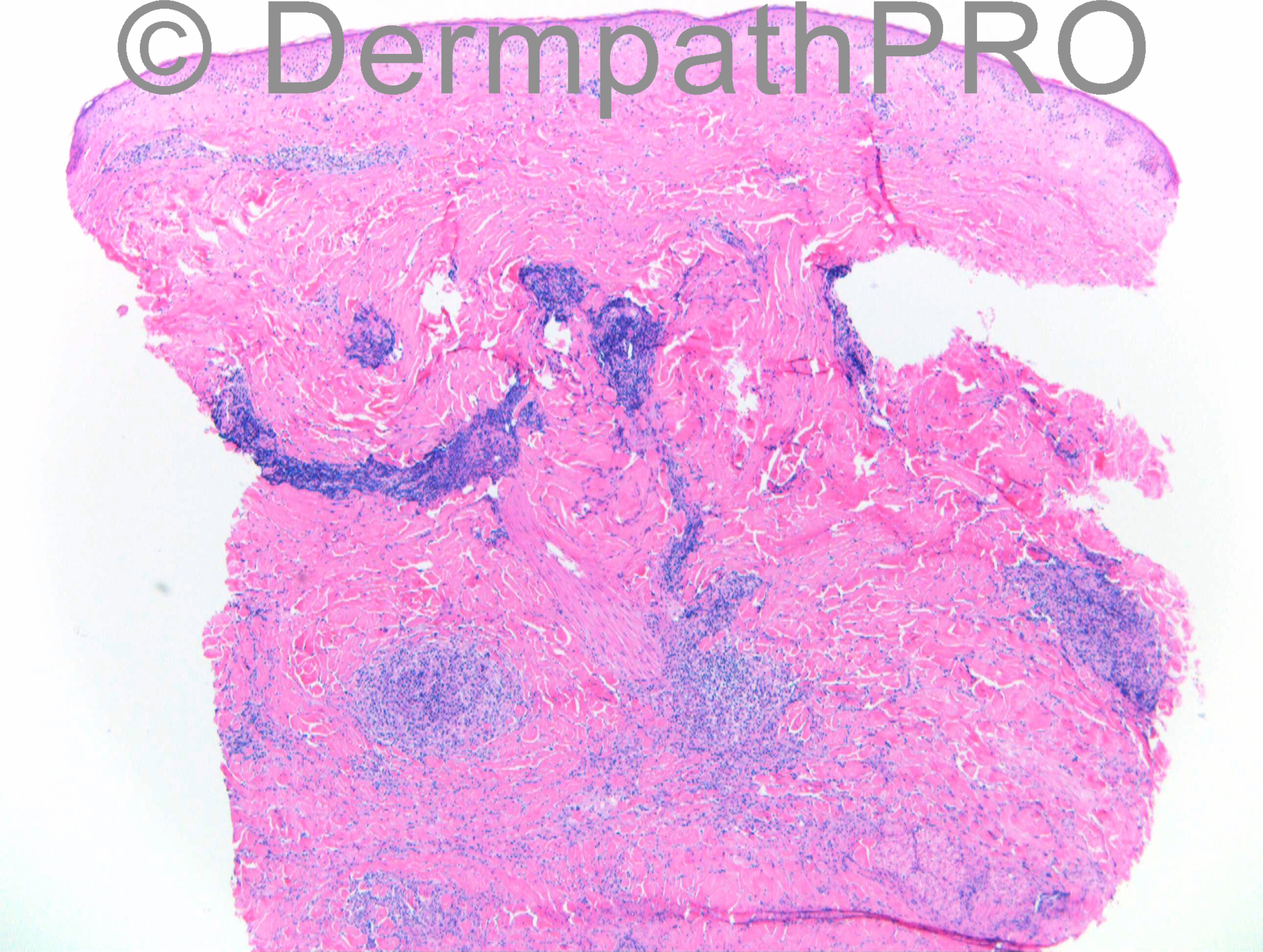
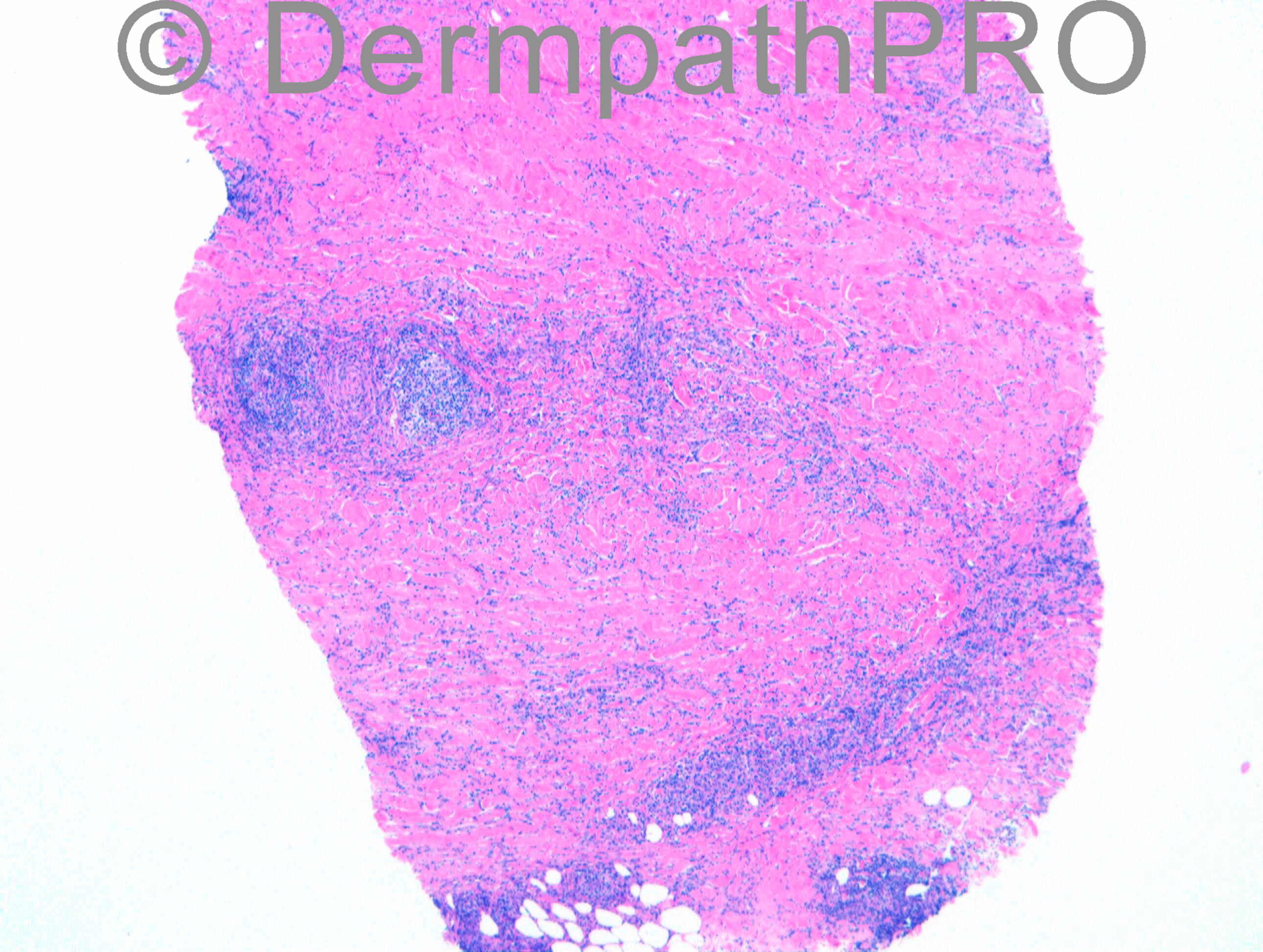
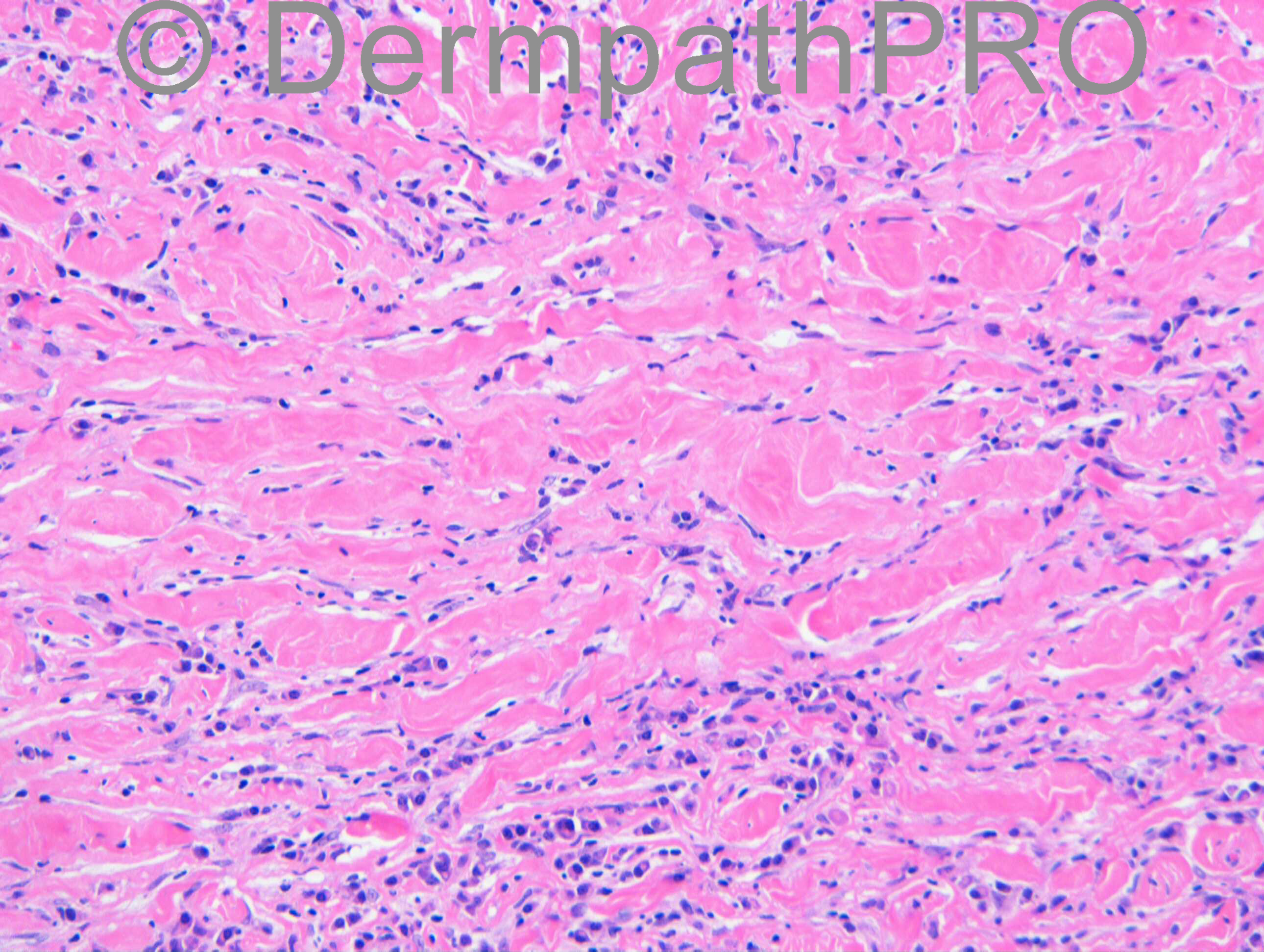
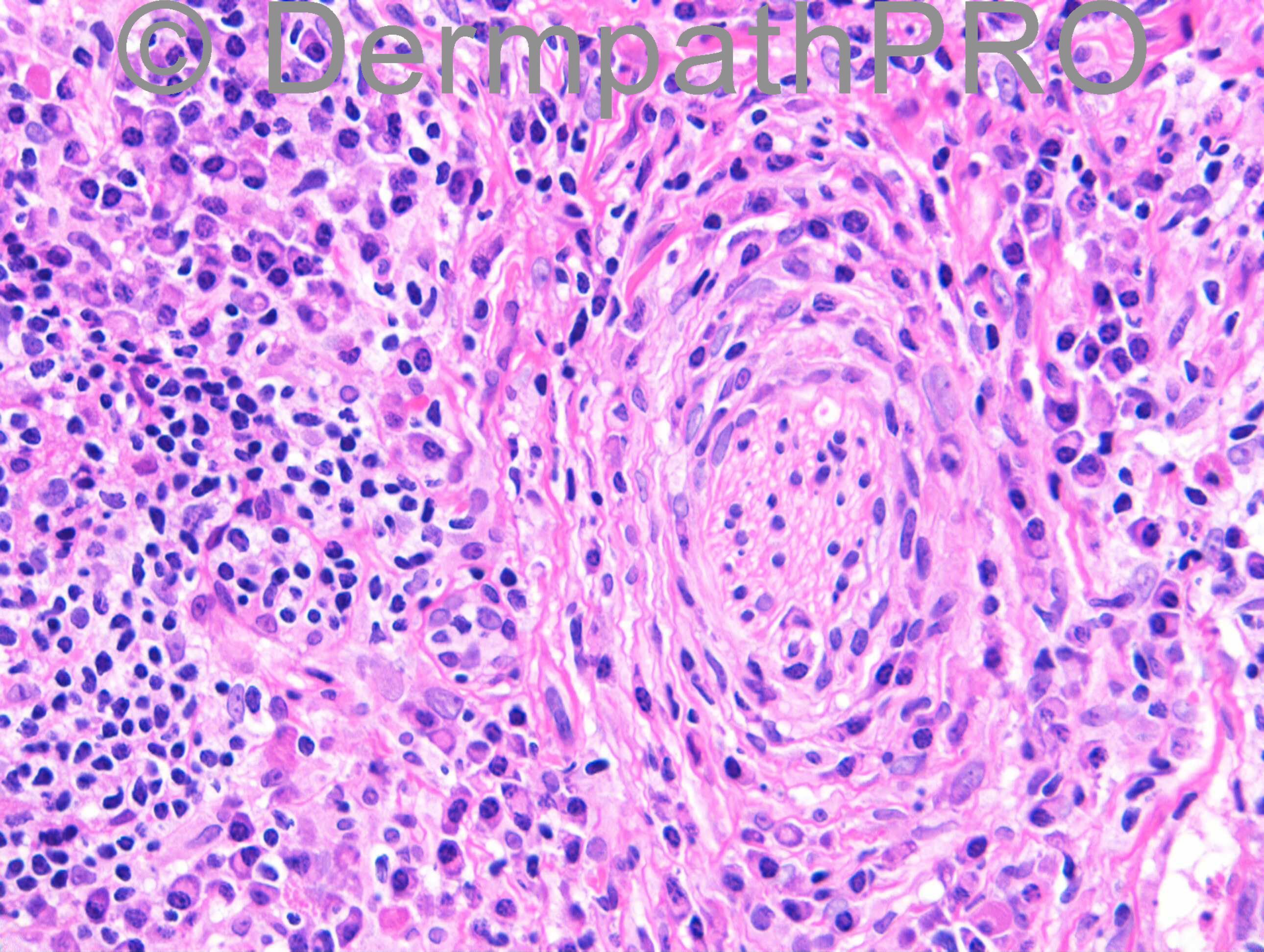
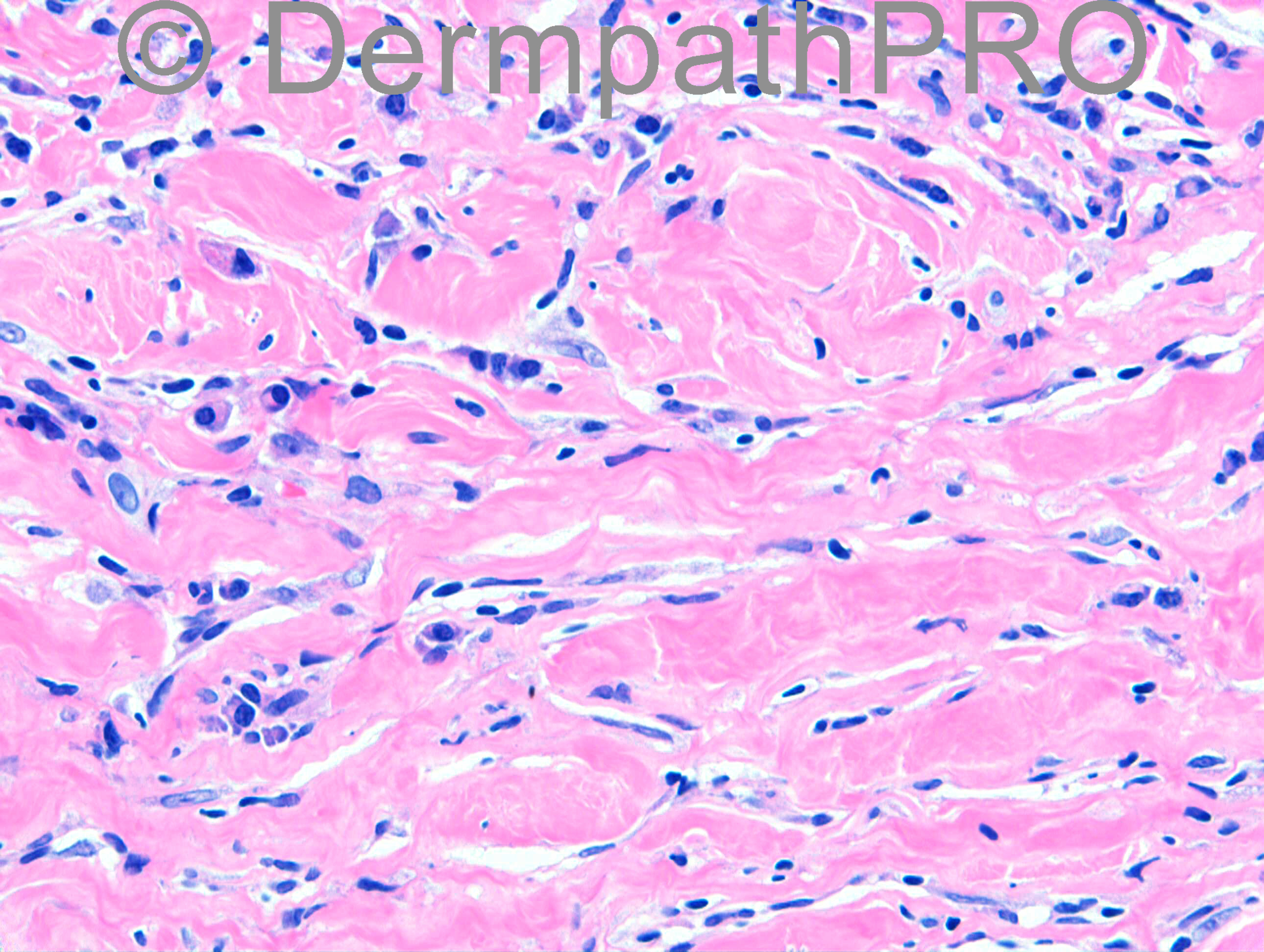
F58. Rheumatoid arthritis. Indurated skin lesion / lump right breast. Also has Diabetes. Punch biopsy in breast clinic.

Case posted by Dr Richard Carr


  Report Record

User Feedback


Robledo F. Rocha

Posted

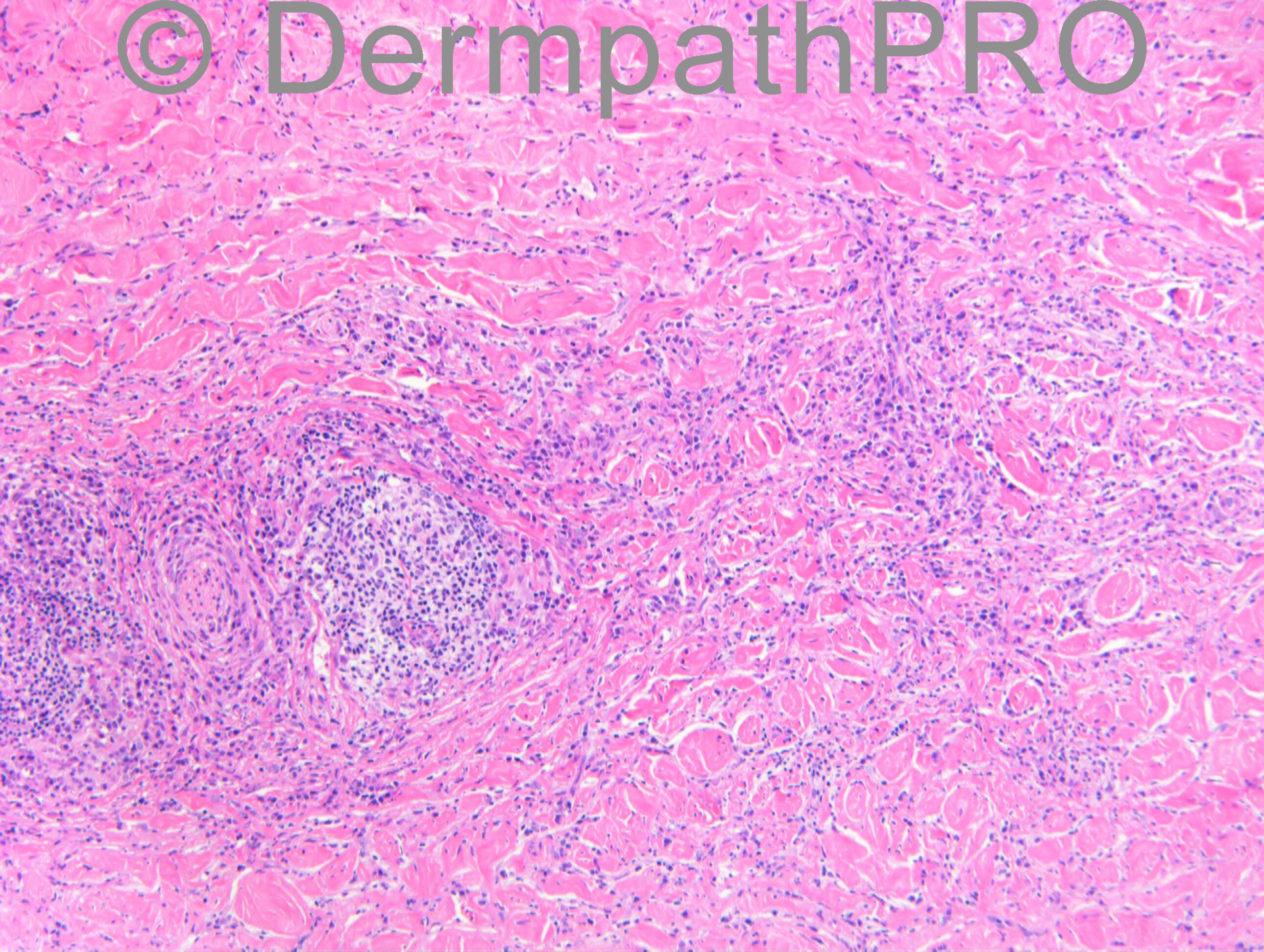
Morphea. Beautiful [url="https://dermpathpro.com/index.html/_/latest-news/perineural-in%EF%AC%82ammation-in-morphea-r52"]perineural inflammation[/url].

Share this comment


Link to comment
share_externally

Sasi Attili

Posted

Beautiful case. Favour inflammatory morphoea

Share this comment


Link to comment
share_externally

Dr. Richard Carr

Posted

Good responses and I agreed with the majority of inflammatory morphoea, my report as follows:

Many thanks for asking me to review this fascinating biopsy. The upper
dermis shows relatively mild perivascular lymphocytic and plasma cell
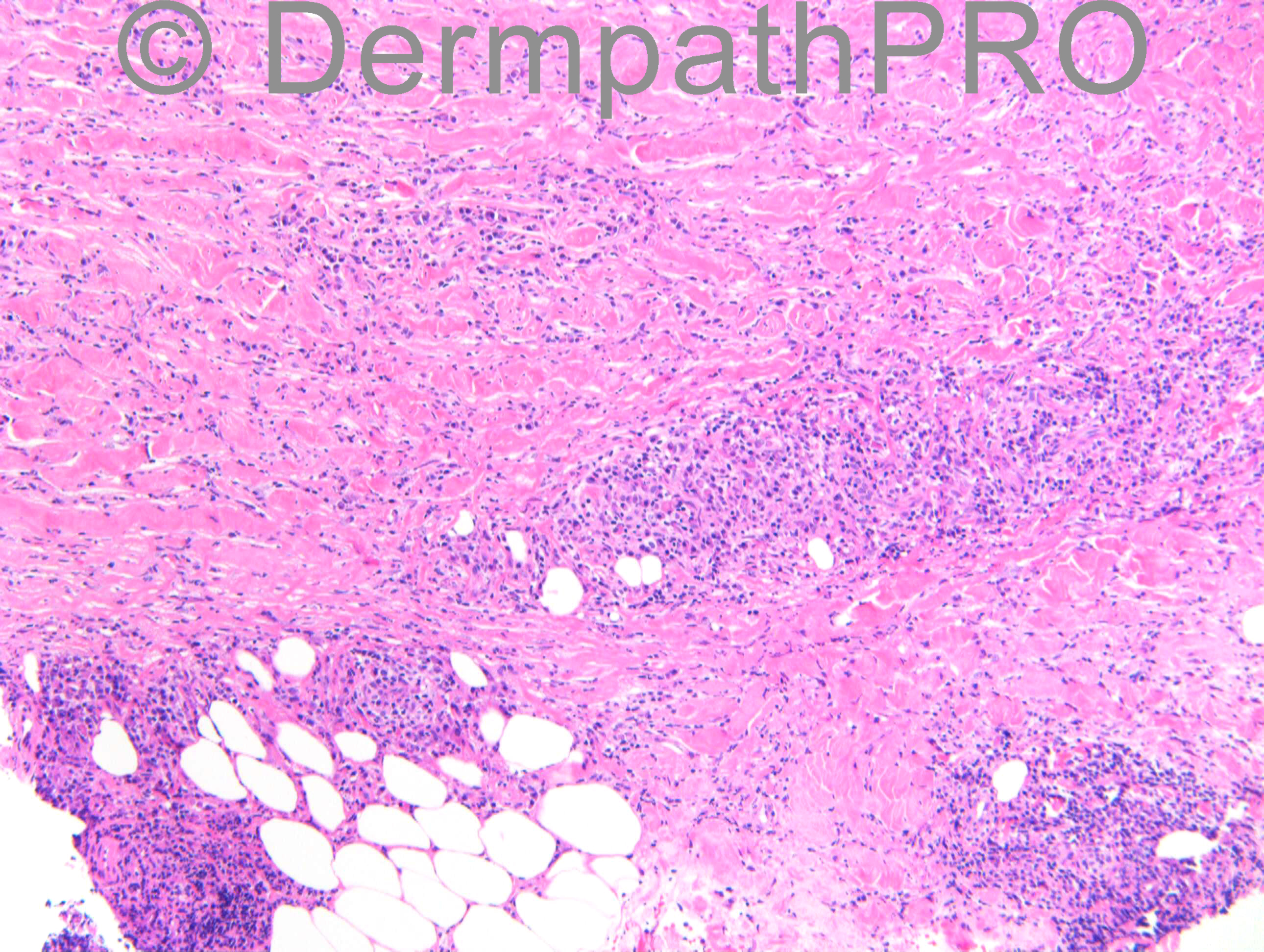
infiltrate. The striking feature is in the lower two-thirds of the
dermis, which is markedly expanded and has a mixed perivascular and
interstitial infiltrate of lymphocytes, histiocytes and numerous plasma
cells. Eosinophils are relatively sparse. I interpreted the collagen
bundles as being rather swollen and highly suggestive that this is a
case of inflammatory morphea. The differential would include Lupus
Profundus, although mucin is lacking. I think it would be prudent to
exclude Lyme Borreliosis, but histologically this appearance is very
good for the severe inflammatory (almost pseudolymphomatous) end of the
spectrum of morphea.

I agree the biopsy does have a zig-zag like appearance but I thought the lack of convincing necrobiosis and lack of palisading histiocytes or well formed granulomas as well as the clinical location argued strongly against necrobiosis lipoidica compared with morphoea and thefore did not mention this differential in my opinion but I think from the histolgoical perspective it is a good consideration. Regarding nerve involvement I was aware from two unpublished cases presented by Dr Eduardo Calonje that this could be a striking feature of morphoea (hence I focussed down on it in this case) but I was not aware this had been published so thanks to Robledo.

Share this comment


Link to comment
share_externally


Join the conversation

You can post now and register later. If you have an account, sign in now to post with your account.

Guest
Add a comment...

×   Pasted as rich text.   Paste as plain text instead

  Only 75 emoji are allowed.

×   Your link has been automatically embedded.   Display as a link instead

×   Your previous content has been restored.   Clear editor

×   You cannot paste images directly. Upload or insert images from URL.

Loading...

×
×
  • Create New...