Jump to content
In this section we have spot diagnoses posted on a daily basis since June 2010, now over 4000! You can review the archived cases and read the suggested diagnoses by users and the final comment by the contributors.
Case are uploaded each week day by 10 am UK time with the correct diagnosis will generally be posted at 8 pm UK time. Why not view the most recent spot diagnosis and proffer a diagnosis?

Case Number : Case 1188 - 12th January Posted By: Guest

Please read the clinical history and view the images by clicking on them before you proffer your diagnosis.
Submitted Date :
   (0 reviews)

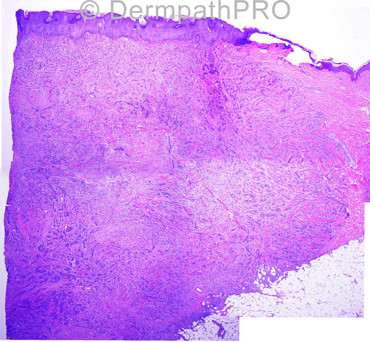
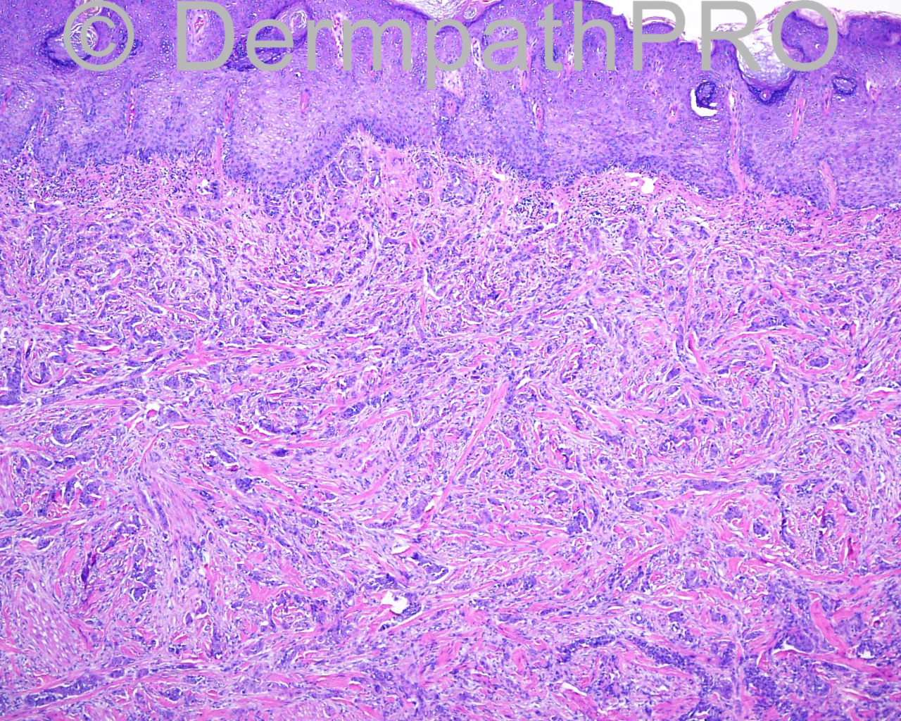
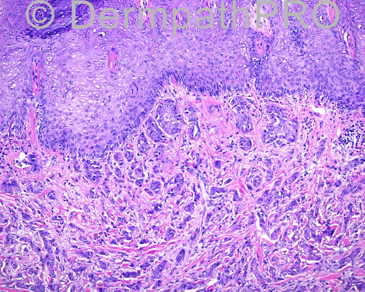
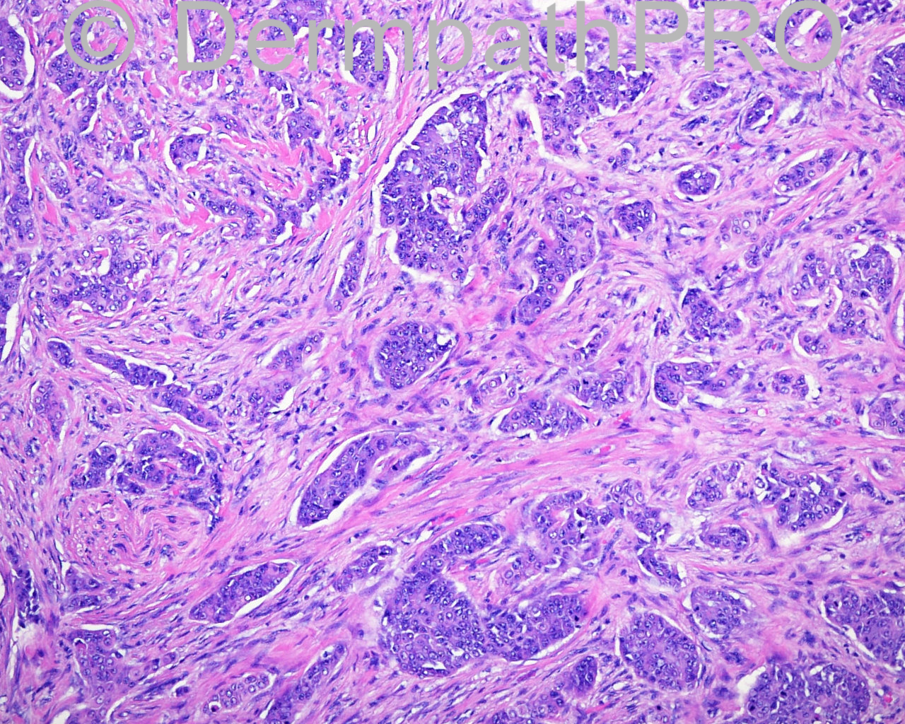
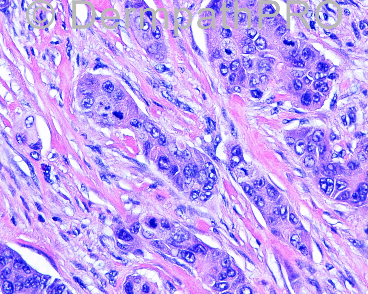
The patient is a 74 year old white man with a previous biopsy performed at another facility. An excision with margin exam is taken from the left lateral lower abdomen.

Case posted by Dr Mark Hurt


  Report Record

User Feedback


Dr. Mona Abdel-Halim

Posted

A suspected epidermal origin is perceived in images 2 and 4, if this is the case, porocarcinoma comes first. If no epidermal connection, will like to exclude metastatic adenocarcinoma.

Share this comment


Link to comment
share_externally

Guest Giovanni Falconieri

Posted

Agree, I suspect from primary in stomach

Share this comment


Link to comment
share_externally

Robledo F. Rocha

Posted

My first impression is metastatic adenocarcinoma. Nuclear pleomorphism is too high for a tumor from prostate and the lack of intracytoplasmatic mucin makes gastrointestinal origin less likely, so lung must be considered.

Share this comment


Link to comment
share_externally

Dr. Mona Abdel-Halim

Posted

So it is metastatic hepatocellular carcinoma

Share this comment


Link to comment
share_externally

Guest Jim Davie MD

Posted

Agree with metastatic carcinoma. Favor metastatic colon adenocarcinoma at first glance.

[size=4]With positive granular staining for HSA, the differential diagnosis may swing in favor of metastatic pseudoglandular hepatocellular carcinoma, although [/size][size=4]HSA (hepatocyte "specific" antigen), despite the name, is not that specific...it can deceitfully stain positive in cholangiocarcinoma, colonic [/size][size=4]adenocarcinoma and gastric adenocarcinoma. Additional stains like CK7, CK20, AFP, etc. as part of a panel can separate these differential possibilities.[/size]

[size=2]Hep Par 1 Antibody Stain for the Differential Diagnosis of Hepatocellular Carcinoma: 676 Tumors Tested Using Tissue Microarrays and Conventional Tissue Sections. [/size][i][size=2]Mod Pathol 2003;16(2):137–144[/size][/i]
[size=2] [url="http://www.nature.com/modpathol/journal/v16/n2/full/3880729a.html"]http://www.nature.co...l/3880729a.html[/url][/size][size=2] (Free Full Article link)[/size]

Share this comment


Link to comment
share_externally

Metastatic carcinoma, be it adeno- or hepatocellular.

Share this comment


Link to comment
share_externally

Thank Dr Hurt for such an interesting case. I agree with all the discussions above. But as for the primary origin of the tumor, per your staining results that the CK 7 is strongly postive are rarely seen In hepatocellular carcinoma which is often CK7 and CK20 negative tumor . Like Dr Davie mentioned, Hepar 1 is not specific for hepatocellular carcinoma. I think now there is a more specific marker for hepatocellular carcinoma called Glypican 3 if you want confirm it. Just don't like the strong CK7 positivity here for considering hepatocellular carcinoma as primary.

Share this comment


Link to comment
share_externally


Join the conversation

You can post now and register later. If you have an account, sign in now to post with your account.

Guest
Add a comment...

×   Pasted as rich text.   Paste as plain text instead

  Only 75 emoji are allowed.

×   Your link has been automatically embedded.   Display as a link instead

×   Your previous content has been restored.   Clear editor

×   You cannot paste images directly. Upload or insert images from URL.

Loading...

×
×
  • Create New...