Jump to content
In this section we have spot diagnoses posted on a daily basis since June 2010, now over 4000! You can review the archived cases and read the suggested diagnoses by users and the final comment by the contributors.
Case are uploaded each week day by 10 am UK time with the correct diagnosis will generally be posted at 8 pm UK time. Why not view the most recent spot diagnosis and proffer a diagnosis?

Case Number : Case 1192 - 16th January Posted By: Guest

Please read the clinical history and view the images by clicking on them before you proffer your diagnosis.
Submitted Date :
   (0 reviews)

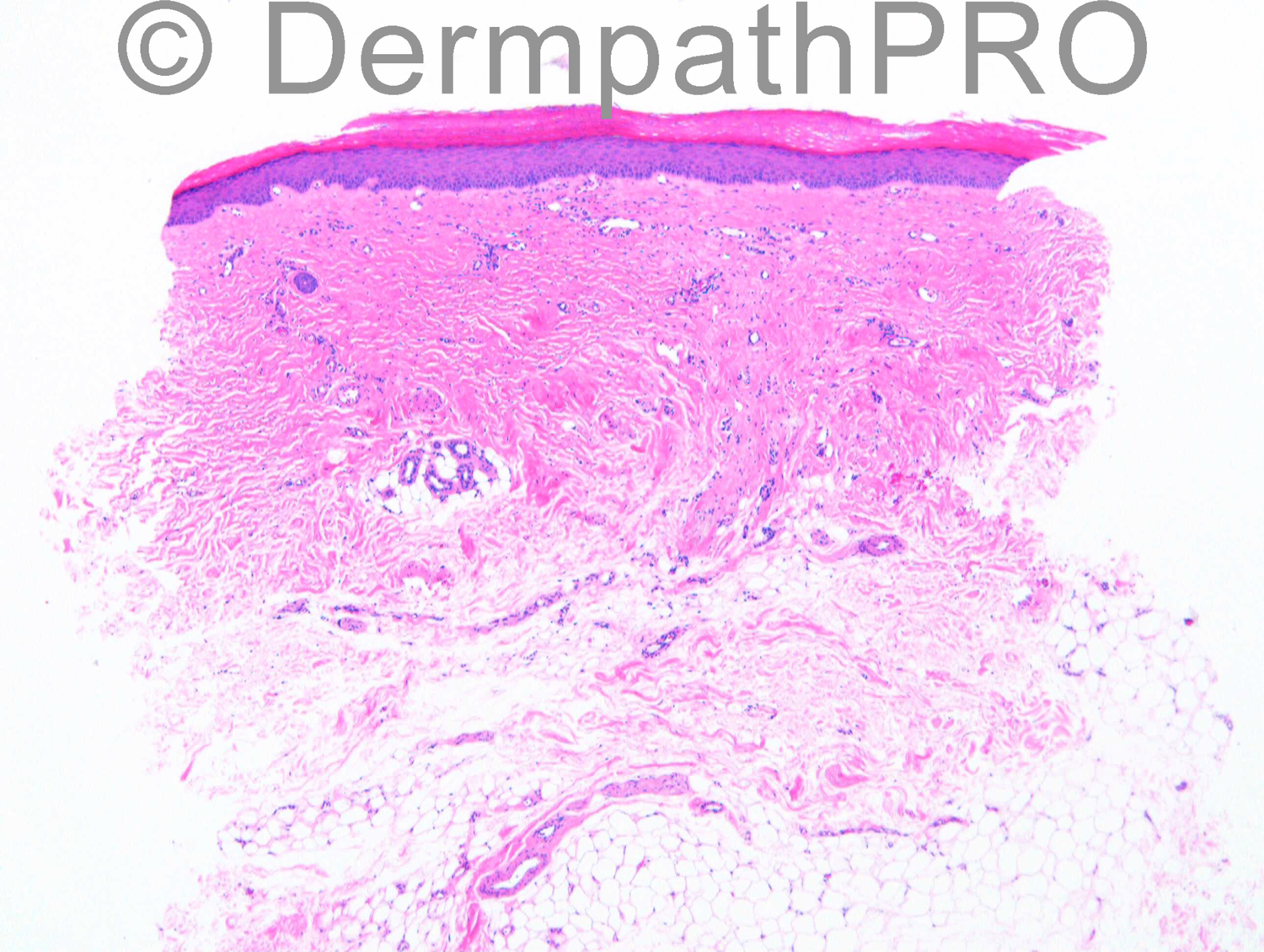
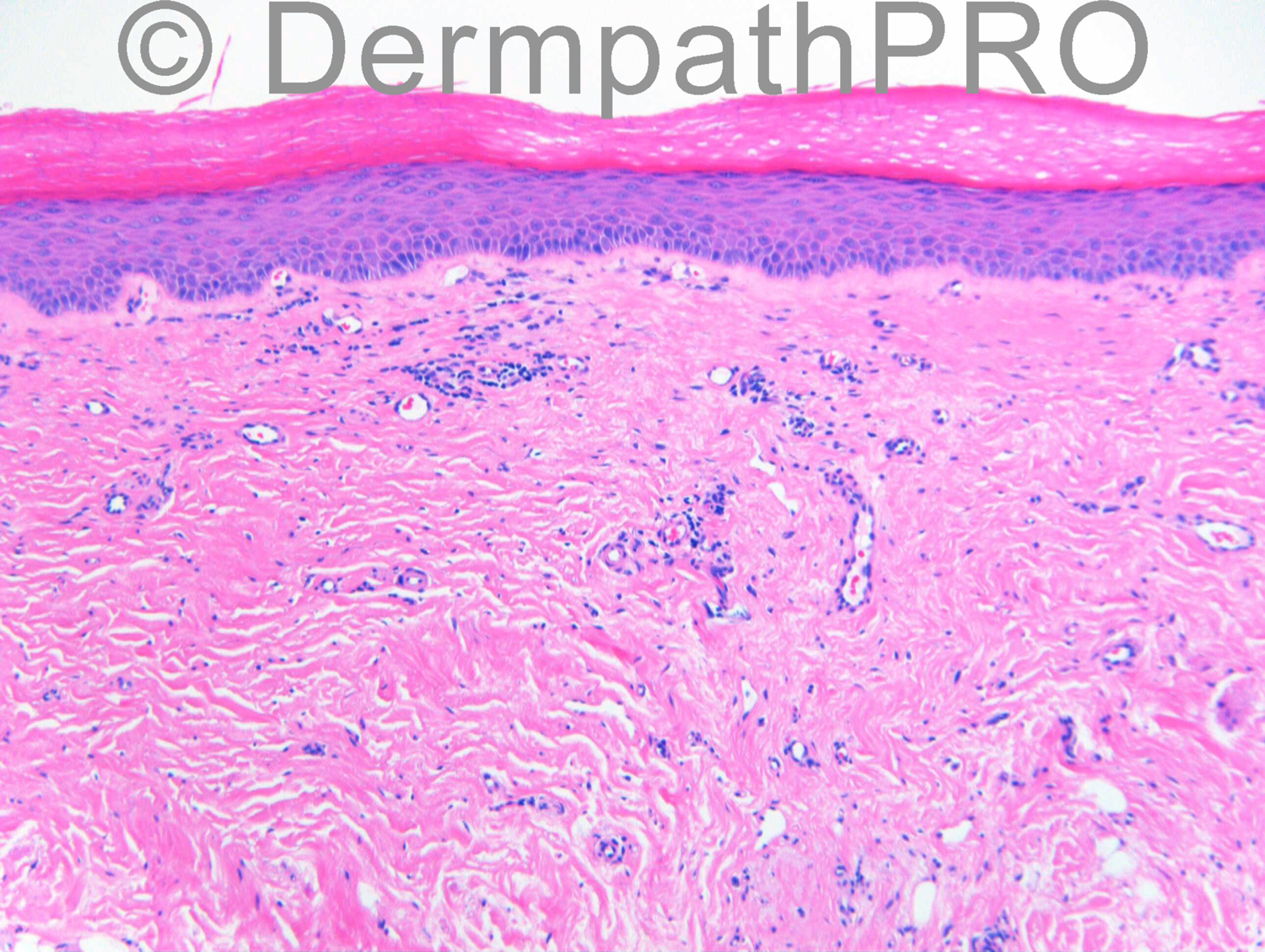
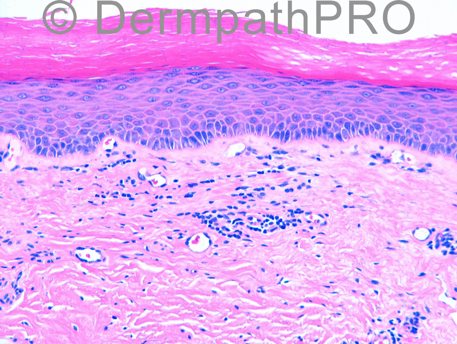
F55. Medial buttock. Clinically lichen sclerosus.

Case posted by Dr Richard Carr


  Report Record

User Feedback


Guest Romualdo

Posted

I would do a Warthin-Starry stain or immunostain for Borrelia. I think this is acrodermatitis chronica atrophicans, despite the location is unusual.

Share this comment


Link to comment
share_externally

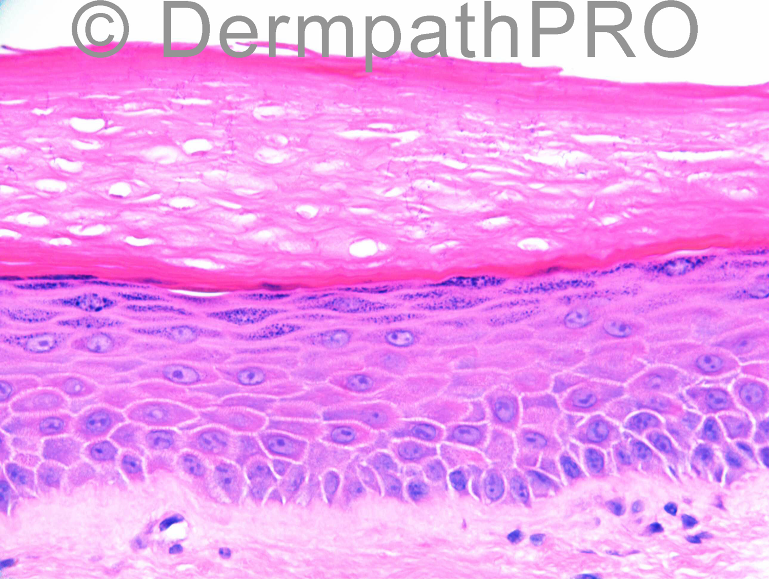
I see some bacterial microorganisms into the cornea. Special stains are necessary and my hypothesis is Erythrasma. There´s a nonintertriginous form of Erythrasma called Disciform erythrasma that can simulate other disease like Lichen sclerosus. Wood´s lamp would also be of help.
http://www.ncbi.nlm.nih.gov/pubmed/6851651

Share this comment


Link to comment
share_externally

Dr. Mona Abdel-Halim

Posted

As Igor said, I am seeing structures in the stratum corneum that worth staining, mostly erythrasma..

Share this comment


Link to comment
share_externally

Dr. Richard Carr

Posted

[img]https://dermpathpro.com/uploads/spot_diagnosis_comment_img/Case%201192%20-%20RAC7023x40_Gram.png[/img]

[img]https://dermpathpro.com/uploads/spot_diagnosis_comment_img/Case%201192%20-%20RAC7023x40b_PAS.png[/img]

Share this comment


Link to comment
share_externally

Robledo F. Rocha

Posted

[left]I must confess that epidermal atrophy, telangiectatic vessels and homogenized collagen bundles in the upper dermis drew my attention and would make me favor a sclerodermoid disorder without special stains. Congratulations to my friend Igor, who first discerned those barely visible Gram-positive bacteria in the stratum corneum of HE-stained images.[/left]

Share this comment


Link to comment
share_externally

Guest Tiberiu Tebeica

Posted

I am back. HNY everybody! Lot easier with special stains. I agree with erythrasma.

Share this comment


Link to comment
share_externally

Dr. Richard Carr

Posted

There was another biopsy of the vulva done at the same time showing features of lichen sclerosus without erythrasma. I don't see how erythrasma could mimic the [u][b]histological[/b][/u] dermal changes of LS seen here. I reported this biopsy as follows:

"Compact hyperkeratosis containing innumerable beaded-filamentous bacteria in keeping with erythrasma. There are also typical superficial dermal changes of quiescent lichen sclerosus with only mild lymphocytic infiltrate."

Share this comment


Link to comment
share_externally


Join the conversation

You can post now and register later. If you have an account, sign in now to post with your account.

Guest
Add a comment...

×   Pasted as rich text.   Paste as plain text instead

  Only 75 emoji are allowed.

×   Your link has been automatically embedded.   Display as a link instead

×   Your previous content has been restored.   Clear editor

×   You cannot paste images directly. Upload or insert images from URL.

Loading...

×
×
  • Create New...