Jump to content
In this section we have spot diagnoses posted on a daily basis since June 2010, now over 4000! You can review the archived cases and read the suggested diagnoses by users and the final comment by the contributors.
Case are uploaded each week day by 10 am UK time with the correct diagnosis will generally be posted at 8 pm UK time. Why not view the most recent spot diagnosis and proffer a diagnosis?

Case Number : Case 1193 - 19th January 2015 Posted By: Guest

Please read the clinical history and view the images by clicking on them before you proffer your diagnosis.
Submitted Date :
   (0 reviews)

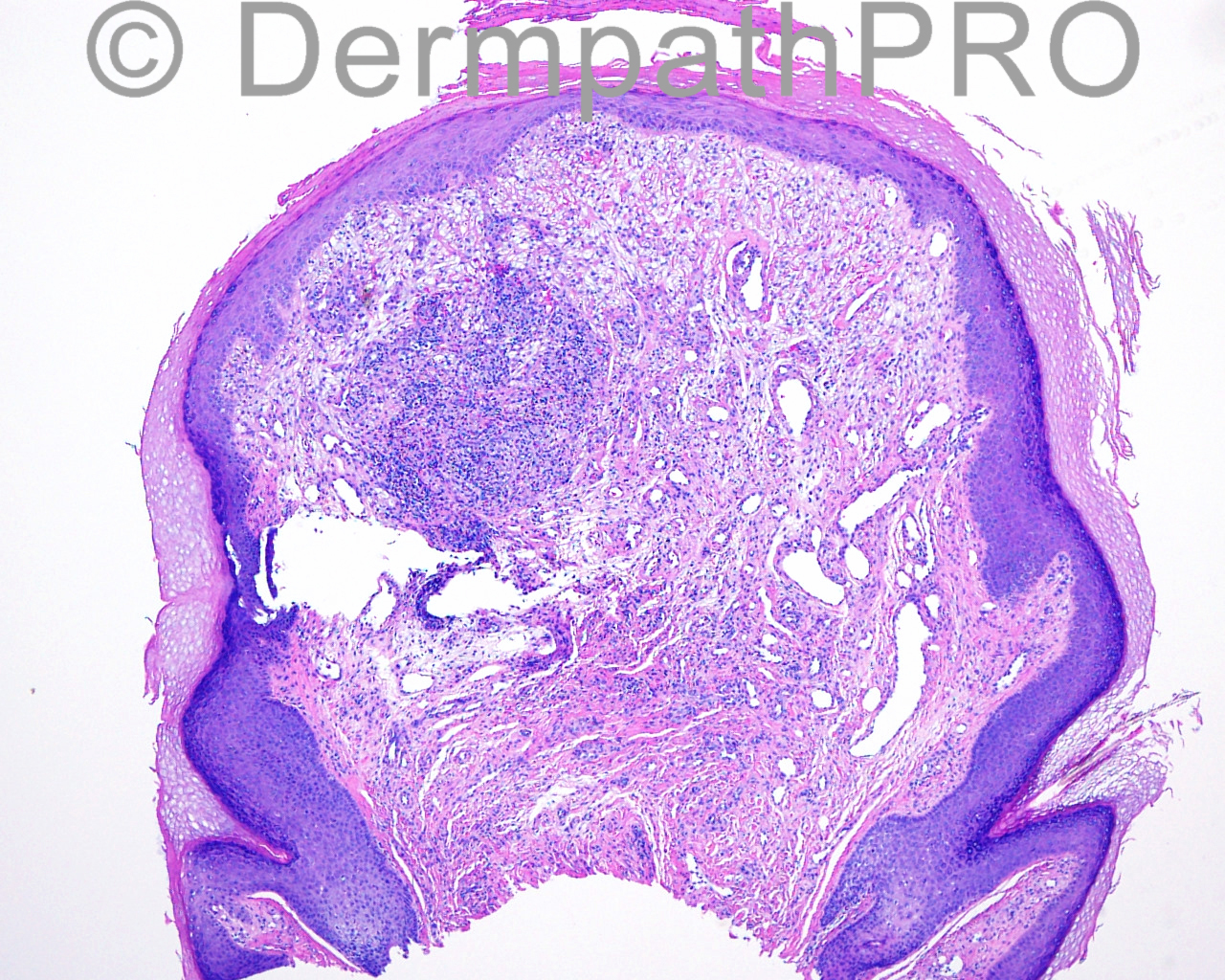
The patient is a 41 year old white woman with a lesion on the right columella which appears like a squamous papilloma, present for two months. A scissor biopsy of a 1 mm hyperkeratotic, palpable, pearly, well marginated, red elevated mass is taken from the right nasal columella.

Case posted by Dr Mark Hurt


  Report Record

User Feedback


Dr. Mona Abdel-Halim

Posted

Yes, fibrous papule of the nose, clear cell variant.

Share this comment


Link to comment
share_externally

Dr. Richard Carr

Posted

I'd be thinking that too but would certainly be inclined to run quite a few IHC stains to see what the cells are.

Share this comment


Link to comment
share_externally

Mark A. Hurt MD

Posted

Well, I did indeed do a bunch of IHC stains. Here is my diagnosis:


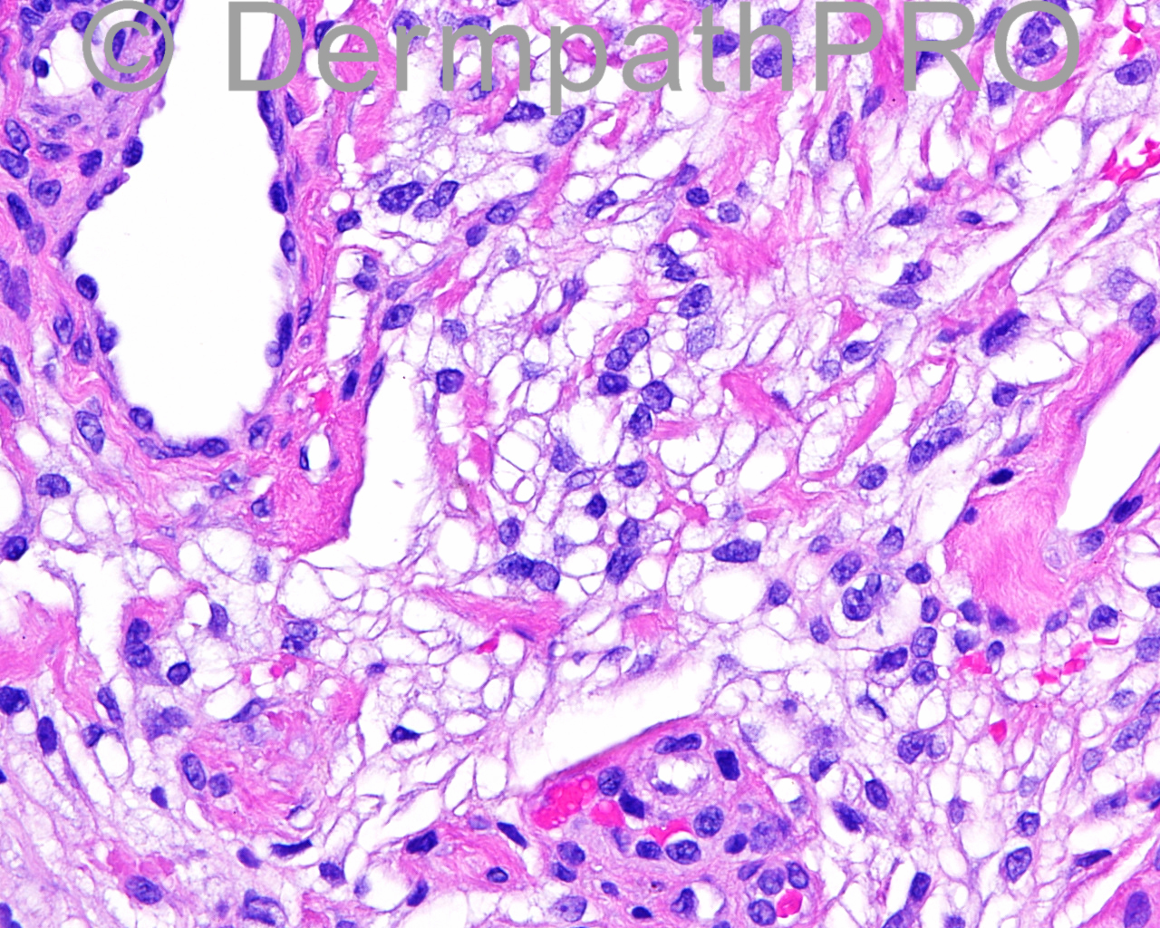
[b]-- FIBROUS PAPULE, CLEAR CELL TYPE[/b]
[indent=1][b]COMMENT: [/b]I do not see any evidence to place this lesion into a melanocytic lesion or neural lesion. I think this is a histological variation of fibrous papule. The findings in this lesion closely parallel those described by Lee, et. al in 2005. I do not identify any evidence of malignancy.[/indent]


[center]Reference:[/center]

Lee AN, Stein SL, Cohen LM. Clear cell fibrous papule with NKI/C3 expression: clinical and histologic features in six cases. Am J Dermatopathol. 2005 Aug;27(4):296-300. PubMed PMID: 16121048.

Micro:

This lesion from the right nasal columella is an exophytic papule that contains a population of clear cells in the dermis. These have a slightly frothy quality to the cytoplasm, but many of them are water clear and they merge with a more conventional amphophilic cytoplasm in some of the similar cells. They are not like xanthocytes in that they do not have a uniform foaminess to them. They are also intermixed with a number of venules of varying sizes throughout the field. The surface epidermis contains a small amount of actinic keratosis with evidence of slightly tattered cornified cells, suggesting some degree of rubbing. These clear cells within the dermal component of the lesion are negative with Melan-A, CD34, renal cell carcinoma antigen, Ki67 (clone 30-9), S100 protein, EMA, actin and HMB45. There is some factor XIIIa positivity throughout the field, but not too many of these clear cells pick up factor XIIIa. The smooth muscle actin marker shows that most of the vessels in the lesion are venules. CD63 and CD68 are both positive in the stroma to some degree, but the CD63 has greater positivity.

Share this comment


Link to comment
share_externally


Join the conversation

You can post now and register later. If you have an account, sign in now to post with your account.

Guest
Add a comment...

×   Pasted as rich text.   Paste as plain text instead

  Only 75 emoji are allowed.

×   Your link has been automatically embedded.   Display as a link instead

×   Your previous content has been restored.   Clear editor

×   You cannot paste images directly. Upload or insert images from URL.

Loading...

×
×
  • Create New...