Jump to content
In this section we have spot diagnoses posted on a daily basis since June 2010, now over 4000! You can review the archived cases and read the suggested diagnoses by users and the final comment by the contributors.
Case are uploaded each week day by 10 am UK time with the correct diagnosis will generally be posted at 8 pm UK time. Why not view the most recent spot diagnosis and proffer a diagnosis?

Case Number : Case 1195 - 21th January Posted By: Guest

Please read the clinical history and view the images by clicking on them before you proffer your diagnosis.
Submitted Date :
   (0 reviews)

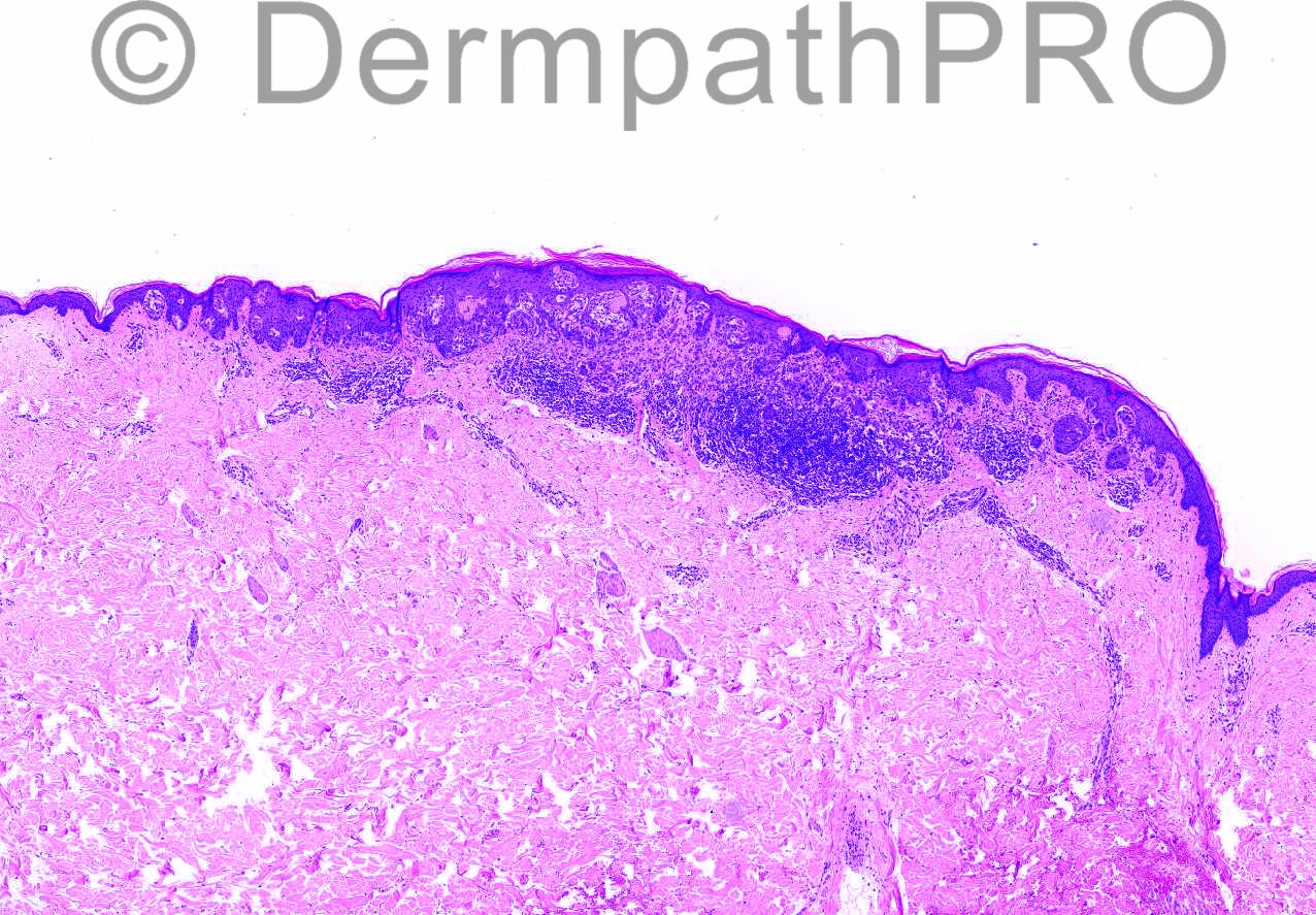
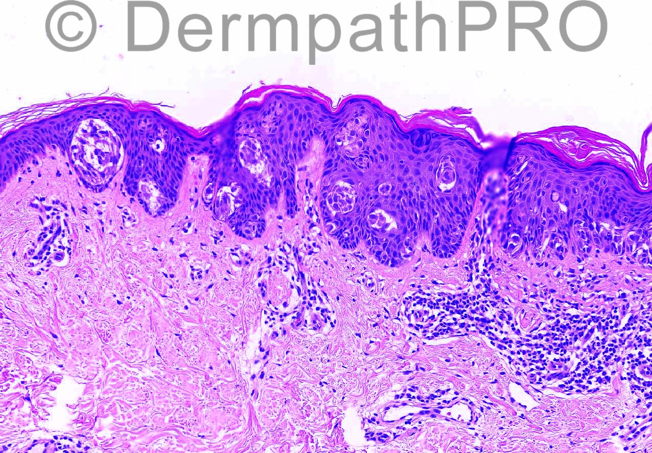
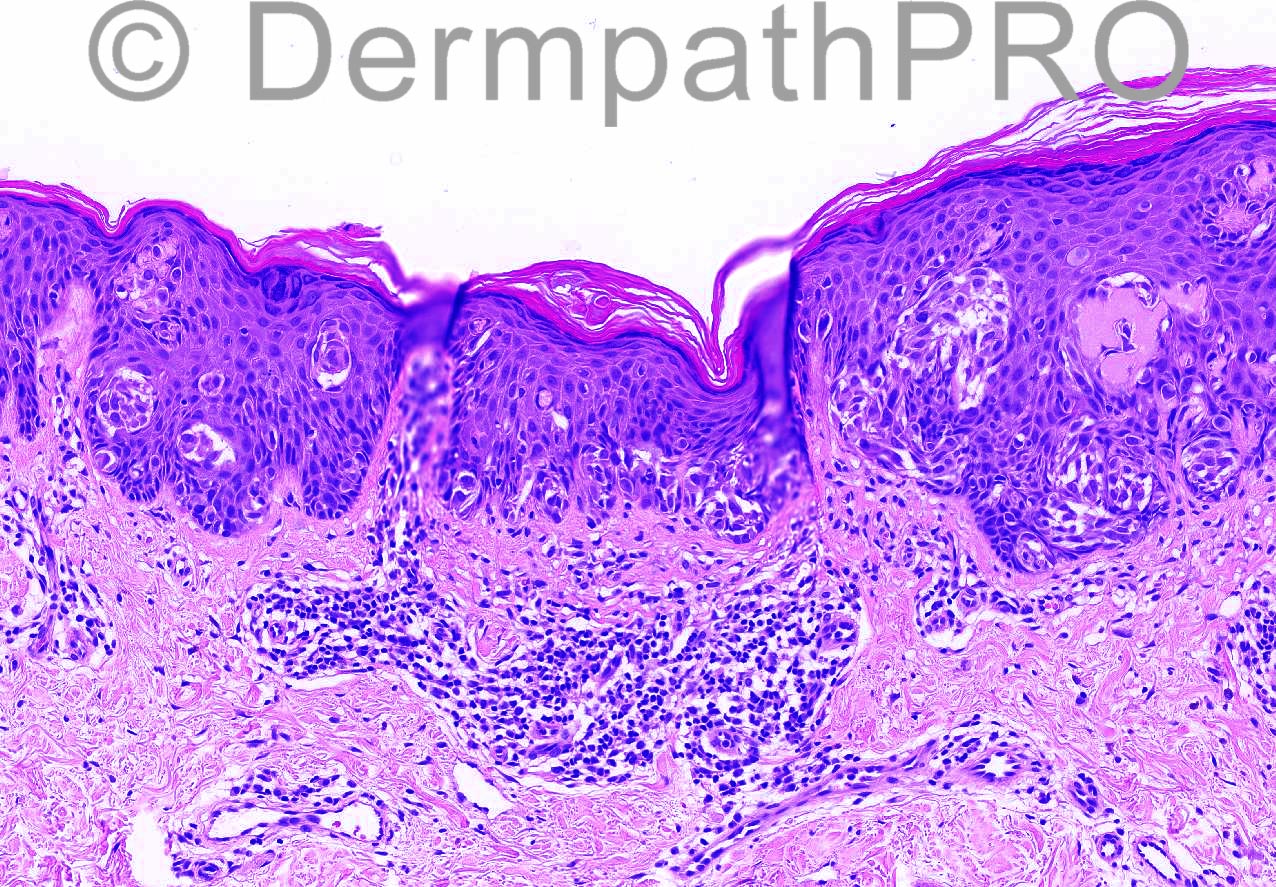
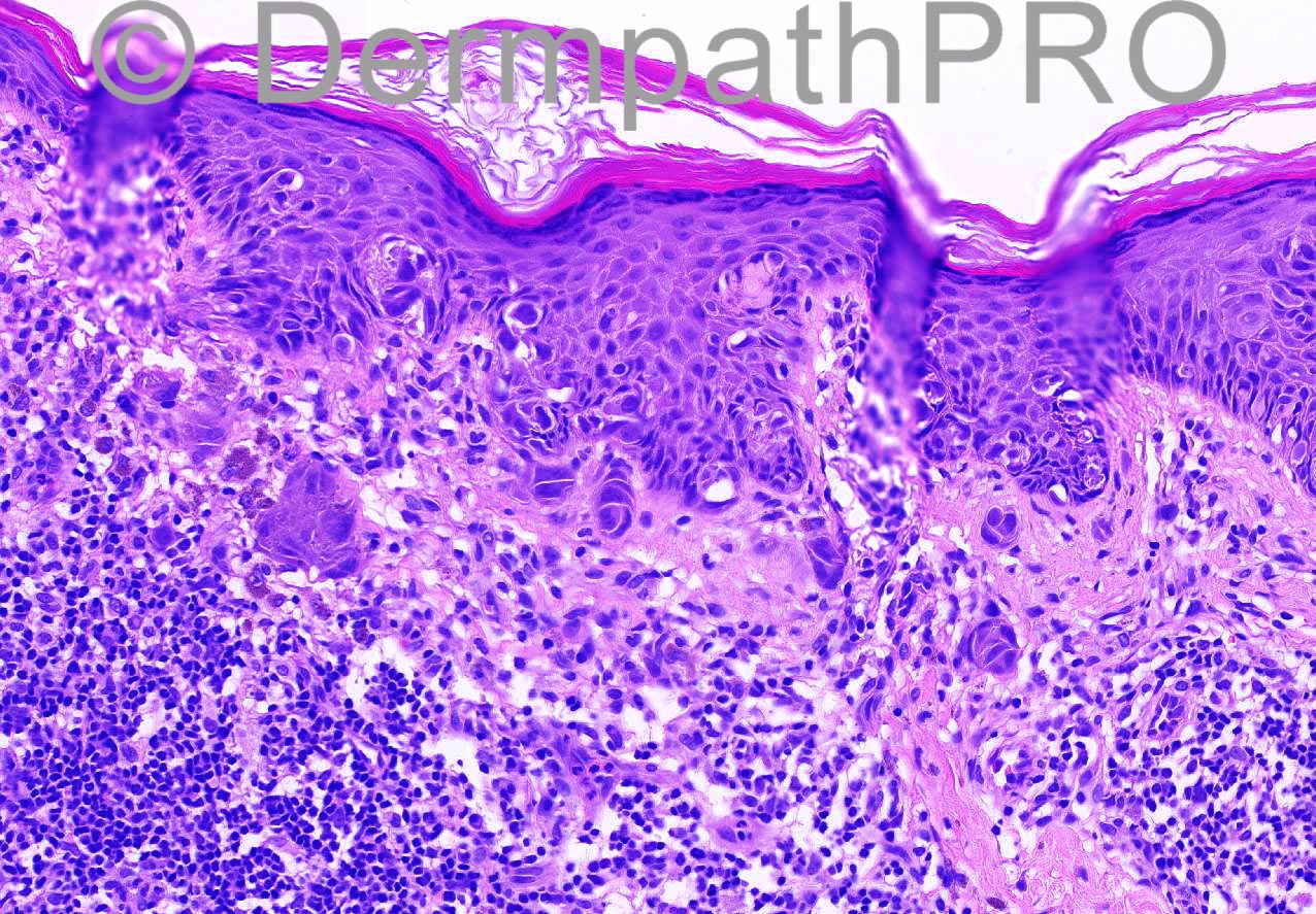
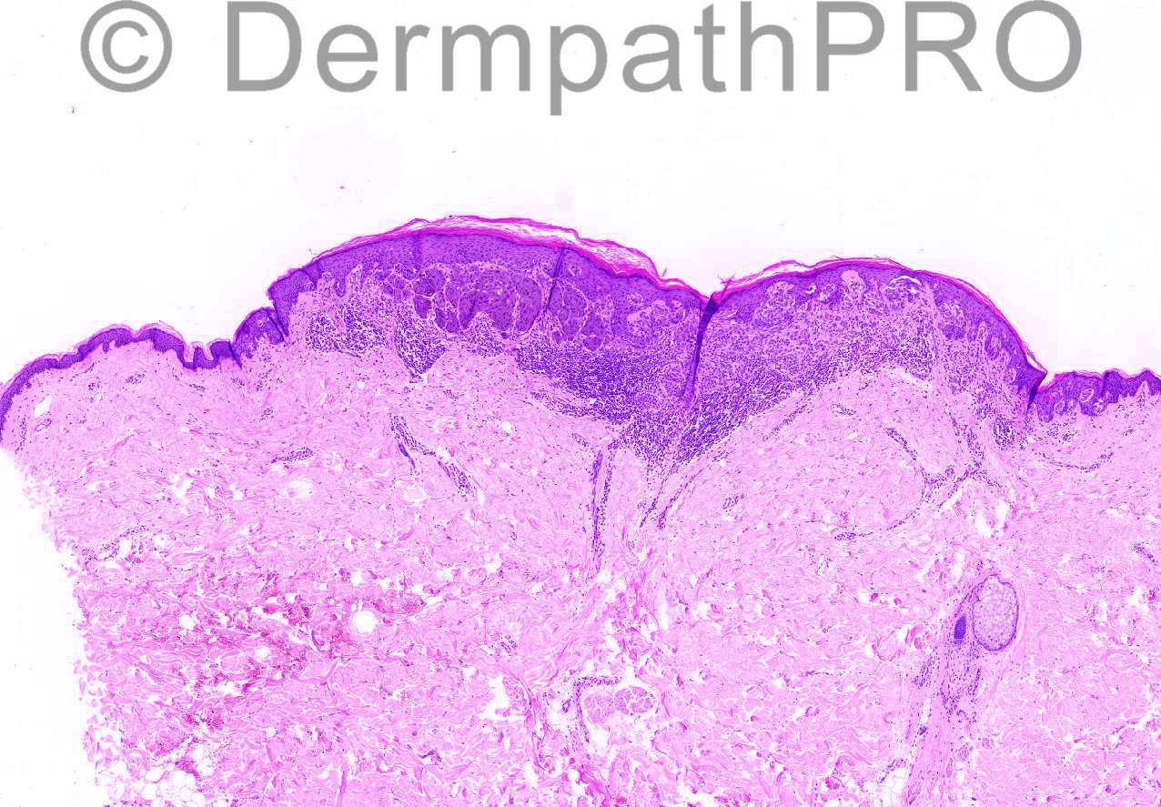
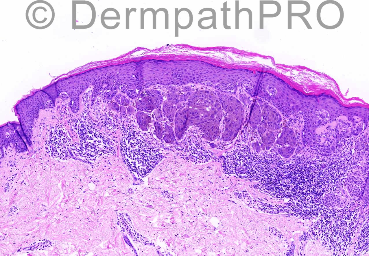
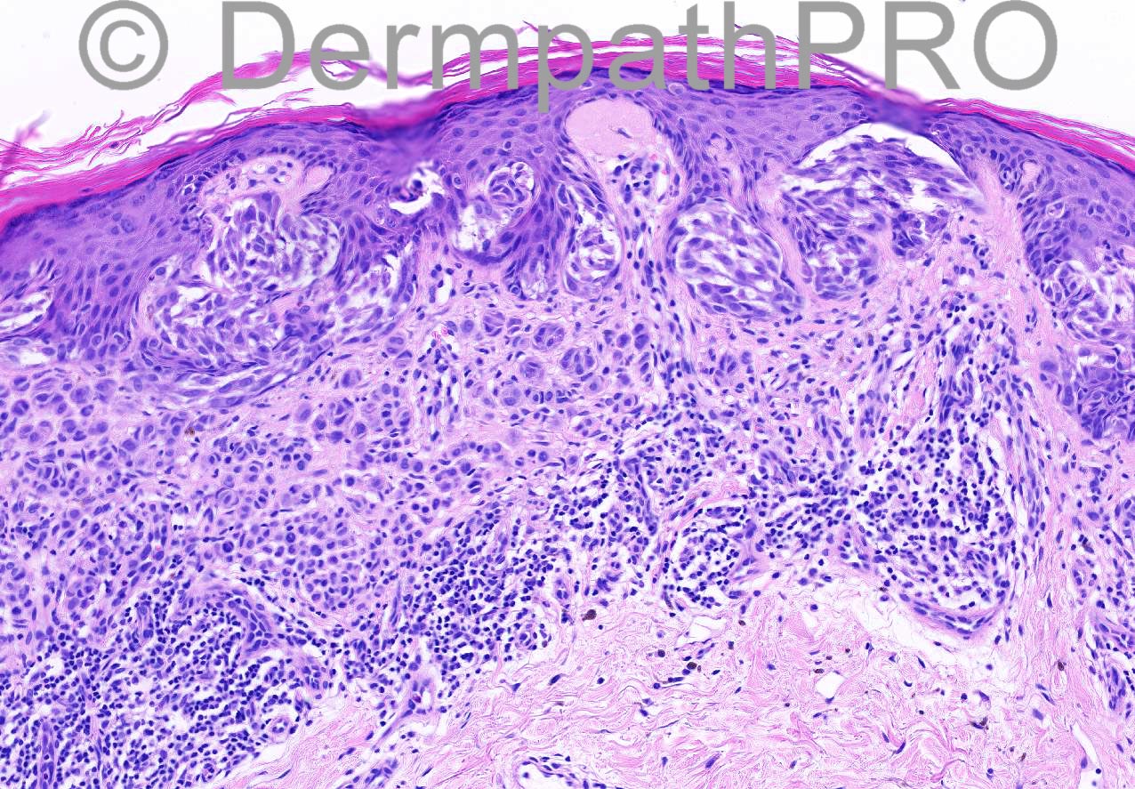
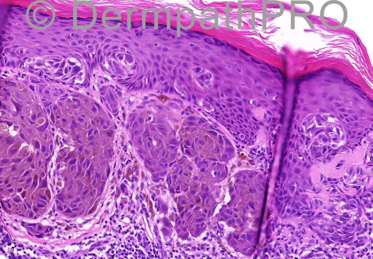
41 year old male, lesion on back – erythematous and pigmented ? BIN


  Report Record

User Feedback


Guest Giovanni Falconieri

Posted

Agree with spitz nevus, with active bottom inflammation consistent with "halo reaction" likely clinically noticed; due to extension of the junctional component I wonder whether this may histologically qualify for a dysplastic "spark" nevus

Share this comment


Link to comment
share_externally

Dr. Mona Abdel-Halim

Posted

I am not an expert when it comes to atypical melanocytic lesions, but I find here some worrisome features: age, large somehow confluent dermal nests in figs 6,7,9 and 10 with atypia (nuclear pleomorphism). When u look at fig 6, u see the marked asymmetry of the dermal component.....Yet there are other- wise typical features of SN. Could it be a melanoma evolving in a Spitz nevus? Or an atypical SN/ Spitzoid neoplasm with uncertain malignant potential. If this is my case, I will definitely ask for additional work up.

Share this comment


Link to comment
share_externally

Robledo F. Rocha

Posted

There's a component on the left of the Image 6, detailed at high power view on Images 7, 9 and 10, which differs from the rest of this spitzoid lesion. This component is comprised of pleomorphic epithelioid cells arranged in nests that vary in size and shape. Unlike the nests of the rest of the lesion, the nests of this component do not show cupping, to wit, the artifactual clefting between the upper edges of the junctional nests and the epidermis above them. Also, this component is hyperpigmented with dusty melanin and have pagetoid spread without epidermal hyperplasia. So, I think this a melanoma arising in a pre-existing Spitz’s nevus.

Share this comment


Link to comment
share_externally

Eman El-Nabarawy

Posted

Melanoma developing in Spitz's nevus.

Share this comment


Link to comment
share_externally

Guest Jim Davie MD

Posted

[size=4]This appears to be a fence-sitter of a Spitzoid neoplasm. Depending on one's choice of (heavily overlapping) criteria I think one can argue convincingly for either dysplastic/atypical compound Spitz nevus vs. Spitzoid melanoma. On H&E alone, I'll opine for dysplastic Spitz arising in pre-existing nevus, cannot rule out Spitzoid melanoma. I would customarily get P16 and Ki67 immunostains, as these may be helpful for differentiating benign-vs-malignant Spitz lesions.[/size]

[size=4] Supporting dysplastic: no definitive mitotic activity in either dermal or epidermal melanocytes, smudgy chromatin with small nucleoli, low-level pagetoid scatter, overall asymmetry, extension of junctional component beyond dermal component (shouldering).[/size]
[size=4]Supporting malignant: Plasma cell component in inflammatory infiltrate, heterogeneity of dermal cytology, and older age bracket (40 years). [/size]

Share this comment


Link to comment
share_externally

Guest Tiberiu Tebeica

Posted

Atypical spitzoid tumor. I think one cannot be certainly sure of this tumor's behavior based solely on histology.

Share this comment


Link to comment
share_externally

Dr. Richard Carr

Posted

Background compound spitz naevus with beutiful Kamino bodies. Separate distinct suspicious epithelioid clone uncertain for malignancy but of low risk for metastasis (thin, small area).

Share this comment


Link to comment
share_externally

Iskander H. Chaudhry

Posted

Thank you all for the detailed comments, I learned a lot from your discussion. We went through exactly the same process thinking whether it is a conventional spitz nevus or melanoma arising from it and based on the age and atypical features we classified it as atypical spitz nevus and recommended wider excision but no sentinel node biopsy.

Share this comment


Link to comment
share_externally


Join the conversation

You can post now and register later. If you have an account, sign in now to post with your account.

Guest
Add a comment...

×   Pasted as rich text.   Paste as plain text instead

  Only 75 emoji are allowed.

×   Your link has been automatically embedded.   Display as a link instead

×   Your previous content has been restored.   Clear editor

×   You cannot paste images directly. Upload or insert images from URL.

Loading...

×
×
  • Create New...