Jump to content
In this section we have spot diagnoses posted on a daily basis since June 2010, now over 4000! You can review the archived cases and read the suggested diagnoses by users and the final comment by the contributors.
Case are uploaded each week day by 10 am UK time with the correct diagnosis will generally be posted at 8 pm UK time. Why not view the most recent spot diagnosis and proffer a diagnosis?

Case Number : Case 1197 - 23rd January Posted By: Guest

Please read the clinical history and view the images by clicking on them before you proffer your diagnosis.
Submitted Date :
   (0 reviews)

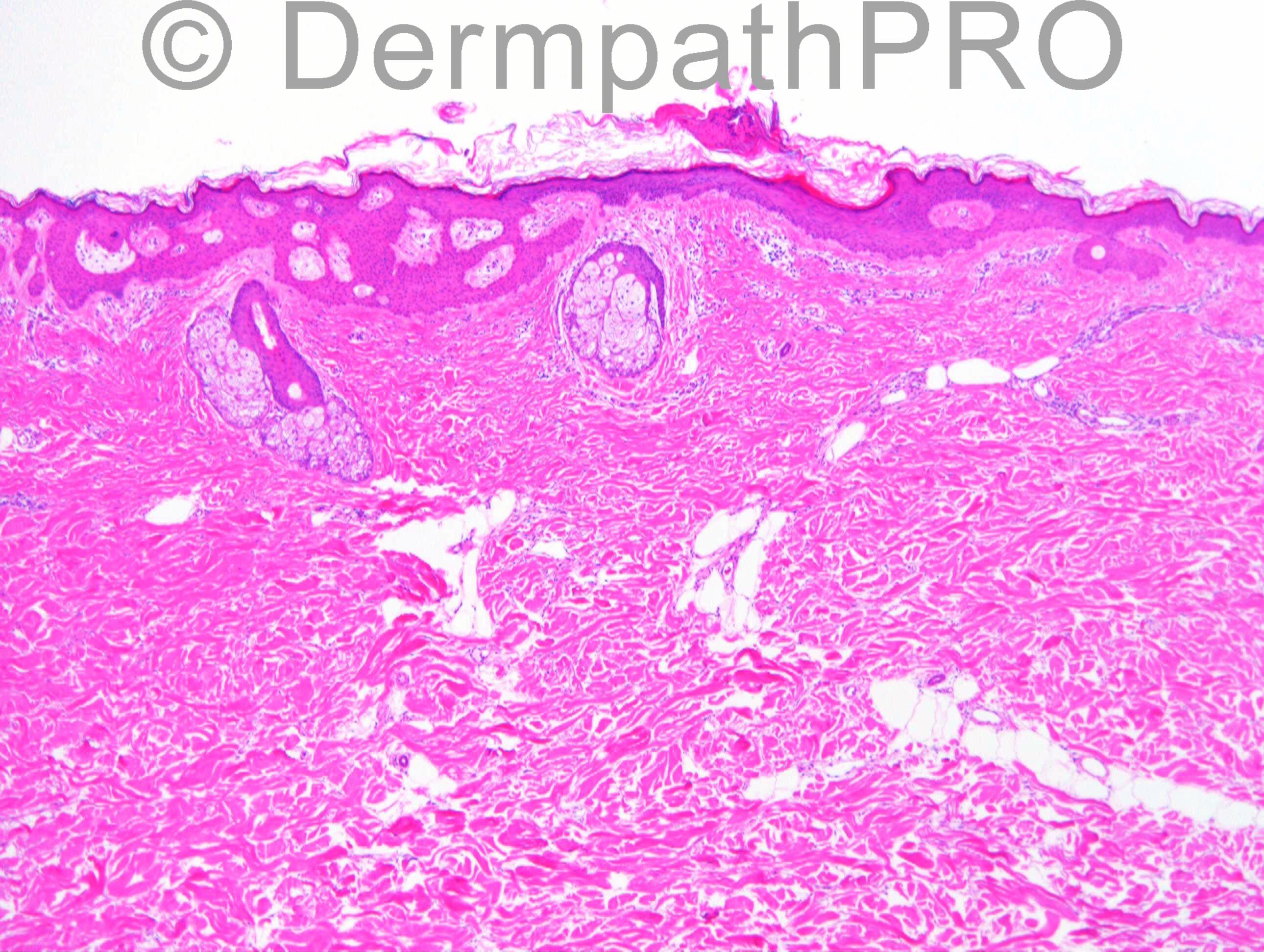
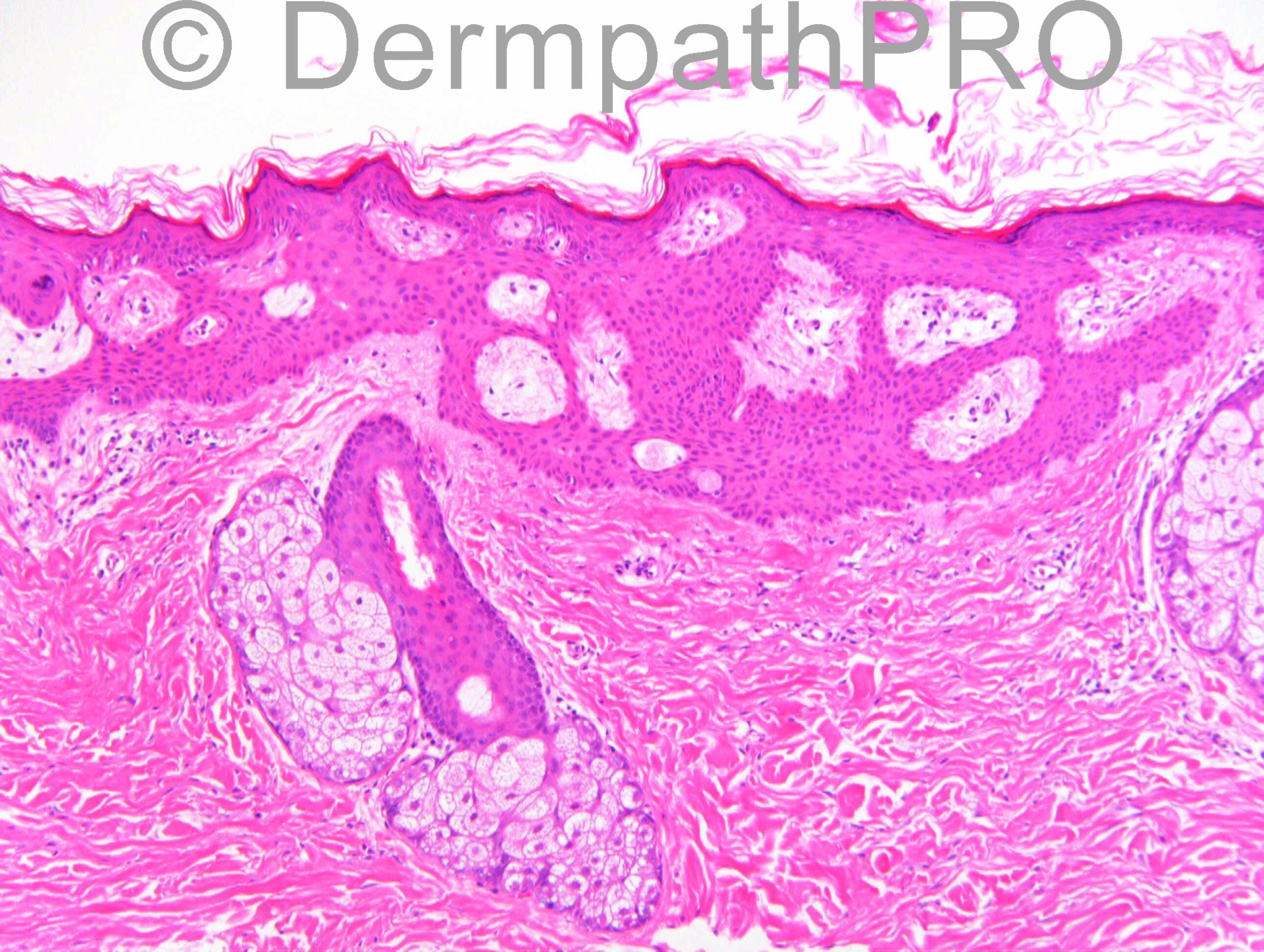
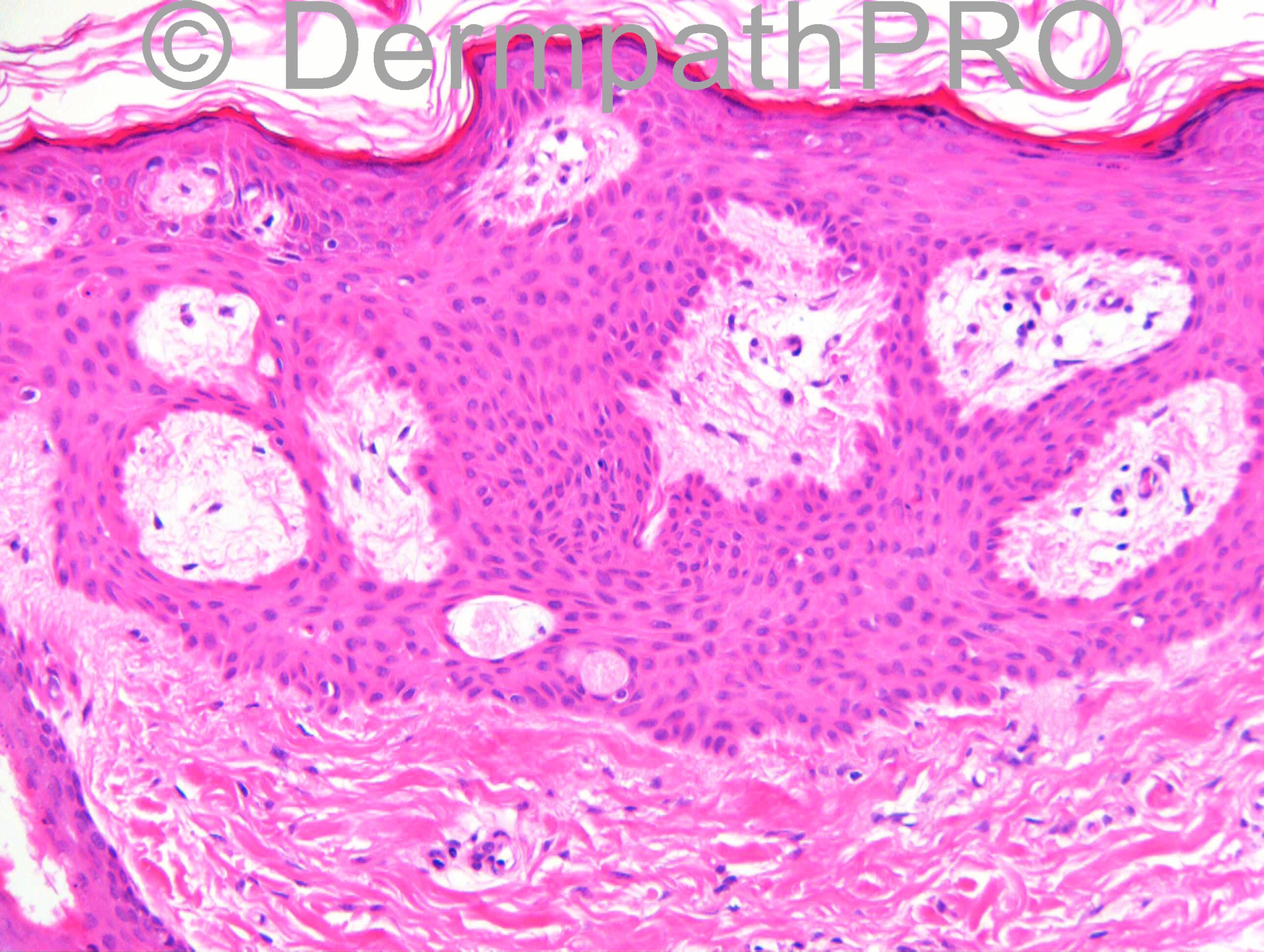
F56. Left upper back. Incidental finding in polar ends of excised dermatofibroma (clinically thought to be BCC).

Case posted by Richard Carr


  Report Record

User Feedback


Agree. Tumour of folicular infundibulum ("infundibuloma").

Share this comment


Link to comment
share_externally

Dr. Mona Abdel-Halim

Posted

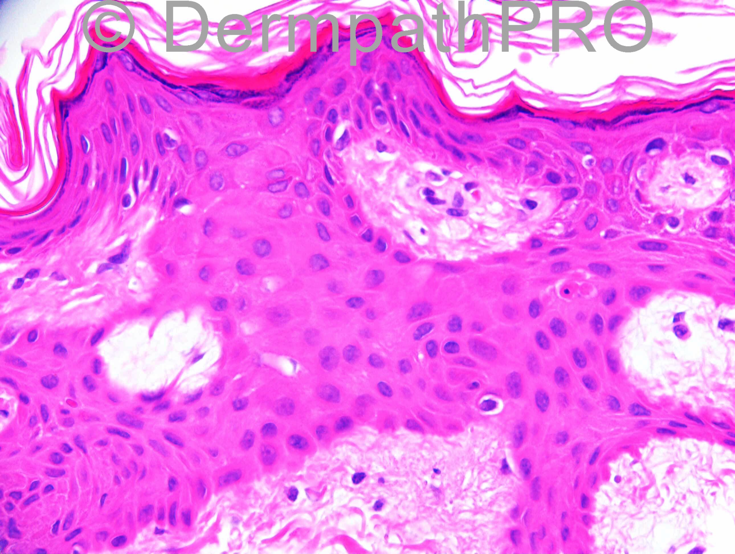
I would consider the history of an excised dermatofibroma and the presence of the incidentally discovered lesion in the polar ends of the excision and would suggest accordingly that this is not an original tumor of the follicular infundibulum but rather an epidermal hyperplastic reaction pattern mimicking it induced by the previously excised dermatofibroma (May be residual fibrohistiocytic cells r still there, not sure from the scanning magnification). I could not find typical pale/clear cells of tumor of follicular infundibulum and also I can not see its characteristic striking peripheral palisading..

Share this comment


Link to comment
share_externally

Guest Tiberiu Tebeica

Posted

Since the coincidence of having a TFI in a DF related scar from the back seems unlikely, until otherwise proven through clinical / grossing correlations I would be tempted to incline more towards Mona's theory and interpret such findings as epithelial induction related to the presence of DF.

Share this comment


Link to comment
share_externally

Eman El-Nabarawy

Posted

Clear cell acanthoma like changes.
I am anxious to know the answer of case number 1195!!

Share this comment


Link to comment
share_externally

Robledo F. Rocha

Posted

There’s no underlying dermatofibroma to regard this as epithelial induction. Also, there aren’t sebocytes clusters for reticulated acanthoma with sebaceous differentiation. So, I think this is tumor of the follicular infundibulum, although isthmic differentiation is not clear-cut.

Share this comment


Link to comment
share_externally

Accessory nipple not following milk line (ectopic)

Share this comment


Link to comment
share_externally

Guest Jim Davie MD

Posted

Agree with tumor of follicular infundibulum.
This is a subtle clonal proliferation with slightly enlarged pale keratinocytes that show [size=4]evenly scattered apoptotic cells, and no significant mitotic activity or atypia. It shows a r[/size][size=4]eticulated, horizontally-arranged, plate-like architecture that extensively involves both epidermis and follicles, and a surface hyperkeratosis resembling that of a SK. The assoicated adjacent dermis appears elastin rich. I don't see evidence of DF or epidermal induction.[/size]

Share this comment


Link to comment
share_externally

Guest Romualdo

Posted

[b]I am curious to know the diagnosis of case 1195![/b]

Share this comment


Link to comment
share_externally

Dr. Richard Carr

Posted

I reported this as an incidental tumour of the follicular infundibulum. Jim covers it particularly well. According to Kazakov's book you can see more basaloid examples with peripheral palisading although for me the images here quite typical of the entity.

Share this comment


Link to comment
share_externally


Join the conversation

You can post now and register later. If you have an account, sign in now to post with your account.

Guest
Add a comment...

×   Pasted as rich text.   Paste as plain text instead

  Only 75 emoji are allowed.

×   Your link has been automatically embedded.   Display as a link instead

×   Your previous content has been restored.   Clear editor

×   You cannot paste images directly. Upload or insert images from URL.

Loading...

×
×
  • Create New...