Jump to content
In this section we have spot diagnoses posted on a daily basis since June 2010, now over 4000! You can review the archived cases and read the suggested diagnoses by users and the final comment by the contributors.
Case are uploaded each week day by 10 am UK time with the correct diagnosis will generally be posted at 8 pm UK time. Why not view the most recent spot diagnosis and proffer a diagnosis?

Case Number : Case 1199 - 27th January Posted By: Guest

Please read the clinical history and view the images by clicking on them before you proffer your diagnosis.
Submitted Date :
   (0 reviews)

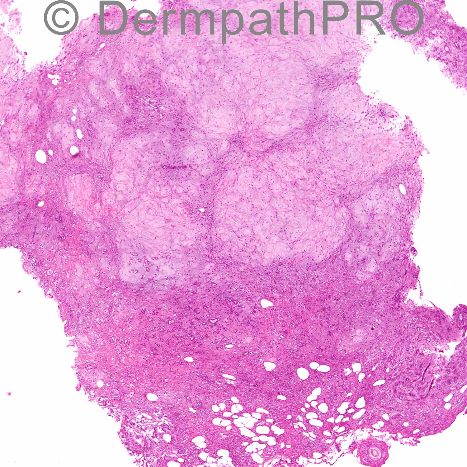
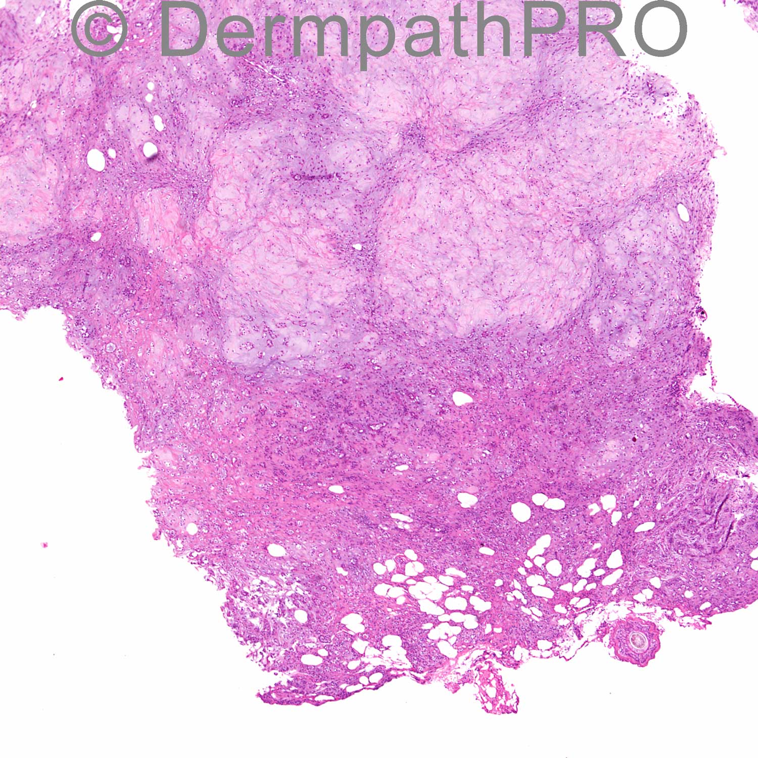
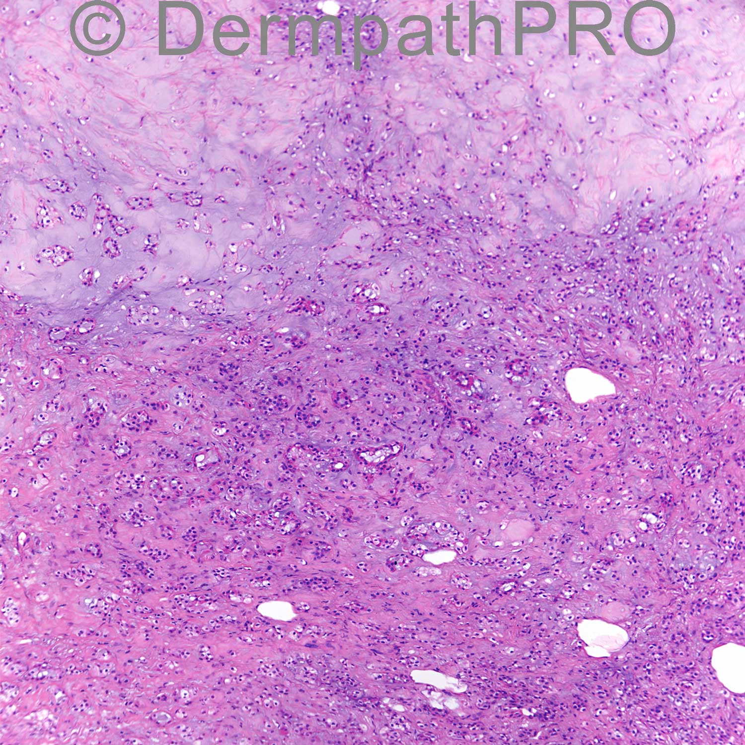
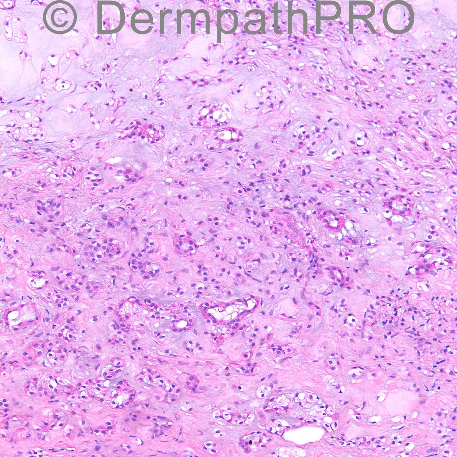
68 year old woman with lesion of right posterior calf. Clinical: r/o dermatofibroma

Case posted by Dr. Uma Sundram


  Report Record

User Feedback


Dr. Mona Abdel-Halim

Posted

I think ductal structures are seen, so I will call it chondoid syringoma or mixed tumor of the skin

Share this comment


Link to comment
share_externally

Robledo F. Rocha

Posted

Myoepithelioma may show focal ductal differentiation, in opposition to those branching tubules that are very often found in chondroid syringoma. Also, myoepithelioma occurs preferentially on the limbs, and chondroid syringoma arises more commonly on the head. Last, this lesion seems to be very large and deep-seated, making me favor myoepithelioma.

Share this comment


Link to comment
share_externally

Robledo F. Rocha

Posted

From Miettinen’s Modern Soft Tissue Pathology:
“Mixed tumors of the skin and soft tissues are analogous to corresponding tumors commonly seen in salivary glands. They are synonymous with [i]chondroid syringomas[/i], a name used particularly in older dermatopathology literature. These tumors have variable epithelial and myoepithelial components and typically have metaplastic cartilage-like elements. In the skin and subcutis, mixed tumors most likely originate in the sweat glands or their ducts; however, it is unclear how the occurrence of similar tumors in intramuscular soft tissues can be explained.
In the current dermatopathology literature, [i]mixed tumor [/i]has been the preferred term, although myoepitheliomas have also been reported. In the literature on soft tissue, the term [i]myoepithelioma [/i]has been preferred. It seems clear that these groups represent a continuum, and therefore they are combined here. Some authors, however, separate myoepitheliomas as tumors that lack any ductal epithelial differentiation and contain only solid epithelioid or spindle cell elements, with or without cartilage-like elements.”

Share this comment


Link to comment
share_externally

Mixed tumour (aka myoepithelioma aka chondroid syringoma). I think more important than the name is to decide whether this is a tumour of skin or soft tissue, as the latter has a higher risk of recurrence and malignancy.

Share this comment


Link to comment
share_externally

Uma Sundram

Posted

I too favored chondroid syringoma (although myoepithelioma is another way to think of this tumor). The tumor was very superficial and we were able to confirm on imaging studies that the deep soft tissues were not involved. Mesenchymal phosphaturic tumors primary to the skin do exist and are exceptionally rare. They are associated with oncogenic osteomalacia and on histology have hemangiopericytoma-like vasculature, osteoclast-like giant cells, and calcified basophilic matrix. These findings were all absent from this case.

Share this comment


Link to comment
share_externally


Join the conversation

You can post now and register later. If you have an account, sign in now to post with your account.

Guest
Add a comment...

×   Pasted as rich text.   Paste as plain text instead

  Only 75 emoji are allowed.

×   Your link has been automatically embedded.   Display as a link instead

×   Your previous content has been restored.   Clear editor

×   You cannot paste images directly. Upload or insert images from URL.

Loading...

×
×
  • Create New...