Jump to content
In this section we have spot diagnoses posted on a daily basis since June 2010, now over 4000! You can review the archived cases and read the suggested diagnoses by users and the final comment by the contributors.
Case are uploaded each week day by 10 am UK time with the correct diagnosis will generally be posted at 8 pm UK time. Why not view the most recent spot diagnosis and proffer a diagnosis?

Case Number : Case 1332 - 31 July Posted By: Guest

Please read the clinical history and view the images by clicking on them before you proffer your diagnosis.
Submitted Date :
   (0 reviews)

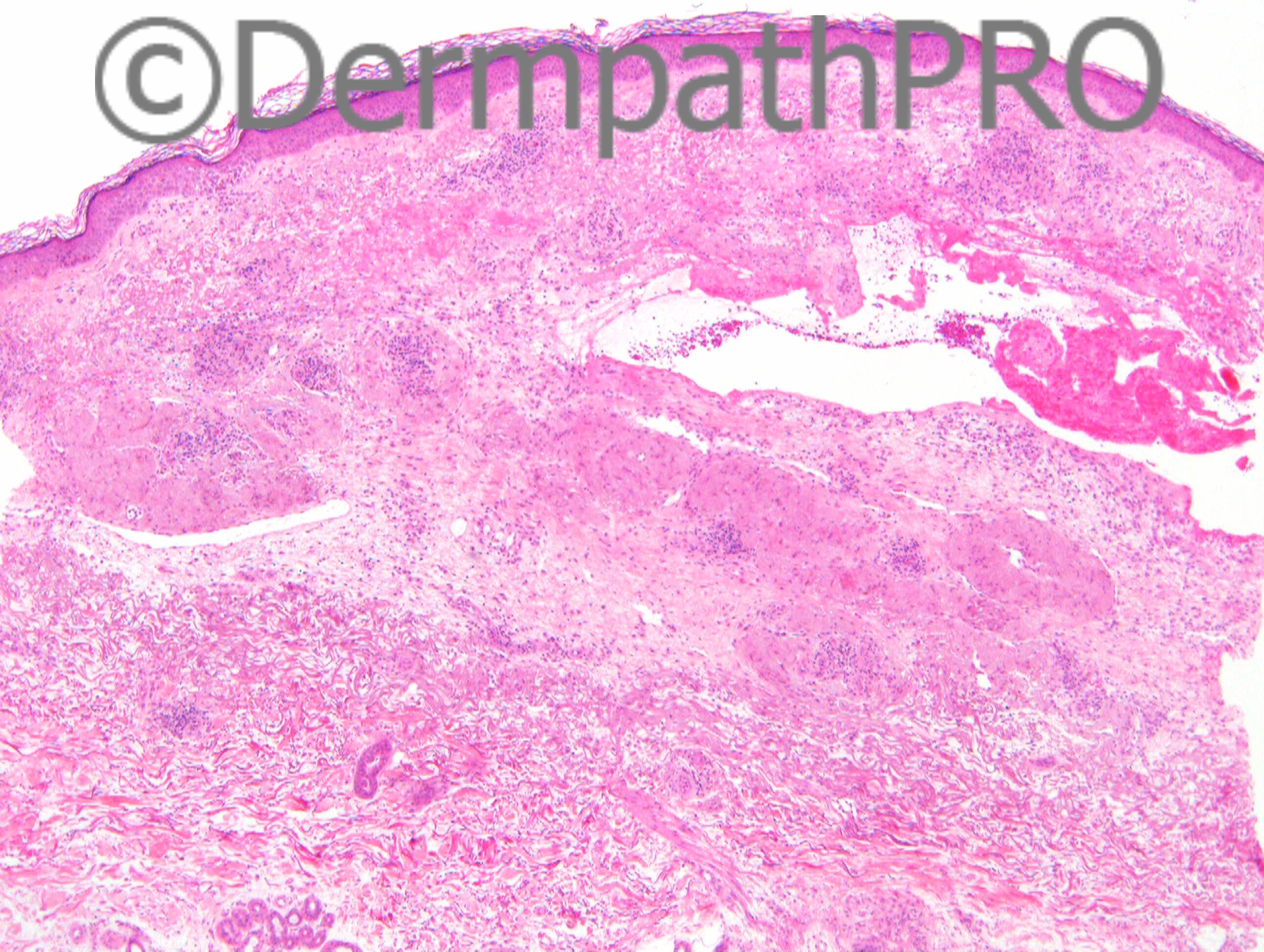
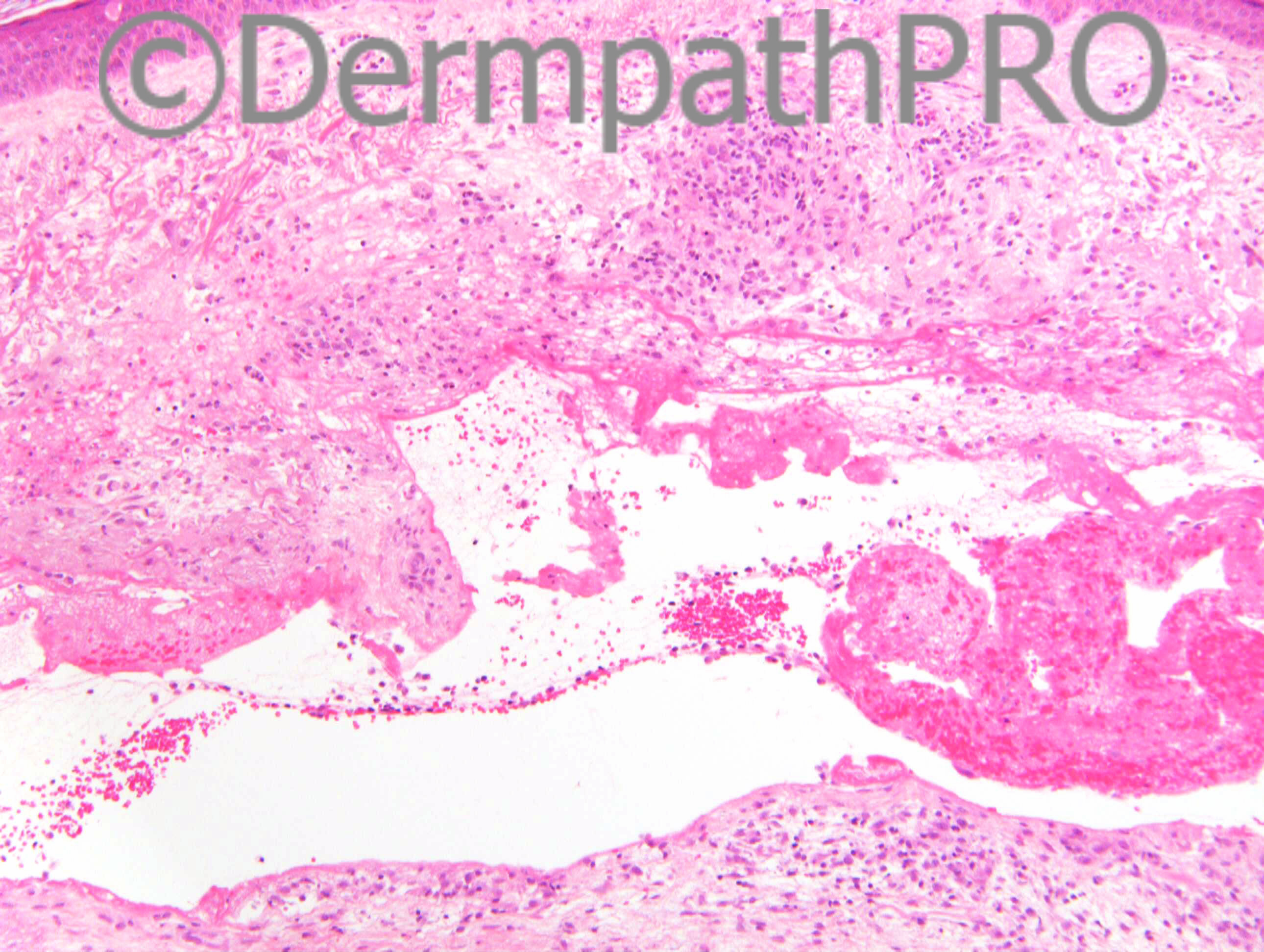
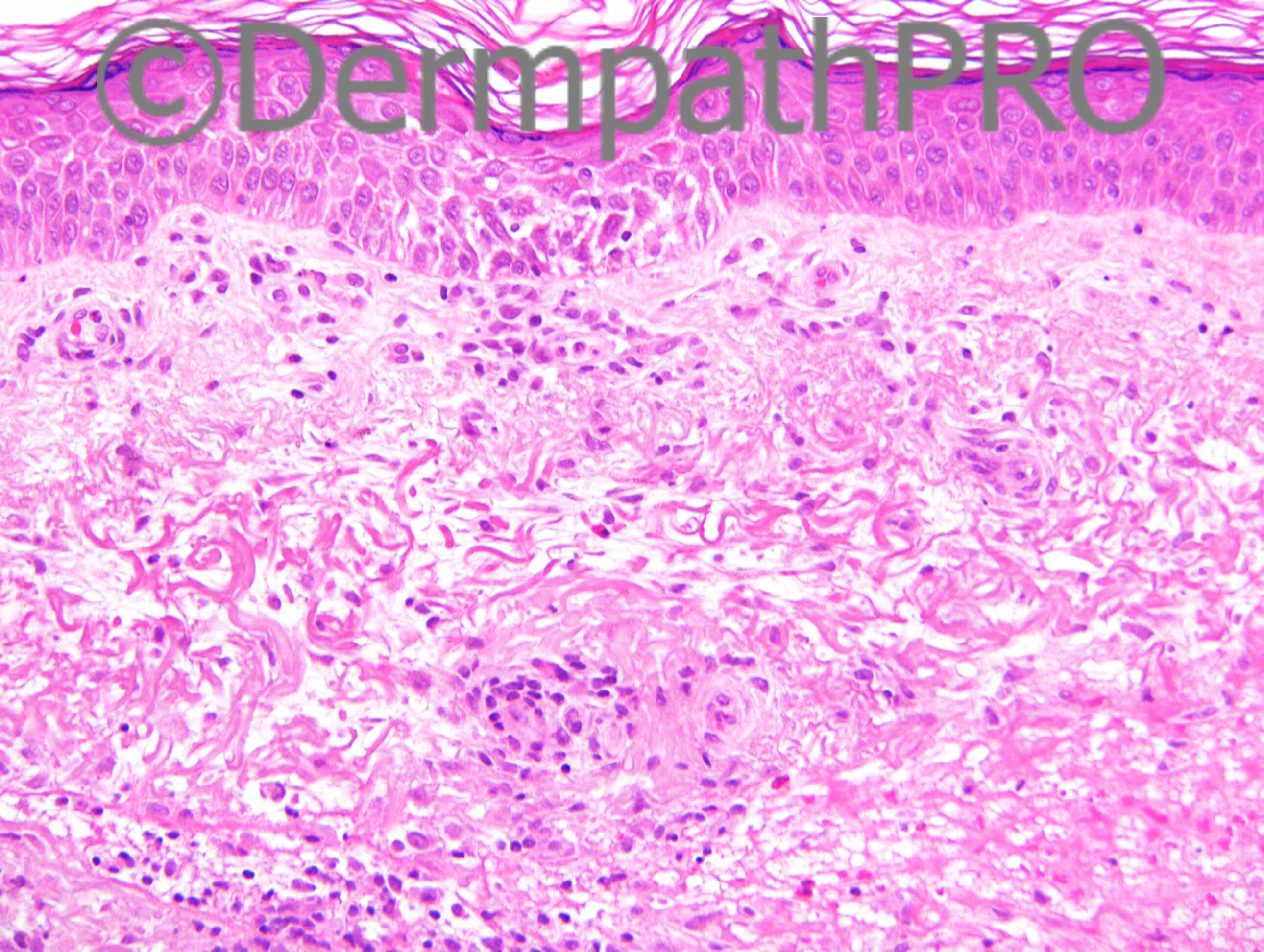
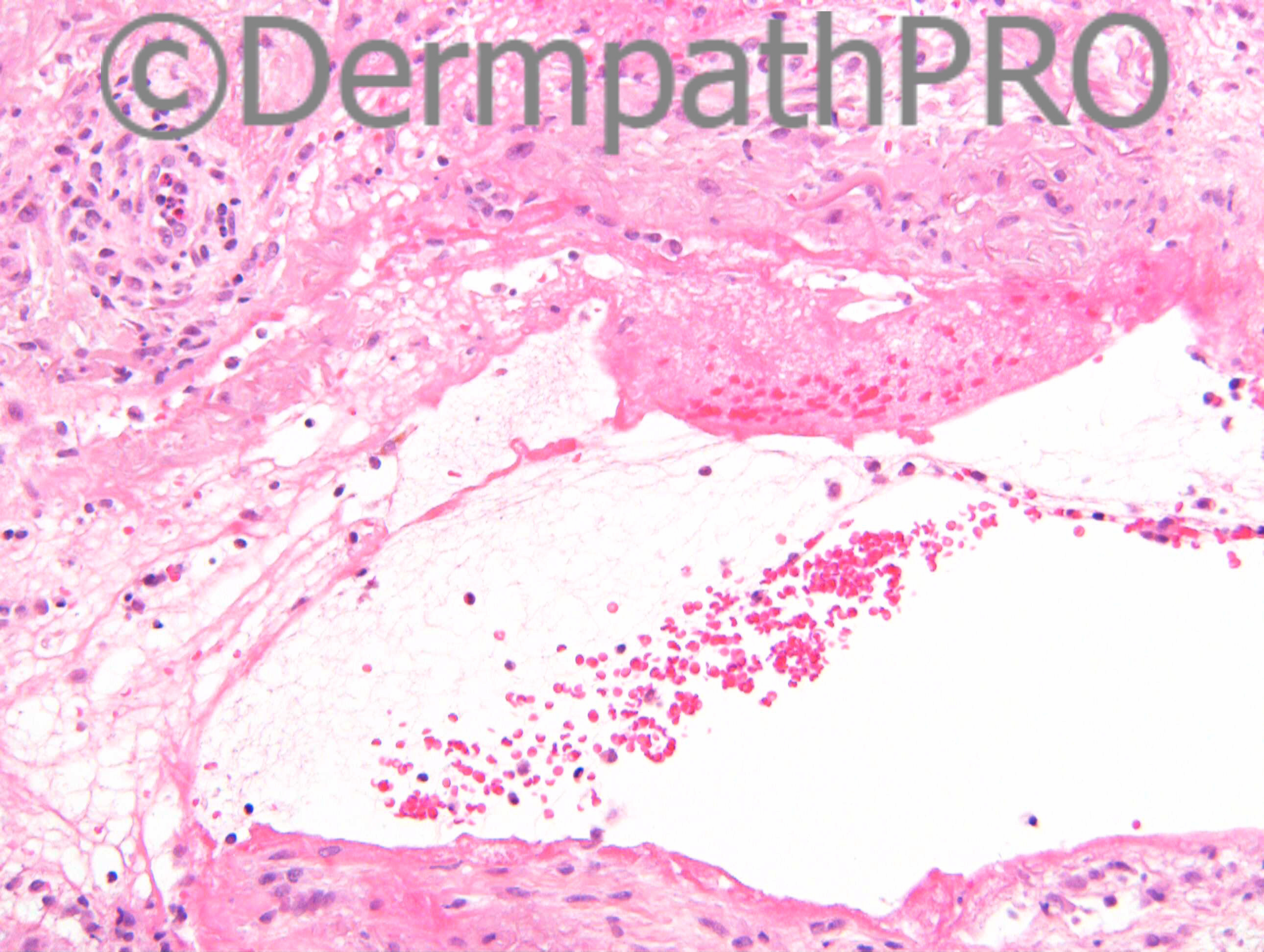
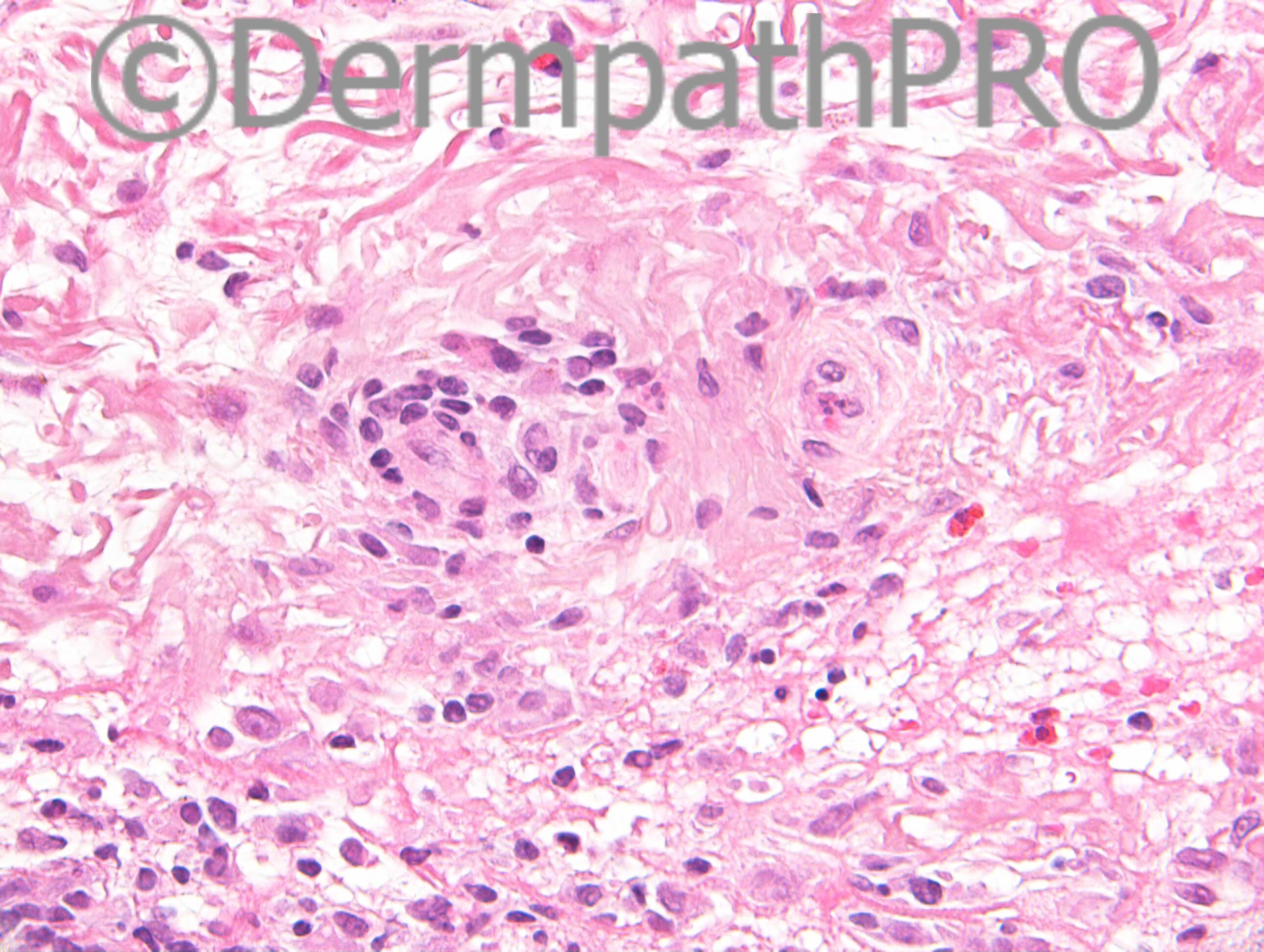
M82. Forearm. 6/12 history of indurated erythematous area on forearm. Resolves after 2-3/7. ?contact dermatitis.

Case posted by Dr Richard Carr


  Report Record

User Feedback


Eman El-Nabarawy

Posted

Interstitial granuloma annulare vs. dermal contact dermatitis.

Share this comment


Link to comment
share_externally

Dr. Richard Carr

Posted

[img]https://dermpathpro.com/uploads/spot_diagnosis_comment_img/Case%201332%20-%20RAC7097x40_Perls_4pm.jpg[/img]

[img]https://dermpathpro.com/uploads/spot_diagnosis_comment_img/Case%201332%20-%20RAC7097x5_EVG_4pm.jpg[/img]

Share this comment


Link to comment
share_externally

Dr. Mona Abdel-Halim

Posted

Have been struggling a lot with this case !!
I think the clue is in the EVG.. The EVG is highlighting proliferation of elastic fibers in the papillary dermis together with amorphous masses of elastotic material in the dermis.
Now after this, looking again on the H&E again, may be we r dealing with solar elastosis with the dermal cleft being bullous solar elastosis... Hemorrhage and extravasated erythrocytes is not uncommon around bullous solar elastosis, Pearl's positivity indicates that the brown deposits seen are hemosiderin depositis secondary to the dermal hemorrhage. I have read in Weedon about chronic heliodermatitis with perivascular lymphohistiocytic infiltrate associated with solar elastosis in chronic actinic damaged skin. It is usually associated with mast cells which I can not c here.
I do not know if I am interpreting it right with the weird clinical presentation!!

Share this comment


Link to comment
share_externally

Guest Romualdo

Posted

After seeing the new photographs and reading Mona's comments I agree with her suggestion of bullous solar elastosis.

Share this comment


Link to comment
share_externally

Dr. Mona Abdel-Halim

Posted

I have made some research and came up with this lovely case presentation of bullous solar elastosis.. I think the clinical photos here mimics the description in the history above given by Dr Carr.. However, I do not think it resolves completely within few days may be just the associated inflammation subsides a bit and re-exacerbates with sun exposure ???

http://www.globalskinpathatlas.com/imagedetail.cfm?topLevelID=77&imageID=595&did=1429

Remember seeing once a pathological demonstration of bullous solar elastosis in a Dermatopathology meeting but have never seen this clinically before !!!!

Marvelous :-))

Share this comment


Link to comment
share_externally

Arti Bakshi

Posted

Wow…have to say Mona's assessment appears spot on! Was struggling with this one!!

Share this comment


Link to comment
share_externally

Eman El-Nabarawy

Posted

Although bullous solar elastosis seems fitting well, I wonder about the rapid resolution of the lésions as regards chronic nature of this condition! Upon searching, I found that early lesions of erythema ab igne are reversible. It's characterized by mid dermal collections of solar elastotic material associated with few hemosiderin(+ ve Pearl) and melanin. Bullous forms are reported but can't find reported dermal clefts! I think the clinical picture will be the discriminating factor.

Share this comment


Link to comment
share_externally

Dr. Richard Carr

Posted

My report:
Marked background solar elastosis, mid to upper dermal fibrin pseudocyts with RBC & iron - suggest traumatic shearing force. Focal mild spongiosis moderate perivascular lymphoytic infiltrate with eosinophils. Consider arthropd, drug and less likely contact dermatitis.

Thanks Mona I think the description of bullous solar elastosis fits quite well. I was thinking that there was probably an underlying inflammatory dermatosis (moderate perivascular lymphocytic infiltrate and scattered eosinophils) that probably led to the local trauma (scratching etc) or a shearing force. In this anatomic location and age skin is often composed of mainly elastic fibres with very little collagen so with the added solar elastosis I suppose it is very susceptible to local trauma.

Share this comment


Link to comment
share_externally


Join the conversation

You can post now and register later. If you have an account, sign in now to post with your account.

Guest
Add a comment...

×   Pasted as rich text.   Paste as plain text instead

  Only 75 emoji are allowed.

×   Your link has been automatically embedded.   Display as a link instead

×   Your previous content has been restored.   Clear editor

×   You cannot paste images directly. Upload or insert images from URL.

Loading...

×
×
  • Create New...