Jump to content
In this section we have spot diagnoses posted on a daily basis since June 2010, now over 4000! You can review the archived cases and read the suggested diagnoses by users and the final comment by the contributors.
Case are uploaded each week day by 10 am UK time with the correct diagnosis will generally be posted at 8 pm UK time. Why not view the most recent spot diagnosis and proffer a diagnosis?

Case Number : Case 1295 - 10 June Posted By: Guest

Please read the clinical history and view the images by clicking on them before you proffer your diagnosis.
Submitted Date :
   (0 reviews)

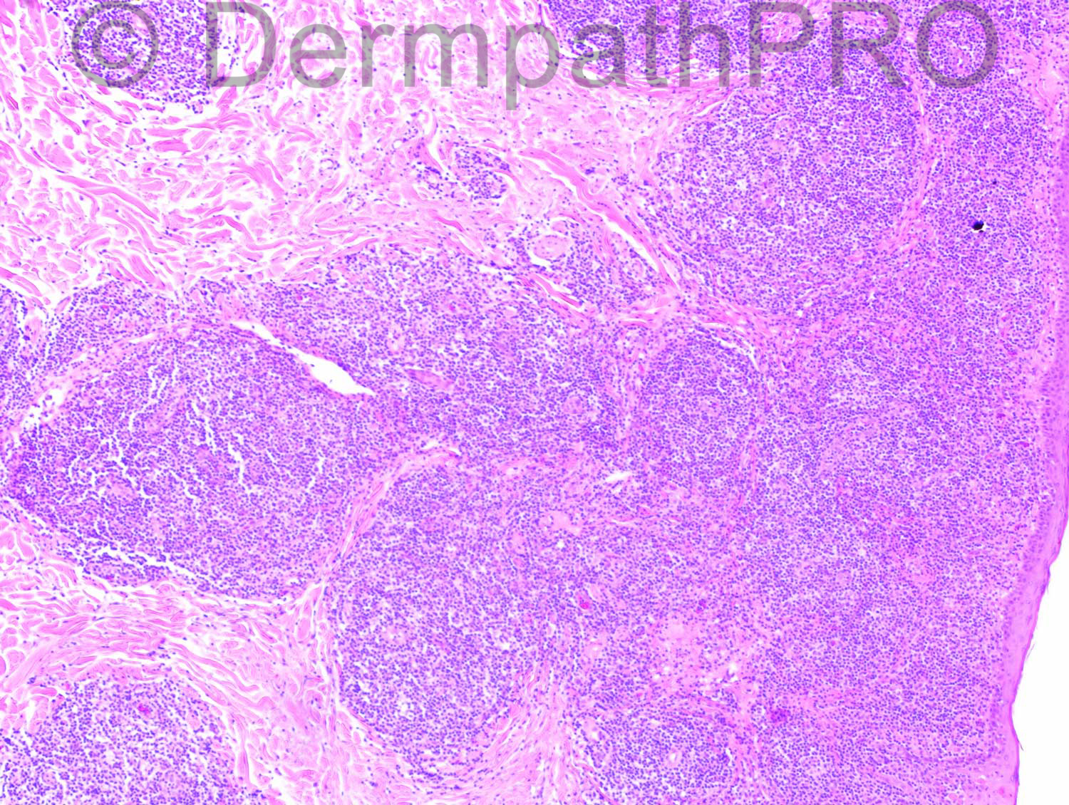
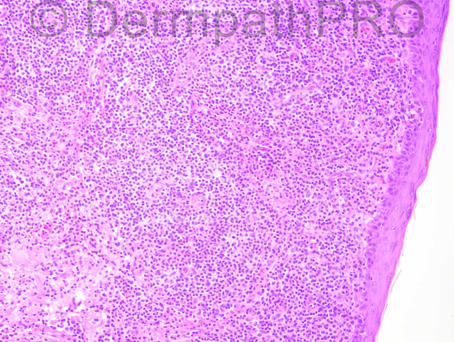
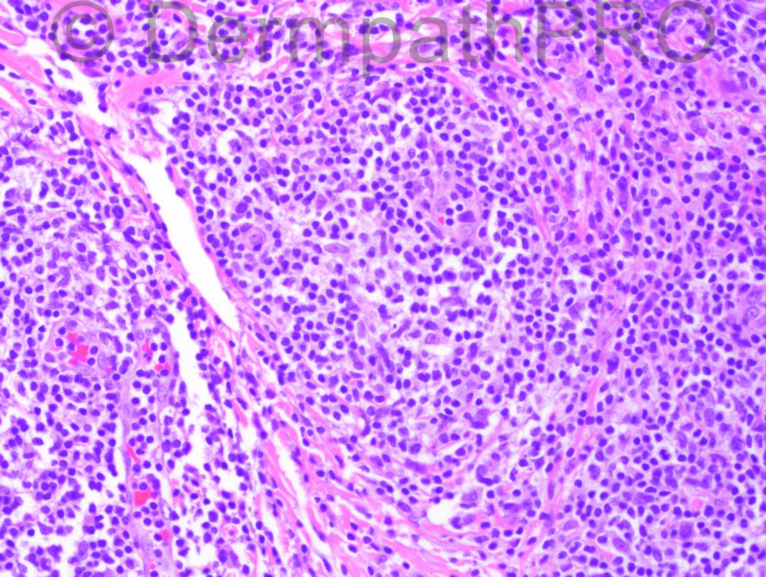
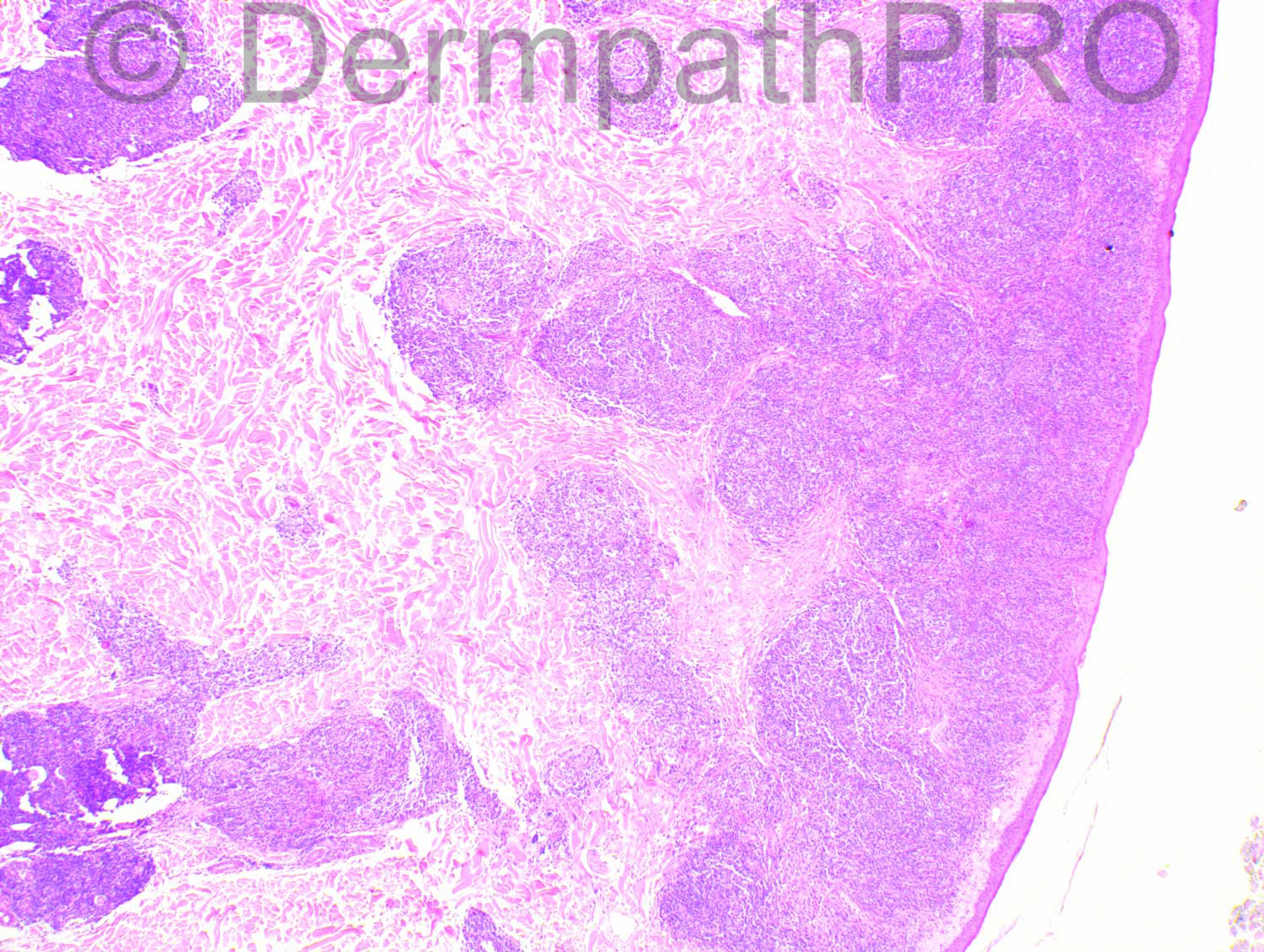
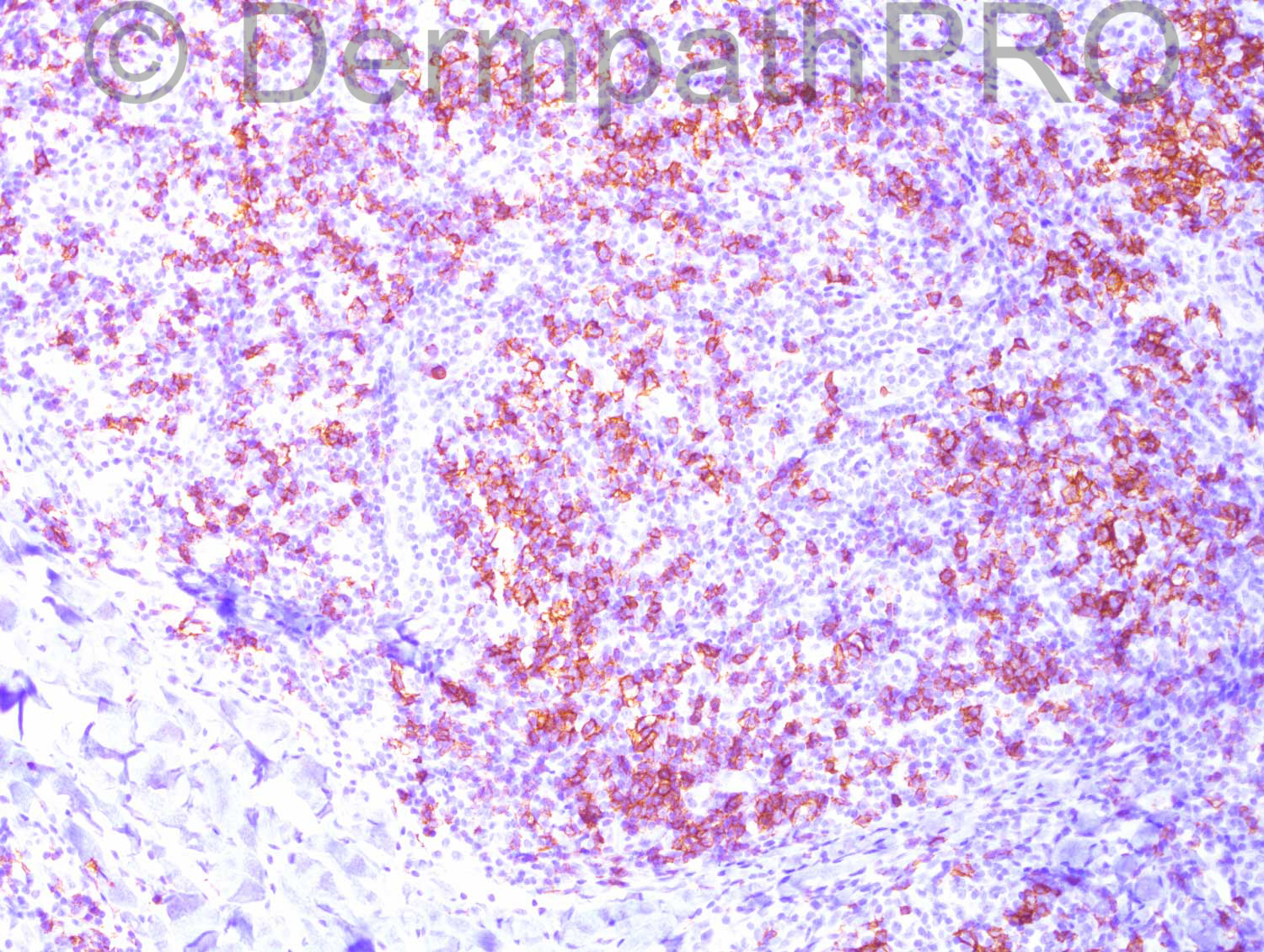
The patient is a 62-year-old female with a chest lesion.

Case posted by Dr Hafeez Diwan

  Report Record

User Feedback


Guest Hazem Hamed

Posted

Follicular pattern with no prominent germinal centres. Smell of follicle centre lymphoma despite the unlabeled immuno images. Marginal zone lymphoma with follicular clonization is the main differential. Reactive lymphoid hyperplasia is less likely based on H&E.

Share this comment


Link to comment
share_externally

Dr. Mona Abdel-Halim

Posted

Same thinking as Dr Hazem... Waiting for the labeling of the immunos !!!

Share this comment


Link to comment
share_externally

Guest Romualdo

Posted

Agree with primary cutaneous follicle center lymphoma as most probable possibility.

Share this comment


Link to comment
share_externally

Dr. Hafeez Diwan

Posted

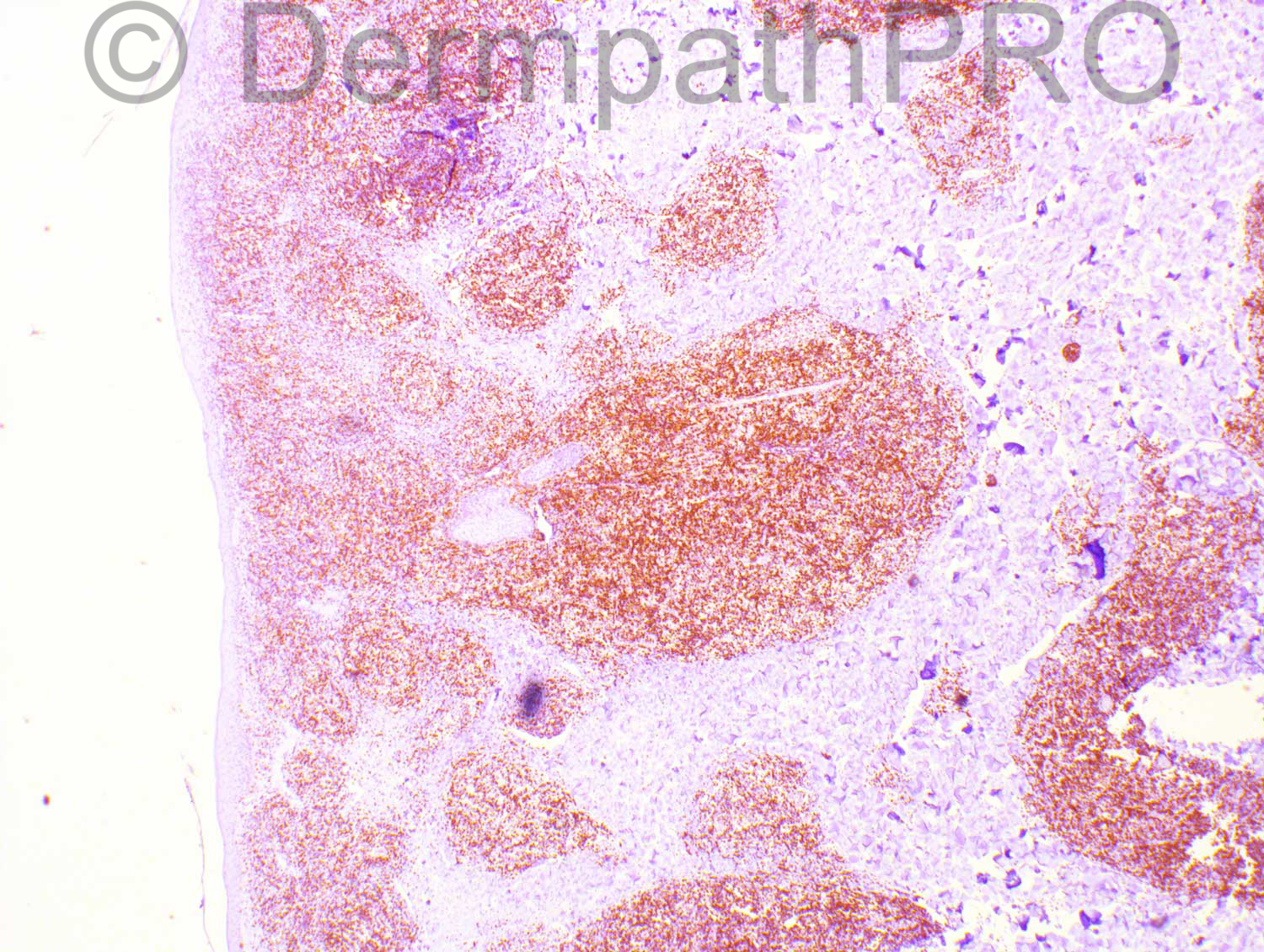
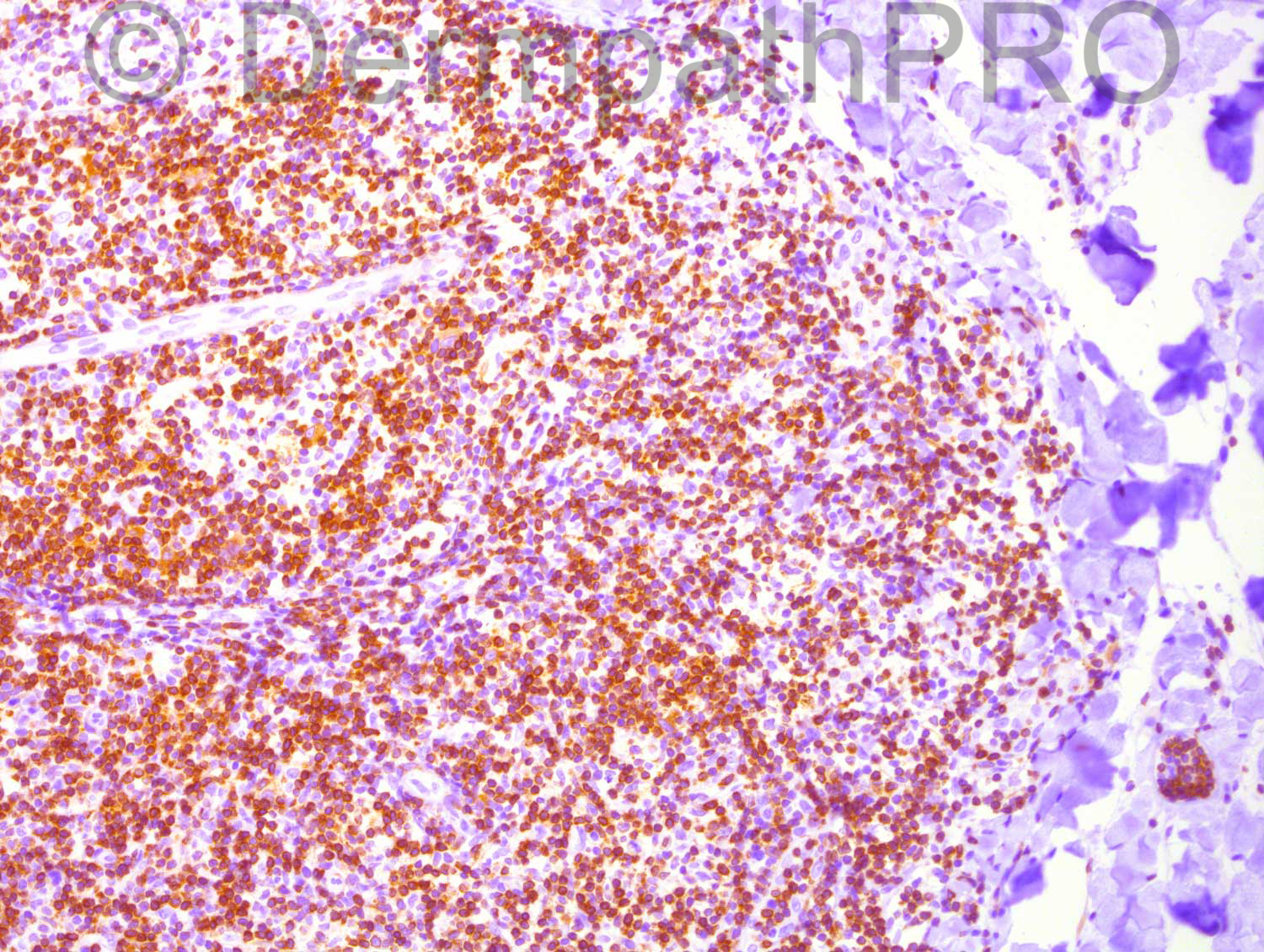
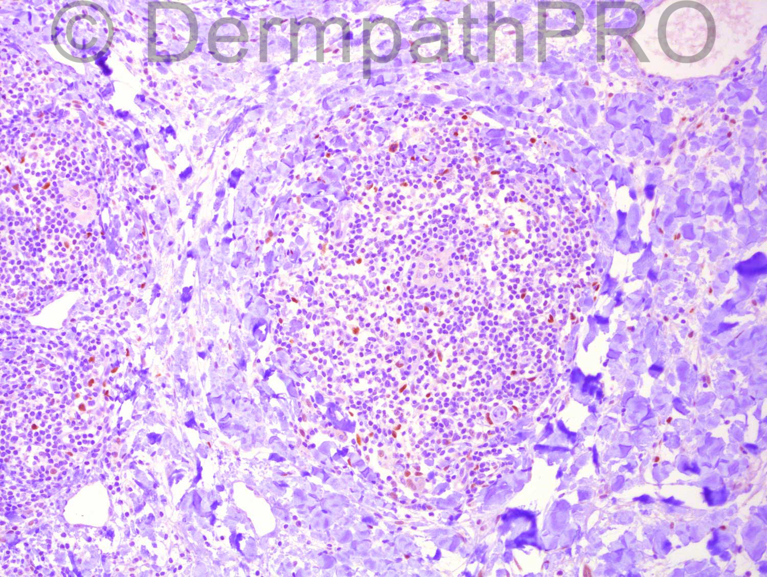
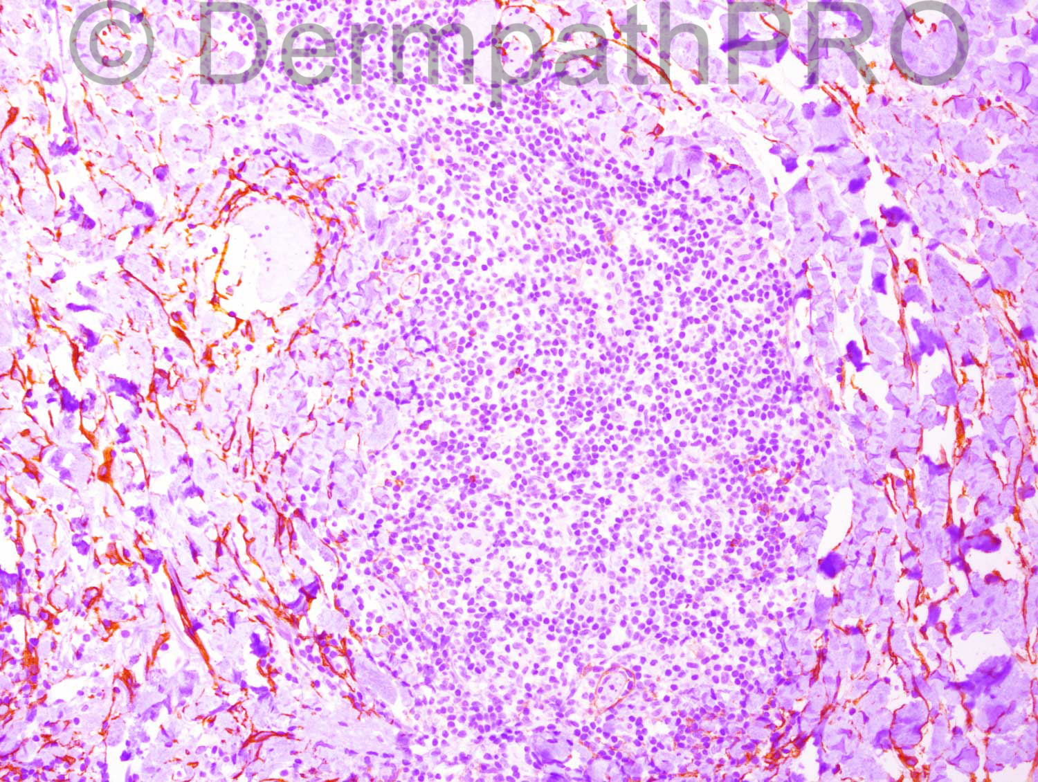
Sorry about the immunos. I included them by mistake. They are: top left and top right - Bcl2; 2nd row left is Bcl6; 2nd row right is CD3; third row, both are CD10; 4th row; both are CD20. I called this cutaneous lymphoid hyperplasia.

Share this comment


Link to comment
share_externally

Guest Hazem Hamed

Posted

Based on negativity for both CD10 and BCL6, we are left with m[font="arial, verdana, tahoma, sans-serif"][color="#1c2837"][size=4]arginal zone lymphoma with follicular colonization and [/size][/color][/font][color=#1C2837][font=arial, verdana, tahoma, sans-serif][size=4]cutaneous lymphoid hyperplasia. [/size][/font][/color]M[font="arial, verdana, tahoma, sans-serif"][color="#1c2837"][size=4]arginal zone lymphoma [/size][/color][/font][font="arial, verdana, tahoma, sans-serif"][color="#1c2837"][size=4]should be considered and excluded before calling this lesion as [/size][/color][/font][color=#1C2837][font=arial, verdana, tahoma, sans-serif][size=4]cutaneous lymphoid hyperplasia. [/size][/font][/color]I would do light chains, CD43, IgM , IgD, FDC meshwork markers (CD21&CD23) and also B cell clonality.

Share this comment


Link to comment
share_externally


Join the conversation

You can post now and register later. If you have an account, sign in now to post with your account.

Guest
Add a comment...

×   Pasted as rich text.   Paste as plain text instead

  Only 75 emoji are allowed.

×   Your link has been automatically embedded.   Display as a link instead

×   Your previous content has been restored.   Clear editor

×   You cannot paste images directly. Upload or insert images from URL.

Loading...

×
×
  • Create New...