Jump to content
In this section we have spot diagnoses posted on a daily basis since June 2010, now over 4000! You can review the archived cases and read the suggested diagnoses by users and the final comment by the contributors.
Case are uploaded each week day by 10 am UK time with the correct diagnosis will generally be posted at 8 pm UK time. Why not view the most recent spot diagnosis and proffer a diagnosis?

Case Number : Case 1297 - 12 June Posted By: Guest

Please read the clinical history and view the images by clicking on them before you proffer your diagnosis.
Submitted Date :
   (0 reviews)

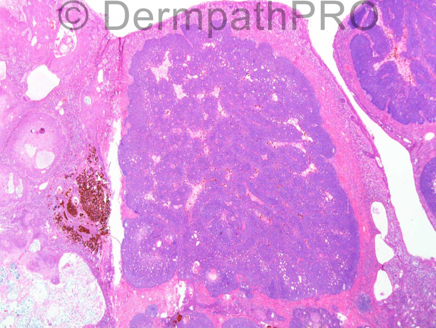
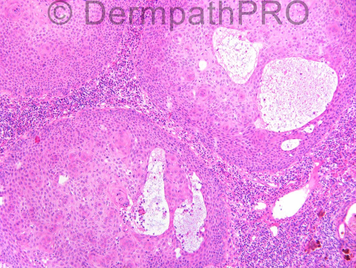
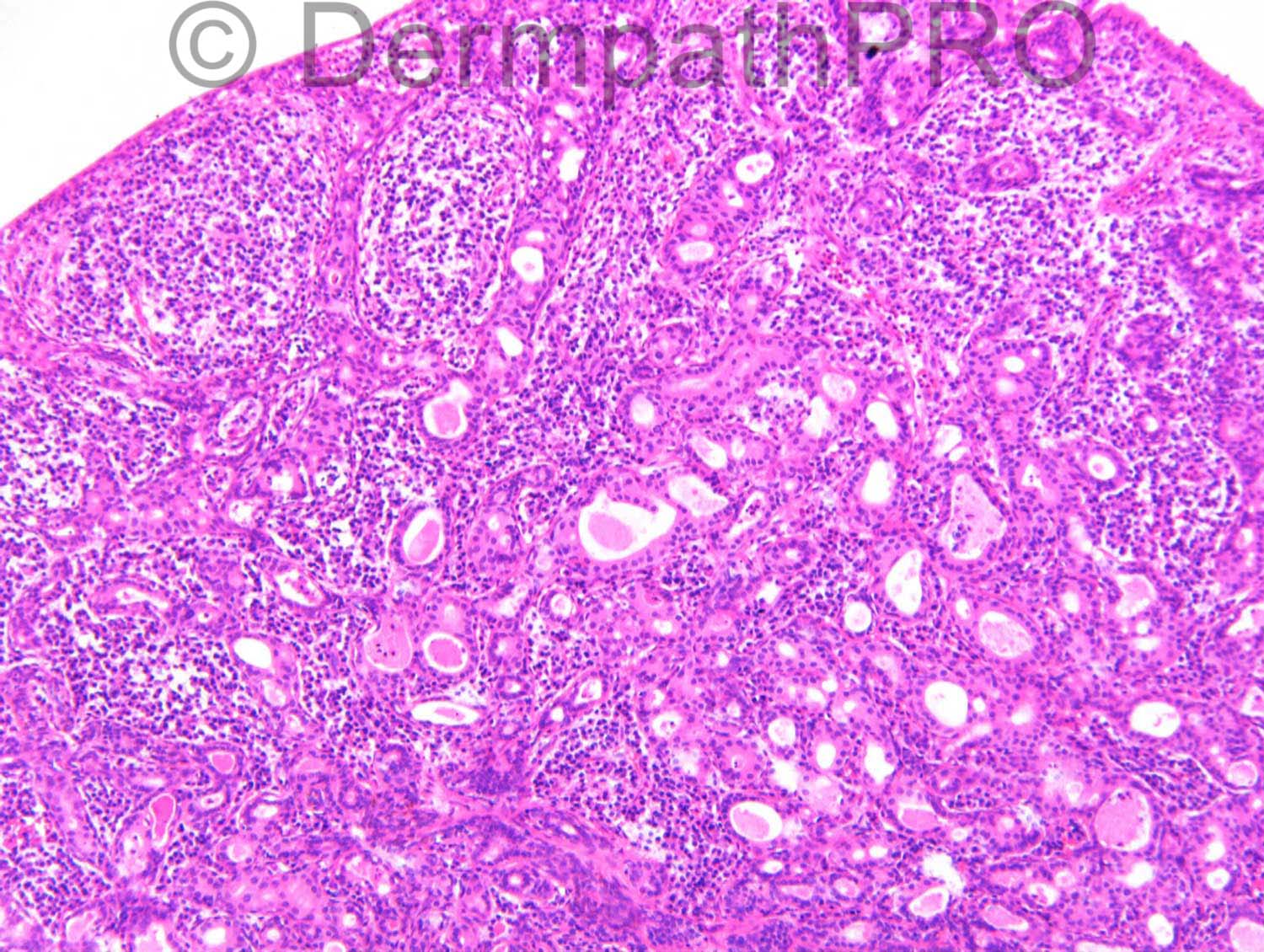
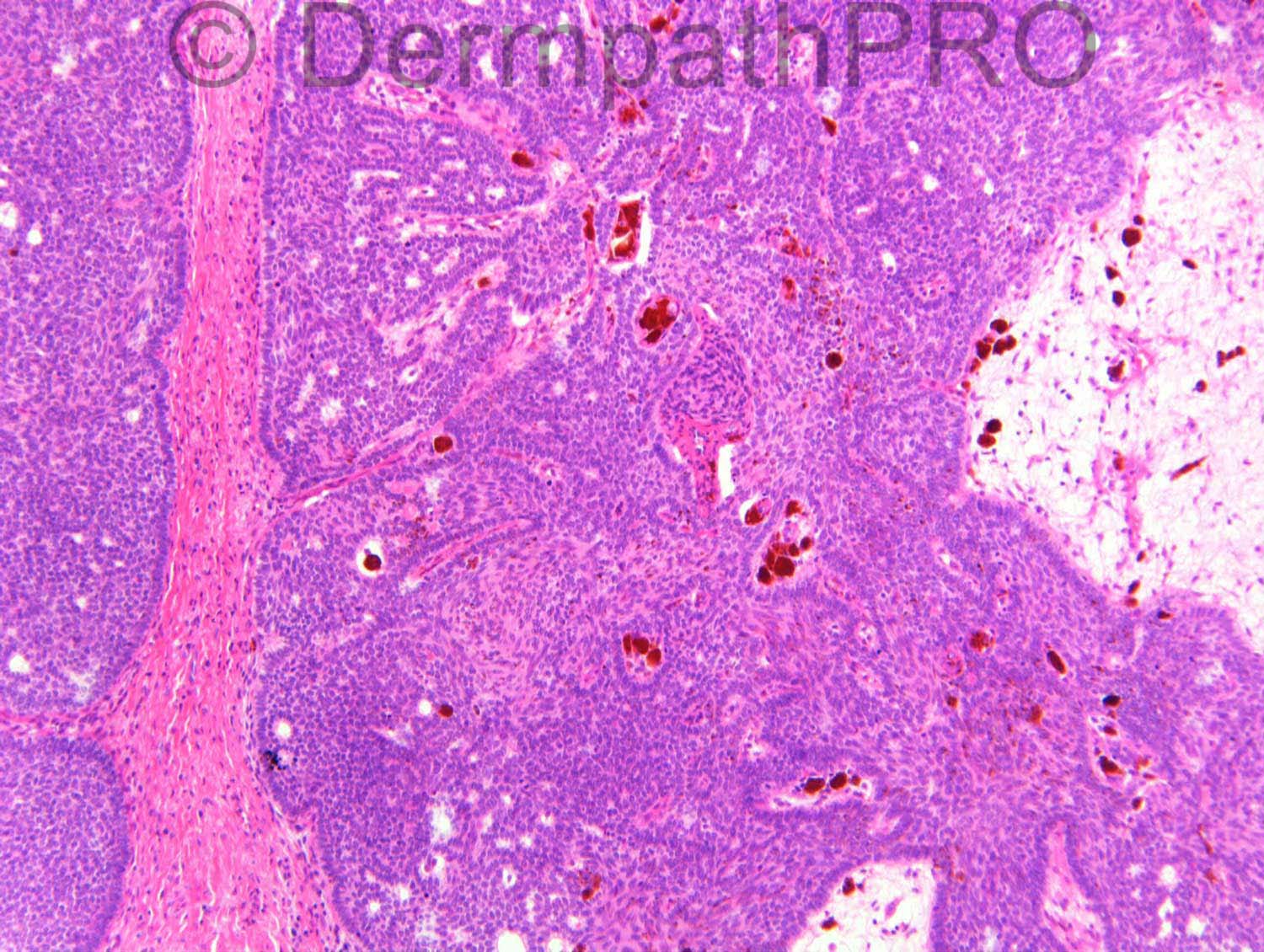
F45. Lateral left eye. 3cm fleshy nodular lesion, pedunculated ?acrochordon. c/o Dr Miroslav Radojkovic

Case posted by Dr Richard Carr

  Report Record

User Feedback


Dr. Mona Abdel-Halim

Posted

Another challenging case!!!
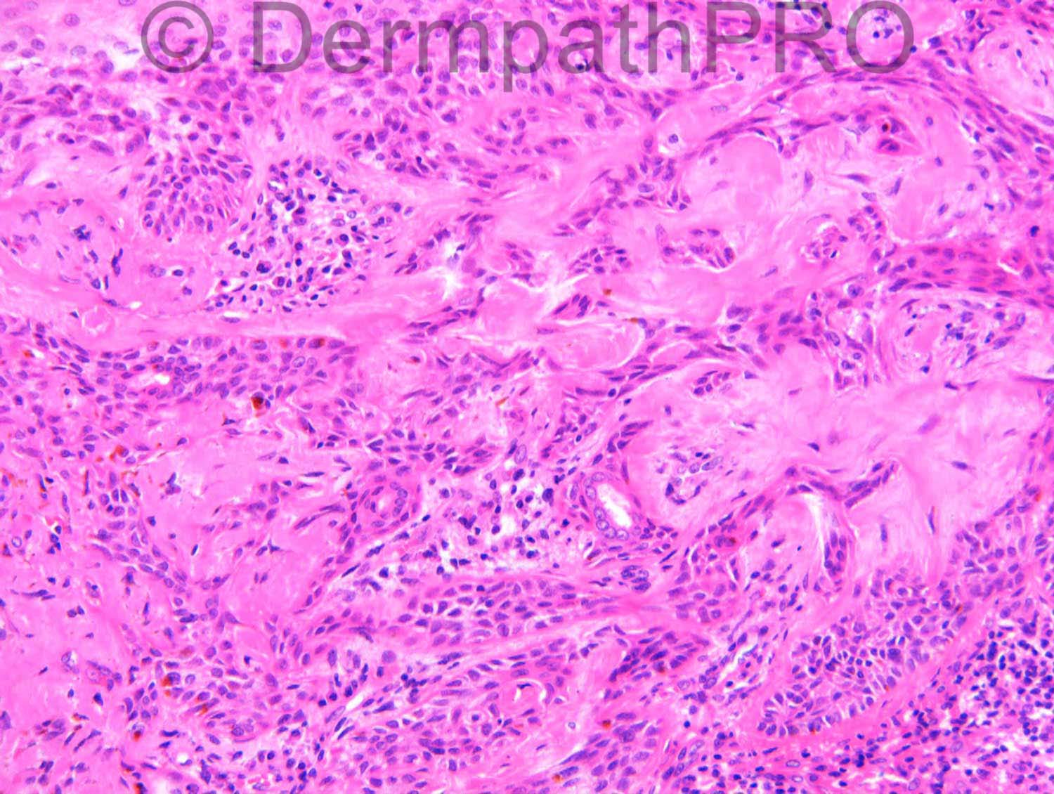
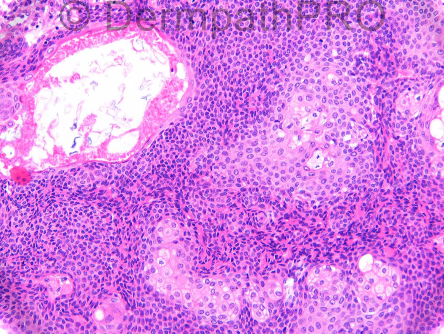
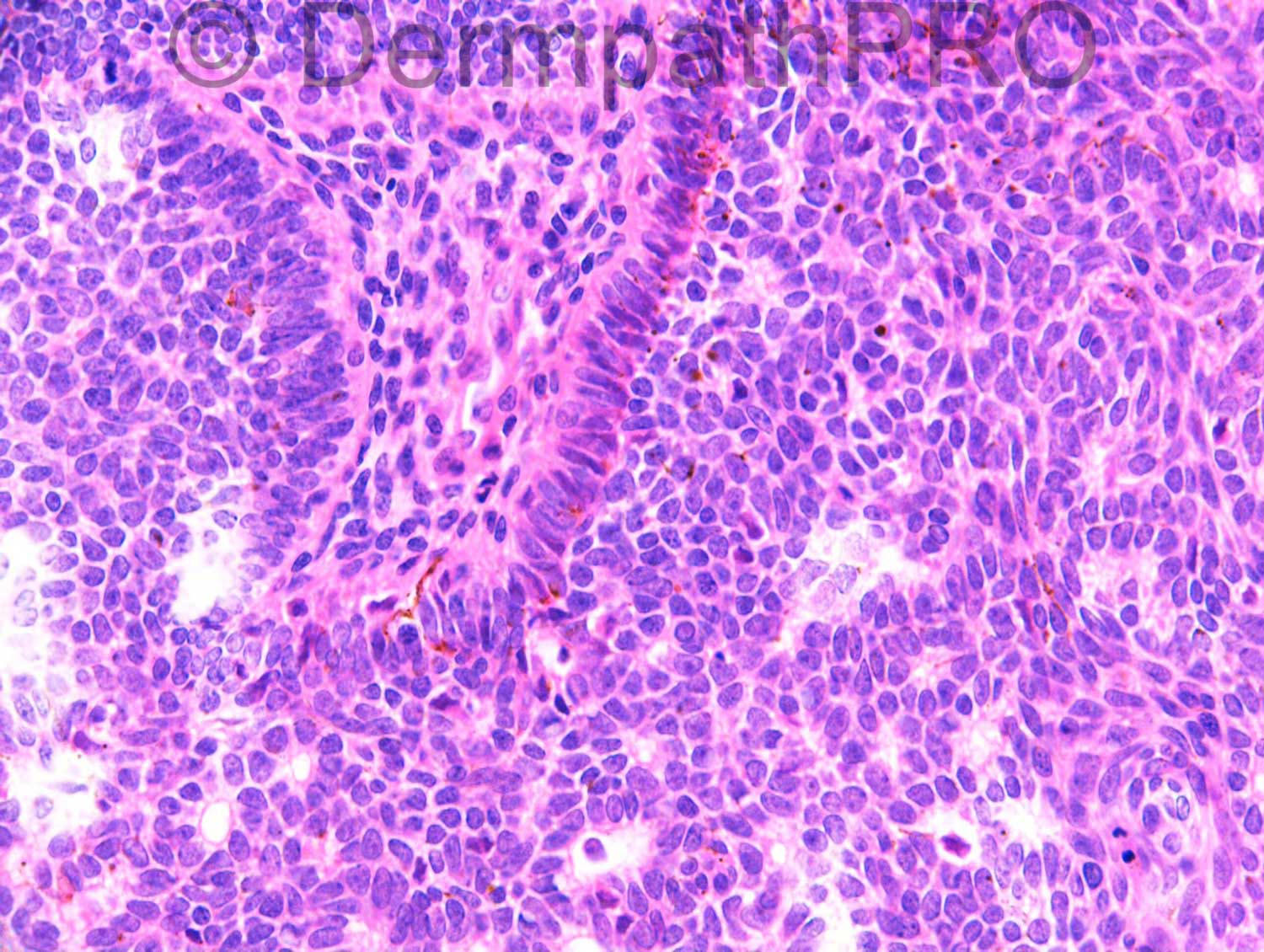
The basaloid component has striking peripheral palisading but no striking mucinous retraction.
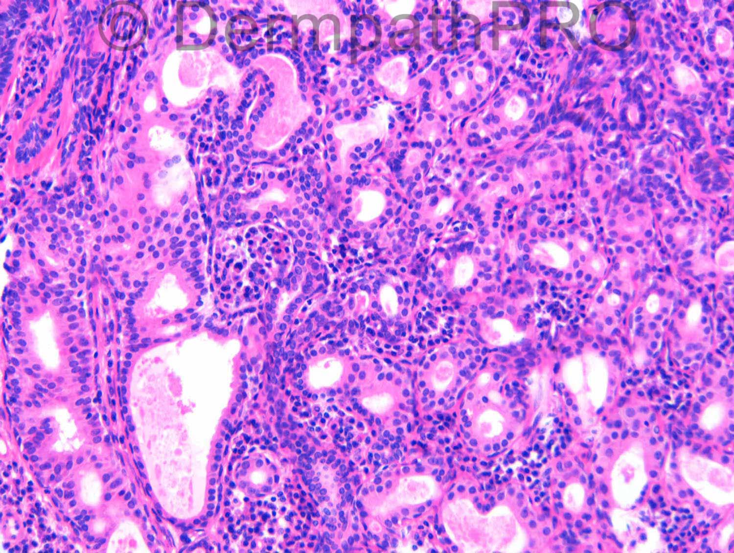
There is focal sebaceous differentiation, apocrine ductal component, a desmoplastic area as well as foci of squamous eddies.
The lesion is very large spanning the whole dermis.
Thinking of trichoblastoma with focal sebaceous differentiation and apocrine ductal differentiation??

Share this comment


Link to comment
share_externally

Guest Hazem Hamed

Posted

Hidradenoma with a focaly hyalinized (desmoplastic) stroma

Share this comment


Link to comment
share_externally

Dr. Richard Carr

Posted

[img]https://dermpathpro.com/uploads/spot_diagnosis_comment_img/Case%201297_RAC7144x10_CD10_4pm.jpg[/img]

[img]https://dermpathpro.com/uploads/spot_diagnosis_comment_img/Case%201297_RAC7144x10_CD34_4pm.jpg[/img]

Share this comment


Link to comment
share_externally

Dr. Mona Abdel-Halim

Posted

The CD34 staining is of BCC pattern not trichoblastoma pattern with almost only vascular positivity and no striking stromal cell positivity.
The CD10 is showing both epithelial and stromal positivity, the epithelial positivity is strong and striking on the left side of the image and only focal (patchy) in the right half. But still we have stromal positivity (a striking peritumoral stromal cell positivity). In BCC there is supposed to be epithelial positivity and weak stromal positivity but in trichoblastomas, CD10 is limited to peritumoral stromal cells. So we have both patterns here... This might be explained by a trichoblastoma showing focal BCC like area BUT what will explain the negative stromal cell positivity with CD34 then??? I did some reading and found this article that might explain this pattern of CD10 by a BCC with follicular differentiation. So are we dealing with a very rare BCC with follicular, sebaceous and apocrine differentiation?????
I need help !!!!!
The article I am referring to is:

http://www.ncbi.nlm.nih.gov/pubmed/19570076

Share this comment


Link to comment
share_externally

Arti Bakshi

Posted

Difficult case….I am struck by the heterogeneity of the constituent cells. There is an admixture of squamoid cells, clear cells, basaloid cells and apocrine cells; plus clear cut ares of ductal differentiation.There is stromal hyalinisation in image 5, but I am not convinced of specialised mesenchymal stroma to consider trichoblastoma. Also I think there is clear cell change rather than sebaceous differentiation in image 6.
Therefore, would go with Hazem's diagnosis of Hidradenoma.
Some of the nodules have basaloid cells with peripheral palisading, which is unusual for Hidradenoma, and does make one think of BCC, but I do not think the rest of the lesion fits with BCC. (unless it is a very rare variant, as Mona has speculated!!)

Share this comment


Link to comment
share_externally

Guest Romualdo

Posted

I think there is a papillary mesenchimal body (last image). I also think there are sebaceous ducts and sebocytes, apocrine differentiaton and areas resembling hidradenoma. If these distinct components are not too focal this neoplasm could be called a composite or combined benign adnexal tumor of the skin.

Share this comment


Link to comment
share_externally

Eman El-Nabarawy

Posted

Based on: the striking peripheral palisading (not explained within the context of hidradenoma), presence of papillary mesenchymal body (highlightend by CD10, I guess), I will go for trichoblastoma with apocrine and sebaceous differentiation.

Share this comment


Link to comment
share_externally

I agree with Romualdo. I think this is what Ackerman would call a "combined" or "mixed" benign adnexal neoplasm. In his atlas of adnexal neoplasms, there is a chapter titled "Proliferations with combined adnexal differentiation", wherein there are examples of combinations of two or more components (follicular, apocrine and/or sebaceous).

Share this comment


Link to comment
share_externally

Dr. Richard Carr

Posted

I reported this as a hybrid large nodular (focally pigmented) trichoblastoma, desmoplastic tricholemmoma, syringocystadenoma papilliferum, tubular apocrine adenoma and sebaceous differentiation. The top right looks infudibular/pilar with mucin pools (i.e. inverted follicular keratosis-like). In my experience these mixed or combined lesions often associate with naevus sebaceous although it was not possible to identify the latter in this case. Regarding CD10 in our experience trichoblastomas often express epithelial staining not surprisingly as the marker can be seen to be positive in inner root sheath cells on normal follicles. The tight peritumoural staining is however specific and fairly sensitive for TE/TB. Absence of tight peritumoural staining with epithelial staining (often peripheral accentuation in contrast to TE/TB) is helpful for the diagnosis of TE-like BCC. That said while most of the the basaloid lesions in naevus sebaceous are clearly benign some cases are morphologically and immunophenotypically indistinguishable from BCC although I have not seen a frankly invasive BCC (as opposed to large rounded) in naevus sebaceous. CD34 is generally not very good (in our experience) for papillary mesechymal cells (may be the antibody clone) but is useful for confirming (desmoplastic) tricholemmoma - there is focal epithelial staining demonstrated here.

Share this comment


Link to comment
share_externally

John Zhang

Posted

On 15/06/2015 at 01:22, Dr. Richard Carr said:

I reported this as a hybrid large nodular (focally pigmented) trichoblastoma, desmoplastic tricholemmoma, syringocystadenoma papilliferum, tubular apocrine adenoma and sebaceous differentiation. The top right looks infudibular/pilar with mucin pools (i.e. inverted follicular keratosis-like). In my experience these mixed or combined lesions often associate with naevus sebaceous although it was not possible to identify the latter in this case. Regarding CD10 in our experience trichoblastomas often express epithelial staining not surprisingly as the marker can be seen to be positive in inner root sheath cells on normal follicles. The tight peritumoural staining is however specific and fairly sensitive for TE/TB. Absence of tight peritumoural staining with epithelial staining (often peripheral accentuation in contrast to TE/TB) is helpful for the diagnosis of TE-like BCC. That said while most of the the basaloid lesions in naevus sebaceous are clearly benign some cases are morphologically and immunophenotypically indistinguishable from BCC although I have not seen a frankly invasive BCC (as opposed to large rounded) in naevus sebaceous. CD34 is generally not very good (in our experience) for papillary mesechymal cells (may be the antibody clone) but is useful for confirming (desmoplastic) tricholemmoma - there is focal epithelial staining demonstrated here.

Thank you Dr. Carr for this very interesting case. I wonder, since this lesion shows apocrine, follicular and sebaceous lineages, would "apocrine mixed tumor" a good term for it?

Share this comment


Link to comment
share_externally


Join the conversation

You can post now and register later. If you have an account, sign in now to post with your account.

Guest
Add a comment...

×   Pasted as rich text.   Paste as plain text instead

  Only 75 emoji are allowed.

×   Your link has been automatically embedded.   Display as a link instead

×   Your previous content has been restored.   Clear editor

×   You cannot paste images directly. Upload or insert images from URL.

Loading...

×
×
  • Create New...