Jump to content
In this section we have spot diagnoses posted on a daily basis since June 2010, now over 4000! You can review the archived cases and read the suggested diagnoses by users and the final comment by the contributors.
Case are uploaded each week day by 10 am UK time with the correct diagnosis will generally be posted at 8 pm UK time. Why not view the most recent spot diagnosis and proffer a diagnosis?

Case Number : Case 1302 - 19 June Posted By: Guest

Please read the clinical history and view the images by clicking on them before you proffer your diagnosis.
Submitted Date :
   (0 reviews)

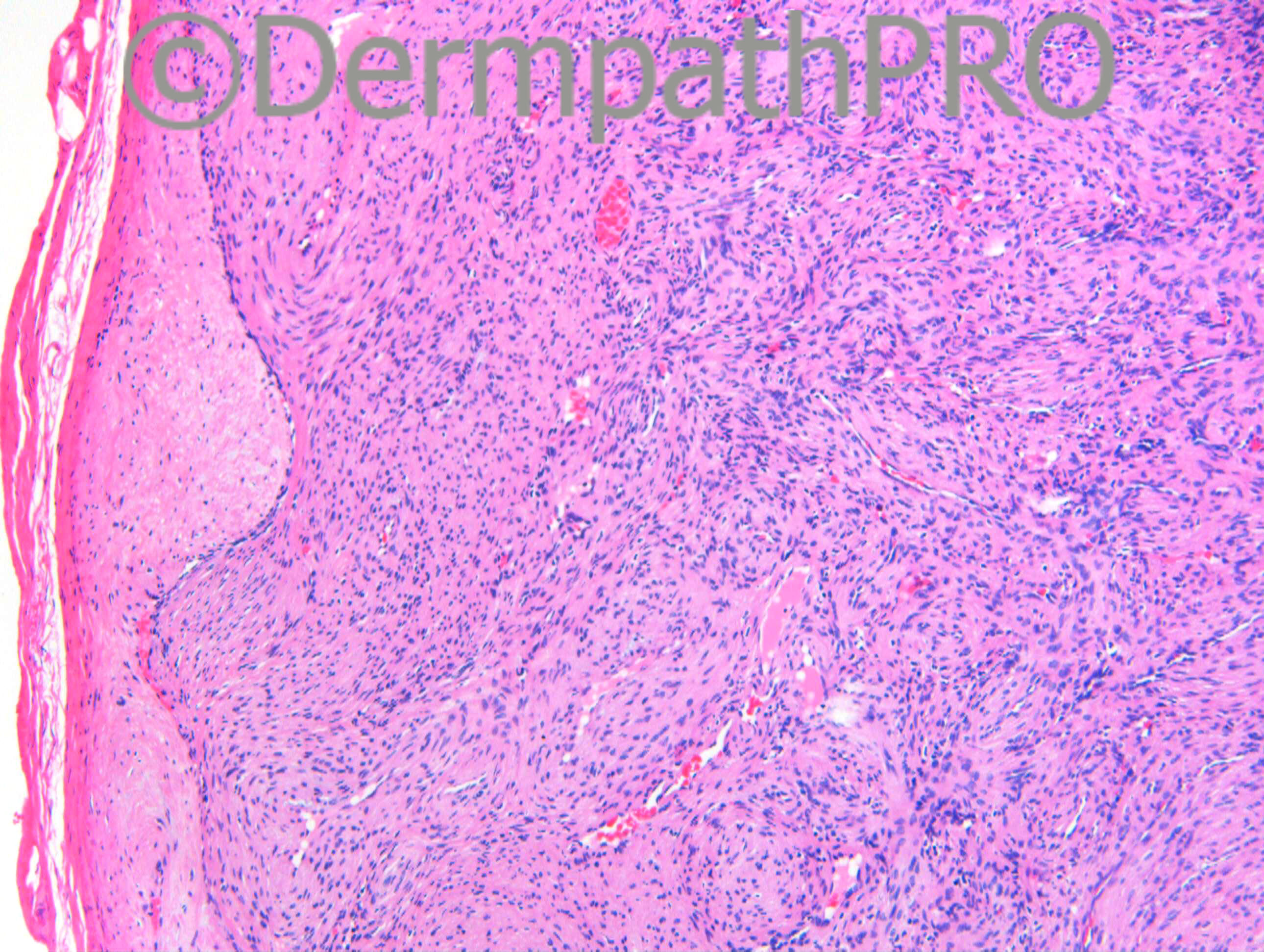
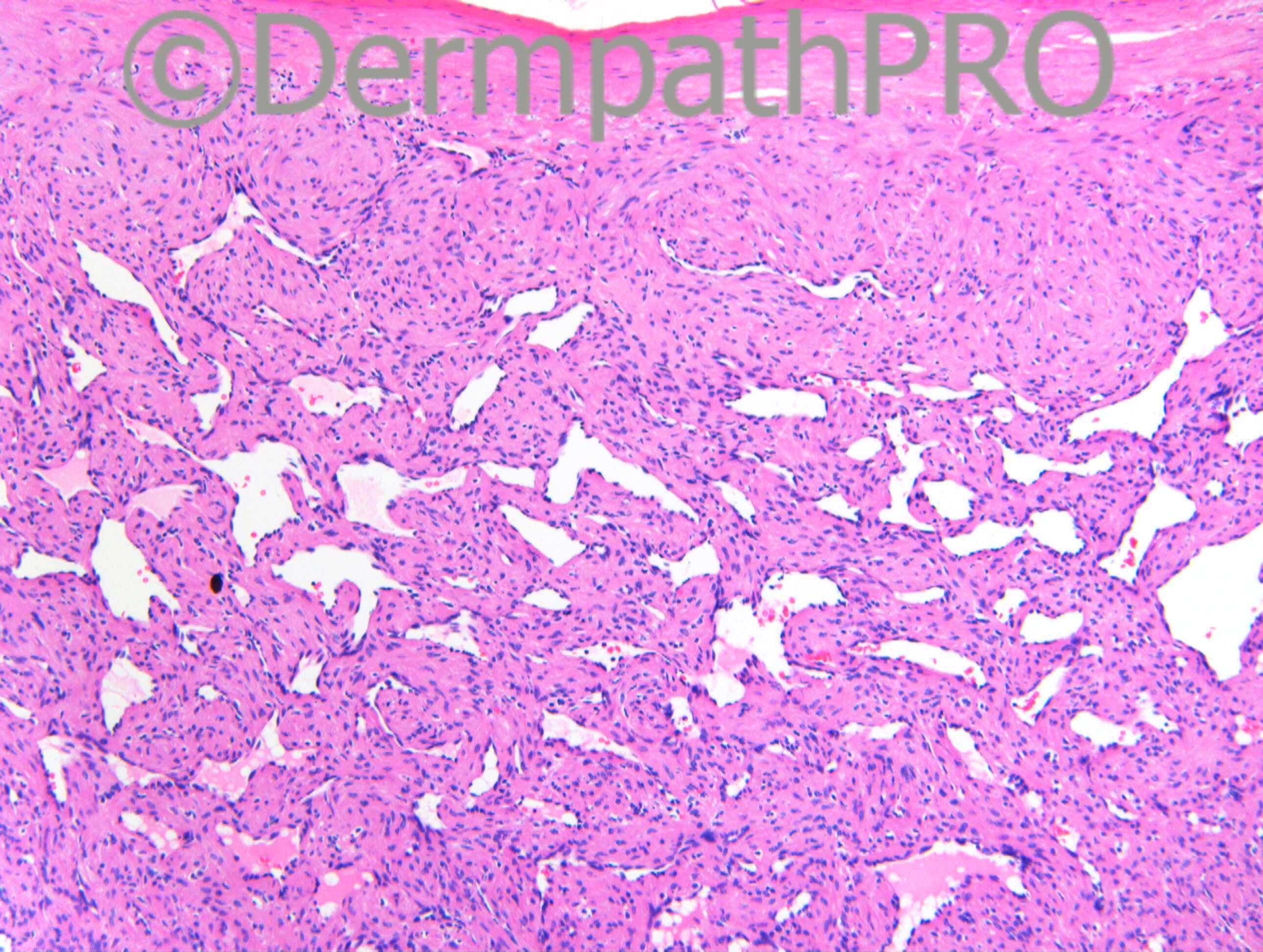
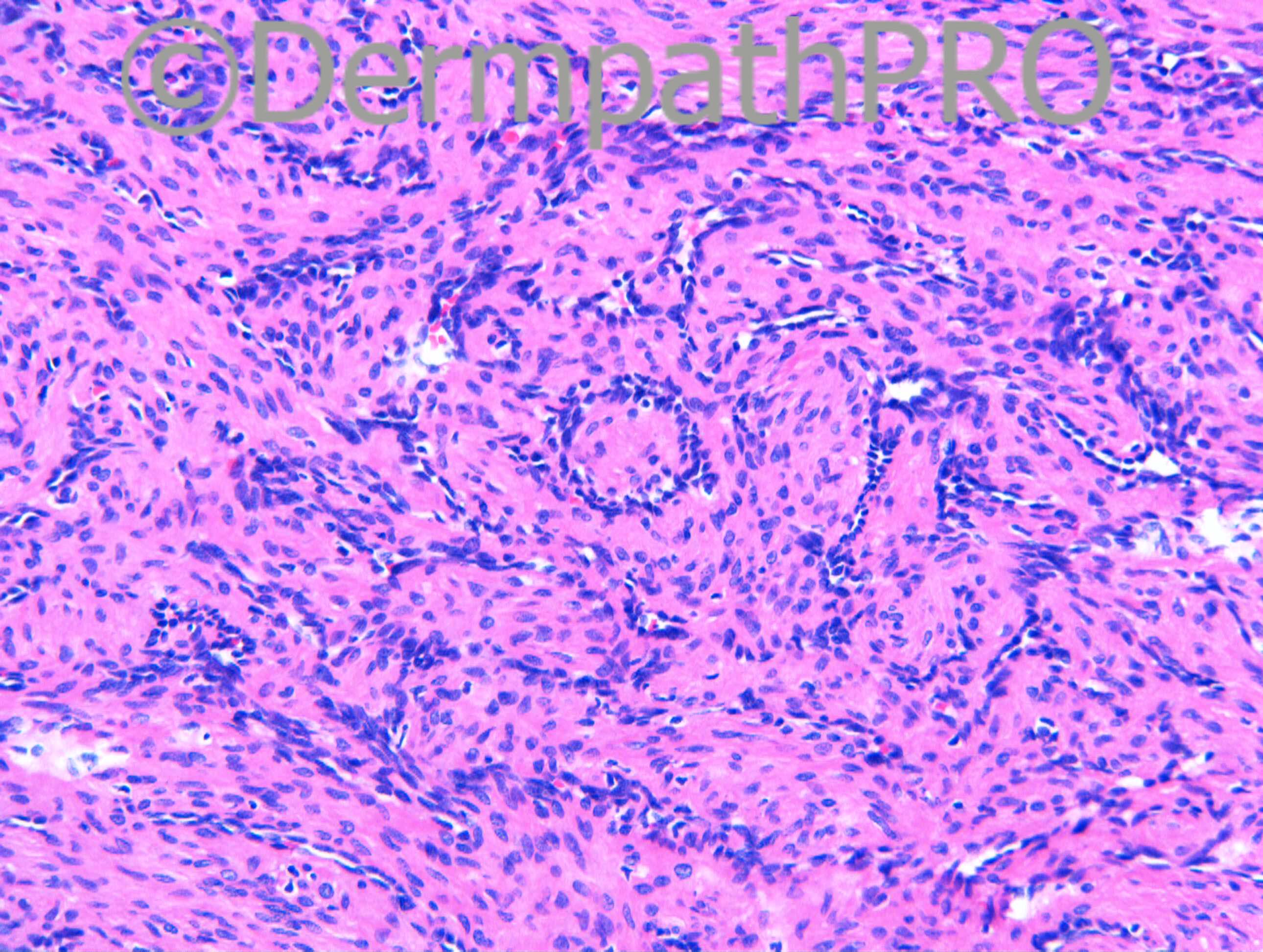
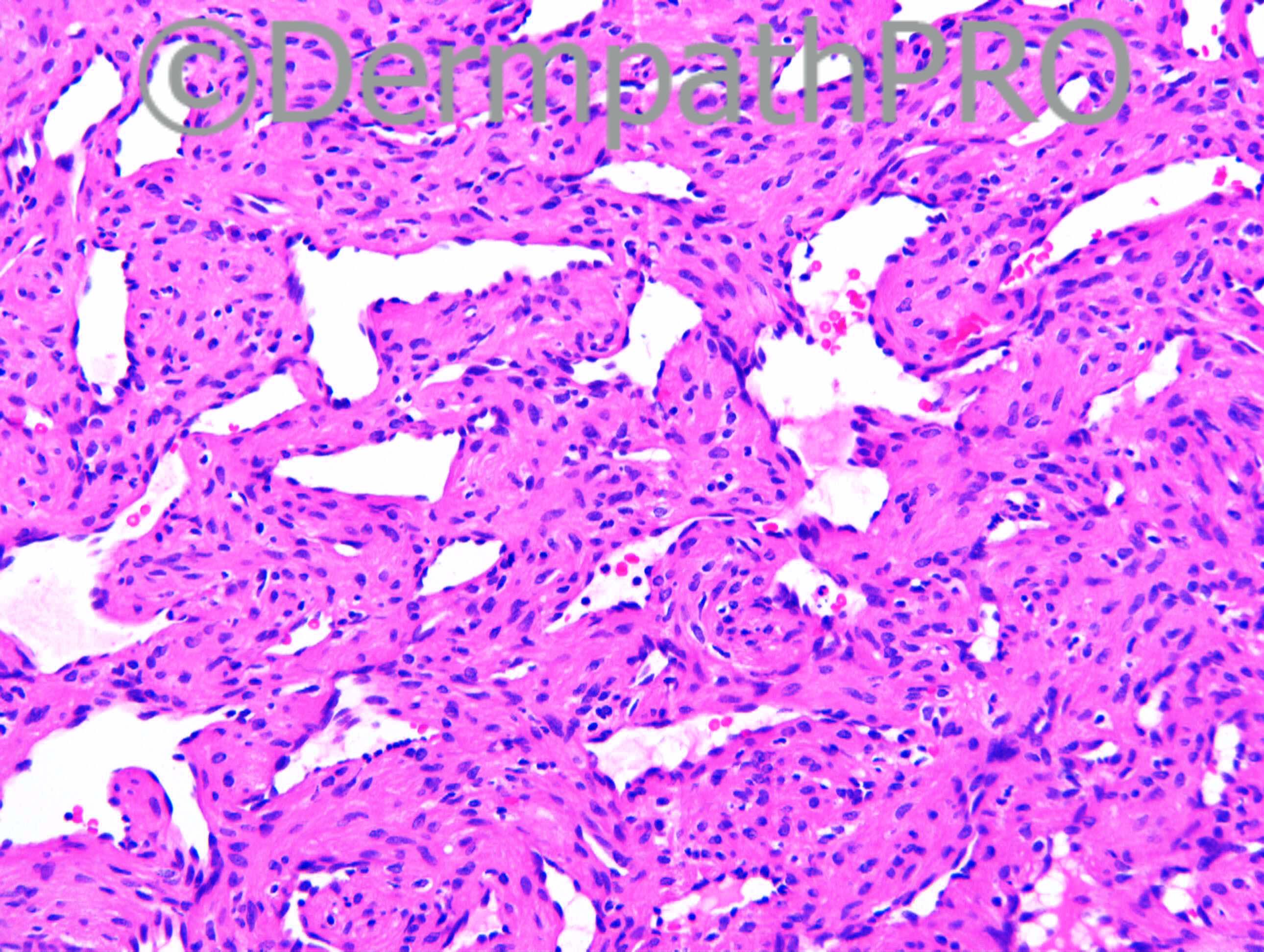
F68. Lump in 4th webspace of right hand

Case posted by Dr Richard Carr


  Report Record

User Feedback


Guest Romualdo

Posted

Although these are related tumors the absence of a biphasic pattern and the distinctly perivascular arrangement of myoid cells are typical of myopericytomas, in this case an intravascular one.

Share this comment


Link to comment
share_externally

Eman El-Nabarawy

Posted

Based on: the age, the onion-ring appearance, myopericytoma Is my diagnosis.Some reports regard myopericytoma as a part of the spectrum of angioleiomyoma.

Share this comment


Link to comment
share_externally

Dr. Mona Abdel-Halim

Posted

According to McKee's pathology of skin: solitary myofibromas may show minimal to absent primitive component. In addition according to McKee's pathology of skin, in recent years the term myopericytoma represent a spectrum that includes tumors classified in the past as glomangiopericytoma, myopericytoma, adult myofibroma and myofibromatosis in adults. So according to recent terminologies, may be it is better to call this lesion myopericytoma as Hazem and Romualdo said.

Share this comment


Link to comment
share_externally

Dr. Richard Carr

Posted

[img]https://dermpathpro.com/uploads/spot_diagnosis_comment_img/Case%201302_RAC7153x10_SMA_4pm.jpg[/img]

[img]https://dermpathpro.com/uploads/spot_diagnosis_comment_img/Case%201302_RAC7153x10b_CD34_4pm.jpg[/img]

[img]https://dermpathpro.com/uploads/spot_diagnosis_comment_img/Case%201302_RAC7153x10b_Desmin_4pm.jpg[/img]

Share this comment


Link to comment
share_externally

Guest Hazem Hamed

Posted

I think this immunoprofile together with the morphology support the diagnosis of myopericytoma

Share this comment


Link to comment
share_externally

Myopericytoma.

Angioleiomyomas are subcutaneous and contain thick-walled muscular vessels interspersed in what would otherwise be a leiomyoma. I don't think that's the case here.

Share this comment


Link to comment
share_externally

Dr. Richard Carr

Posted

I thought this was an angioleiomyoma / myopericytoma overlap. I asked Thomas Mentzel to comment as I have only seen one or two of these rare tumours. He commented as follows:
Dear Richard,
thanks and I think that myopericytoma is a good differential diagnosis, however, usually these lesions are desmin negative! So probably, it represents rather an angioleiomyoma with some similarities to myopericytoma and we know that there are overlaping features emphasizing the hypothesis of a spectrum of perivascular myoid neoplasms including glomus tumour, myopericytoma, angioleiomyoma and myofibroma.
Best wishes
Thomas

Share this comment


Link to comment
share_externally


Join the conversation

You can post now and register later. If you have an account, sign in now to post with your account.

Guest
Add a comment...

×   Pasted as rich text.   Paste as plain text instead

  Only 75 emoji are allowed.

×   Your link has been automatically embedded.   Display as a link instead

×   Your previous content has been restored.   Clear editor

×   You cannot paste images directly. Upload or insert images from URL.

Loading...

×
×
  • Create New...