Jump to content
In this section we have spot diagnoses posted on a daily basis since June 2010, now over 4000! You can review the archived cases and read the suggested diagnoses by users and the final comment by the contributors.
Case are uploaded each week day by 10 am UK time with the correct diagnosis will generally be posted at 8 pm UK time. Why not view the most recent spot diagnosis and proffer a diagnosis?

Case Number : Case 1228 - 09 March Posted By: Guest

Please read the clinical history and view the images by clicking on them before you proffer your diagnosis.
Submitted Date :
   (0 reviews)

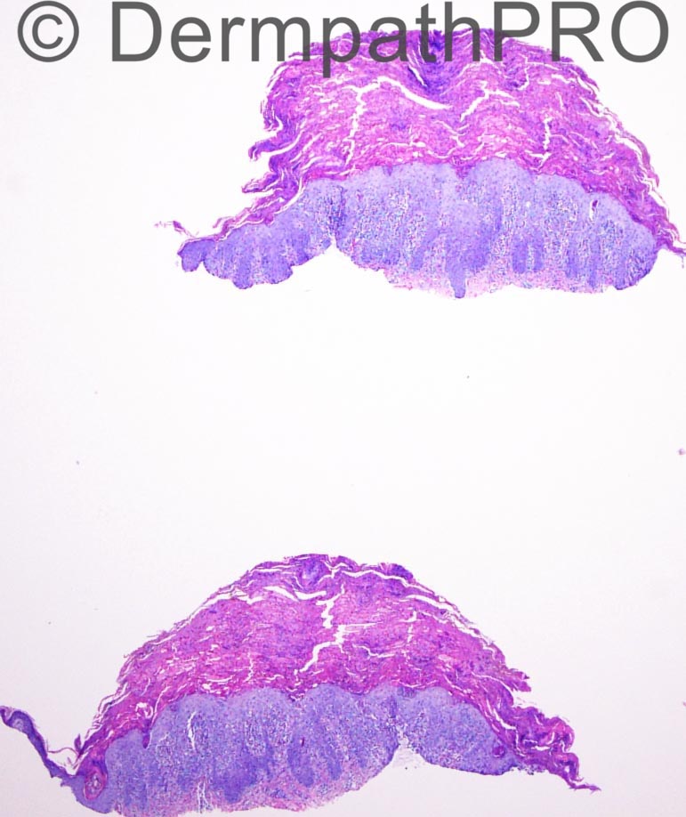
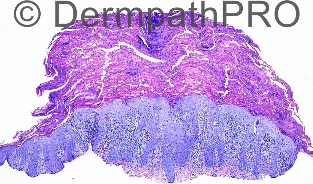
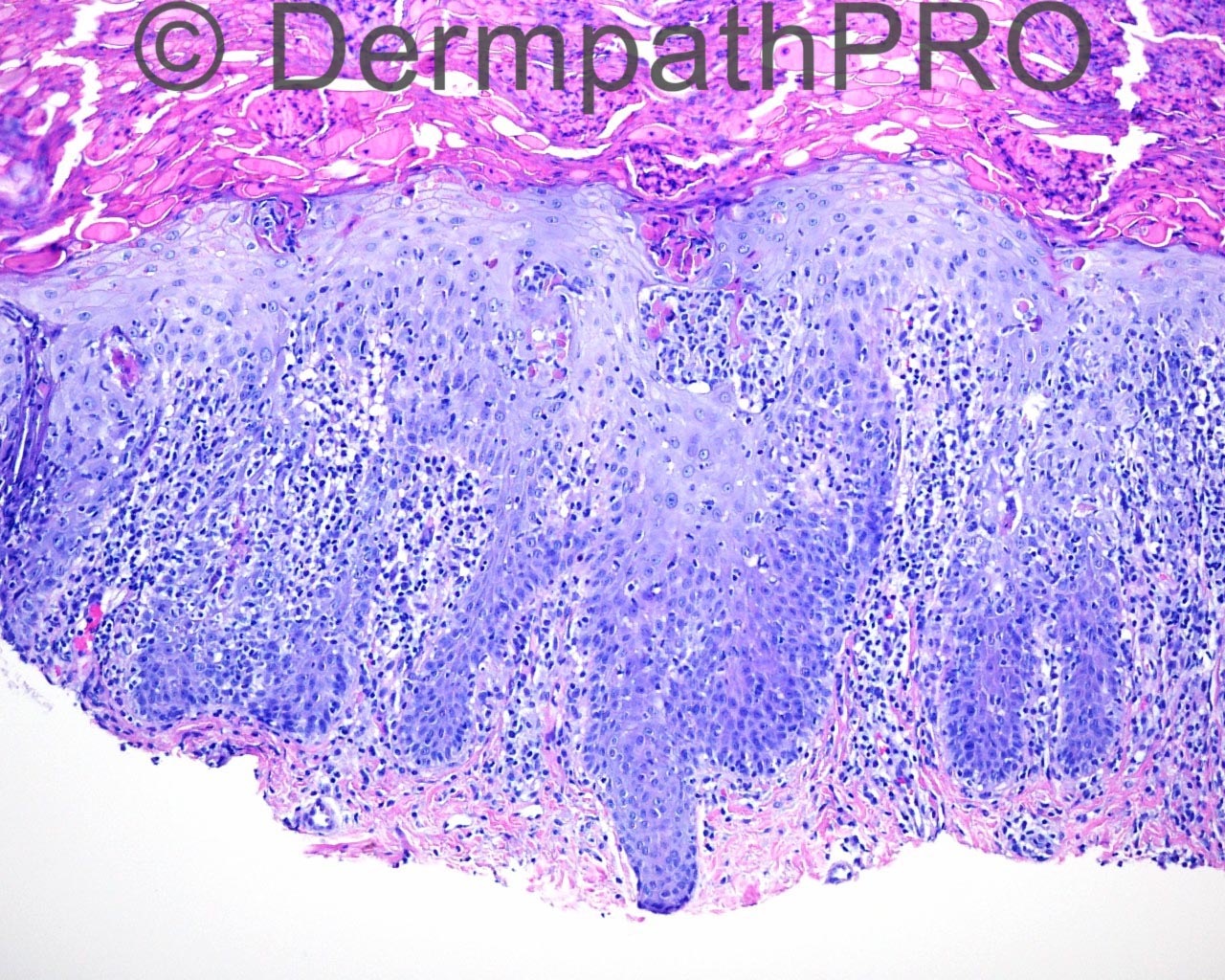
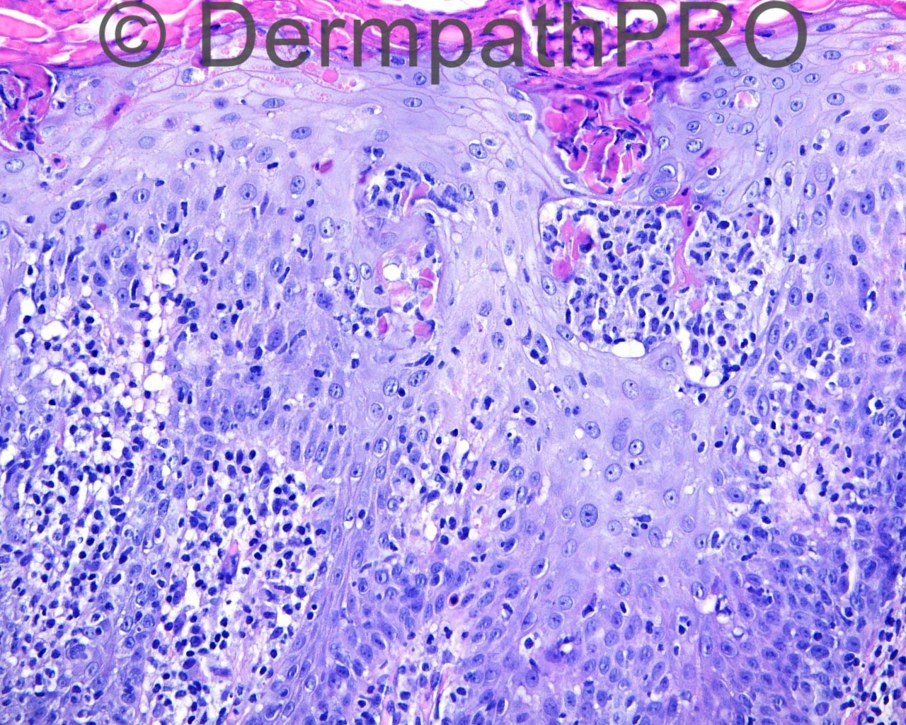
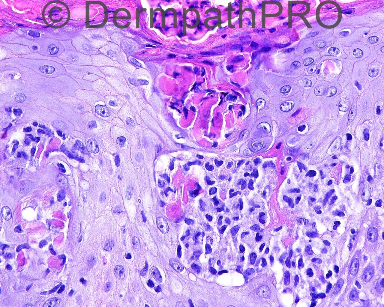
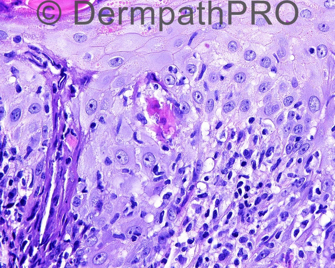
The patient is a 6 year old white boy with crusted papules on the trunk and limbs, present for a few years, taken from the right abdomen.

Case posted by Dr Mark Hurt


  Report Record

User Feedback


biswanath behera

Posted

[b]
[size=3]scale crust, keratinocyte pallor, collection of Langerhan's cells in epidermis, necrotic keratinocytes, upper dermal lymphohistiocytic infiltrate, wiry collagen......Langerhans' cell histiocytosis????[/size][/b]

Share this comment


Link to comment
share_externally

Dr. Mona Abdel-Halim

Posted

Pityriasis lichenoides

Share this comment


Link to comment
share_externally

Dr. Mona Abdel-Halim

Posted

Considering the wiry fibroplasia, I would like to add also in the DDx: papular MF but I think reported cases were not described as crusted but still I think it is worth considering. OR, type B LyP which will fit more with the crusted nature of the papules.

Share this comment


Link to comment
share_externally

Arti Bakshi

Posted

Will go with pityriasis lichenoides. (combination of spongiosis, marked lymphocytic exocytosis and dyskeratosis).
LCH less likely as no eosinophils…., but need immunos!

Share this comment


Link to comment
share_externally

Guest Romualdo

Posted

I agree with Mona and also think this is PLEVA (Mucha-Habermann disease) with associated Langerhans' cell microabscesses.

Share this comment


Link to comment
share_externally

Dr. Richard Carr

Posted

I struggled with this one a lot this morning. Interesting clustered dyskeratotic cells and slightly epidermotropic lymphocytes. I don't think it is LCH as these look like reactive Langerhan's cells to me. My first consideration is pityriasis lichenoides with the other differentials (Type B Lyp and papular MF less likely but requiring clinpath correlation and follow-up to rule out). Nice images.

Share this comment


Link to comment
share_externally

Guest Tiberiu Tebeica

Posted

My first tought is PLEVA (Mucha-Habermann disease). I would consider type D LyP in the ddx, so curious to see if all the intraepidermal lymphocytes are positive for CD8. Anyway, since usually PLEVA shows a CD8+ phenotype, how could one reliably differentiate between these two entities? I don't favor LyP because I don't see enough pagetoid spread of lymphocytes and atypical lymphoid cells, but these are subjective criteria. Moreover, there is plenty of apoptotic keratinocytes, usually present in PLEVA and absent in LyP.

Share this comment


Link to comment
share_externally

Pityriasis lichenoides seems to be the best fit, histologically. But this is a relapsing-remitting disease and the lesions are papulosquamous ("chronic") and/or papulonecrotic ("acute"), neither of which is reflected in the history.

Share this comment


Link to comment
share_externally


Join the conversation

You can post now and register later. If you have an account, sign in now to post with your account.

Guest
Add a comment...

×   Pasted as rich text.   Paste as plain text instead

  Only 75 emoji are allowed.

×   Your link has been automatically embedded.   Display as a link instead

×   Your previous content has been restored.   Clear editor

×   You cannot paste images directly. Upload or insert images from URL.

Loading...

×
×
  • Create New...