Jump to content
In this section we have spot diagnoses posted on a daily basis since June 2010, now over 4000! You can review the archived cases and read the suggested diagnoses by users and the final comment by the contributors.
Case are uploaded each week day by 10 am UK time with the correct diagnosis will generally be posted at 8 pm UK time. Why not view the most recent spot diagnosis and proffer a diagnosis?

Case Number : Case 1232 - 13 March Posted By: Guest

Please read the clinical history and view the images by clicking on them before you proffer your diagnosis.
Submitted Date :
   (0 reviews)

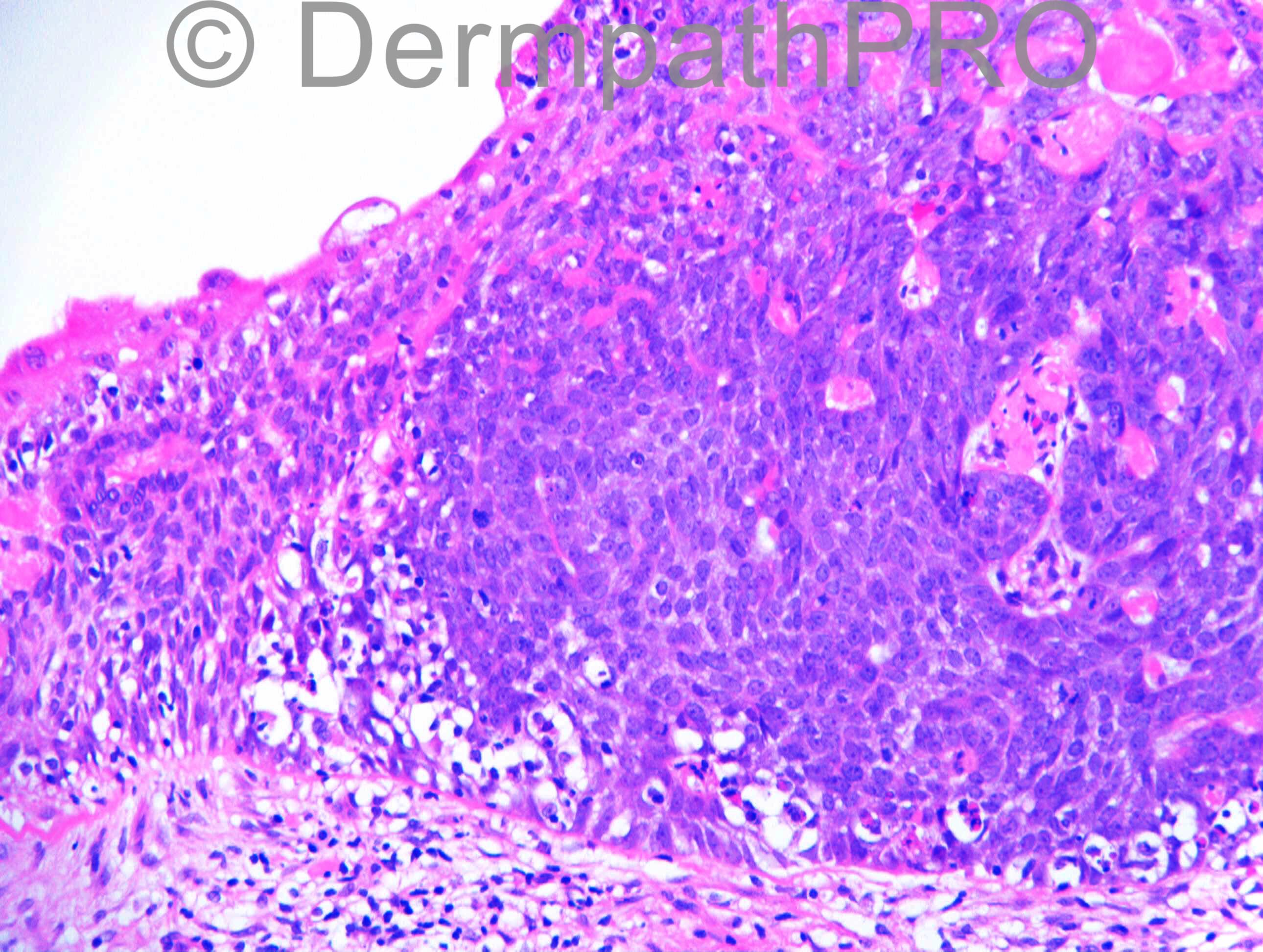
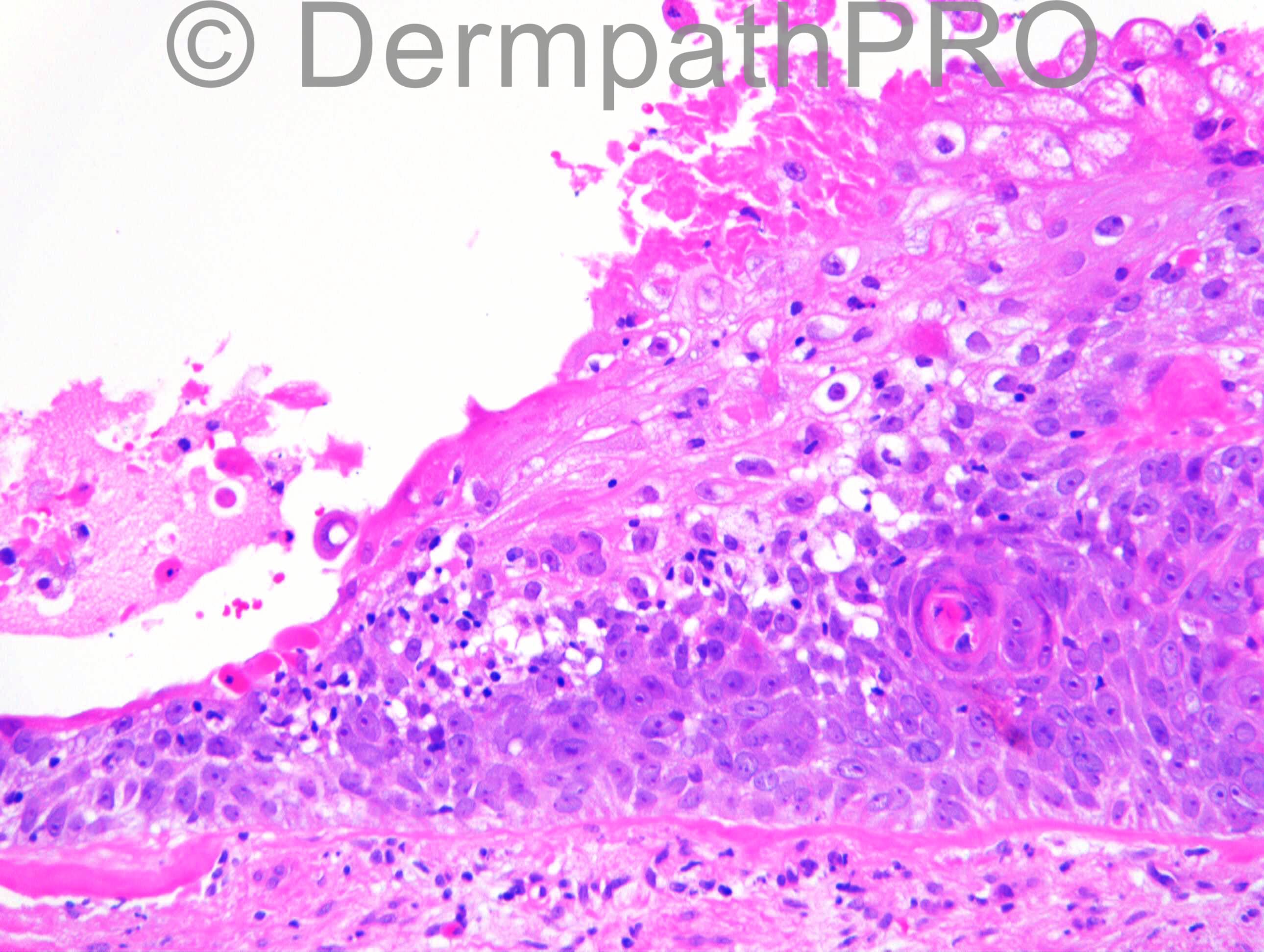
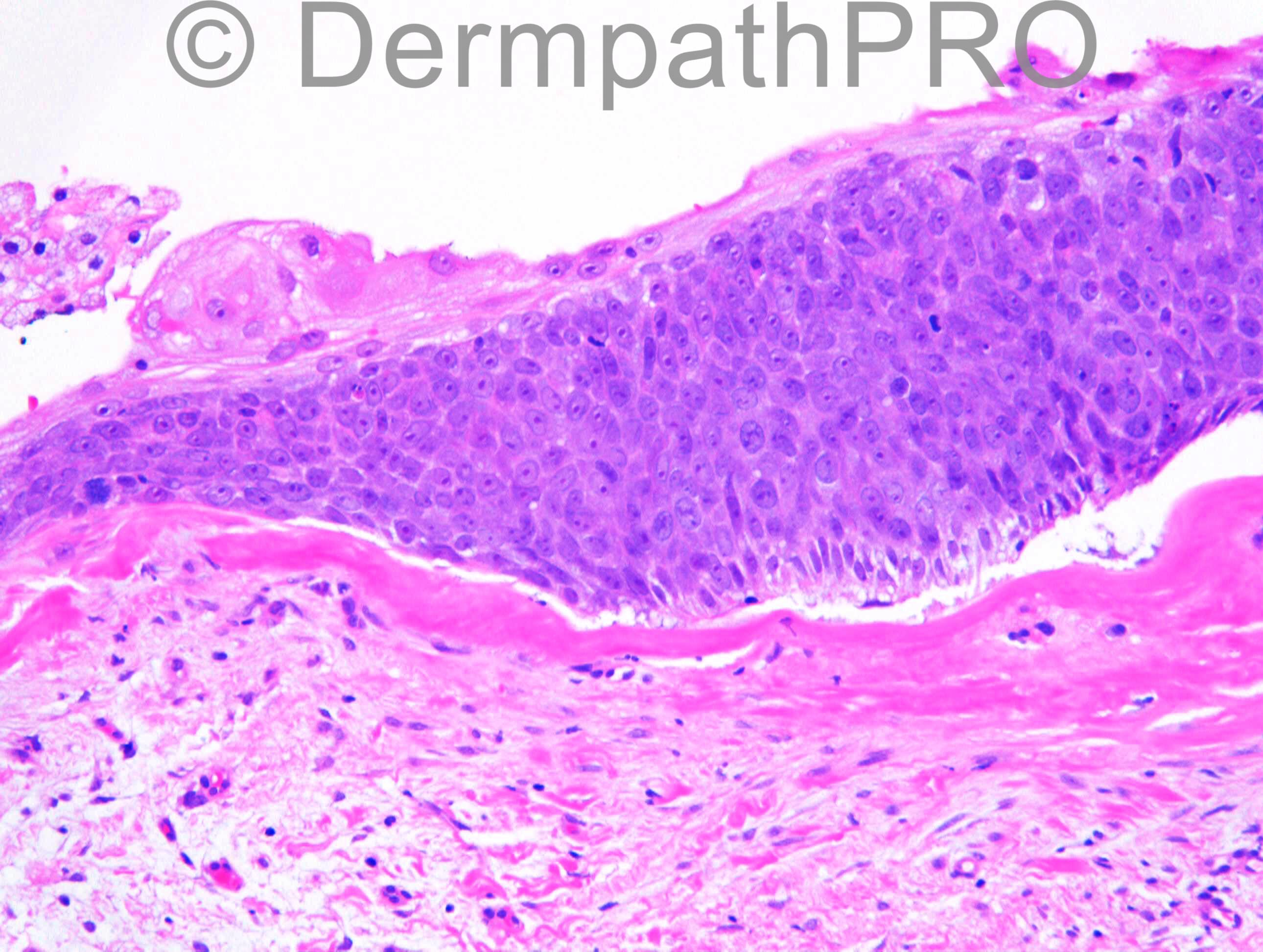
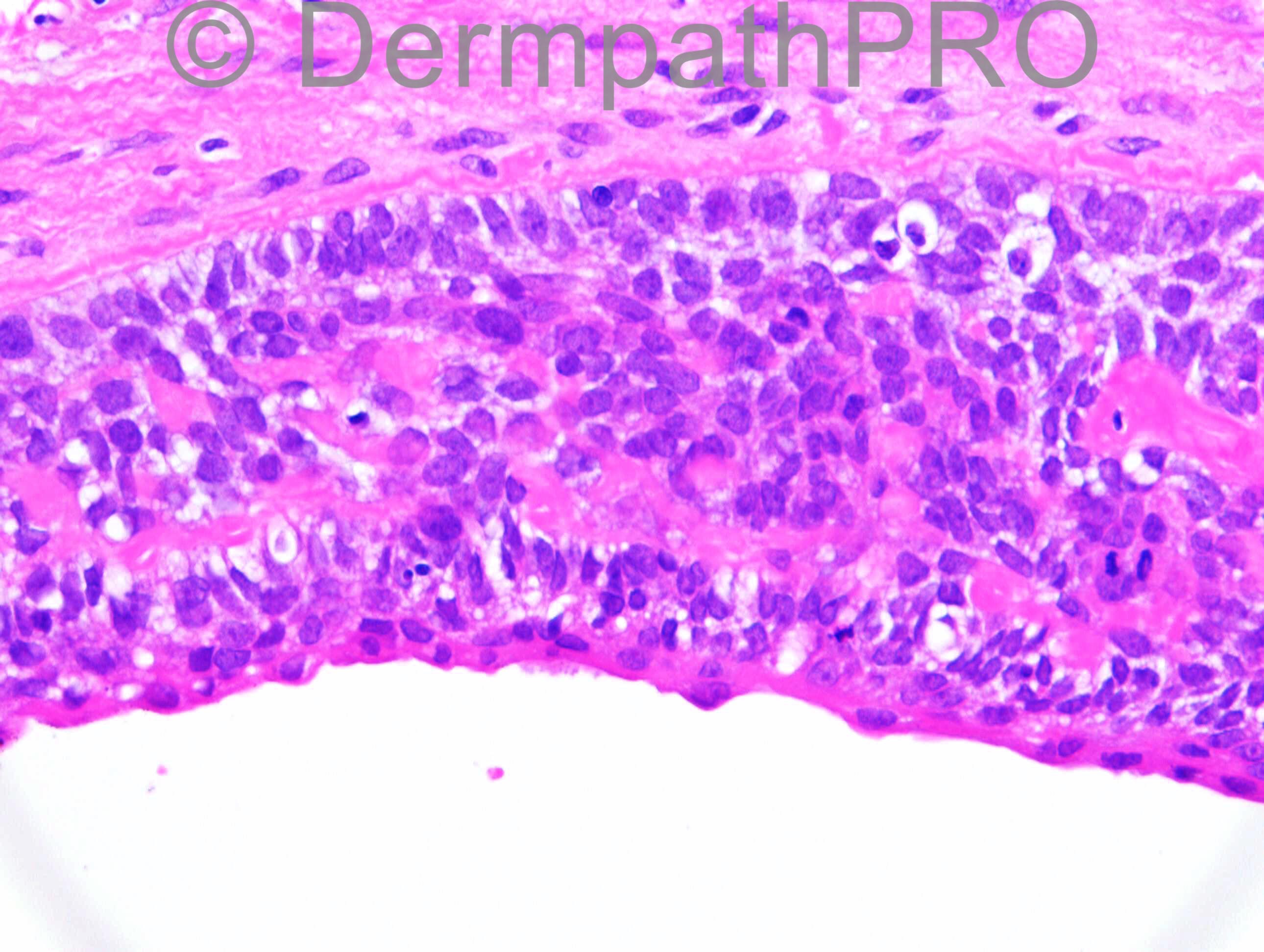
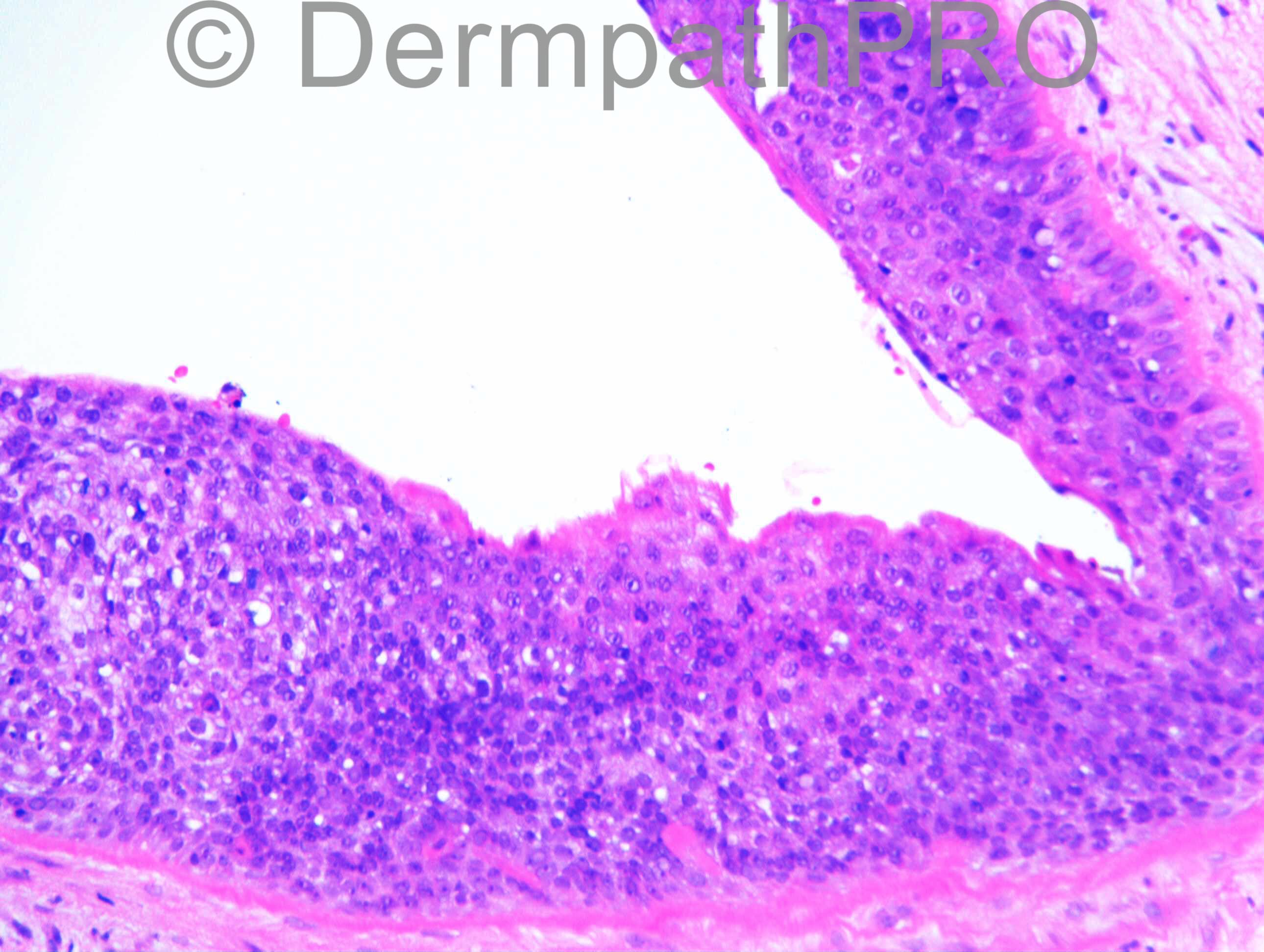
M76. Right buttock.? BCC treated with aldara.

Case posted by Dr Richard Carr

  Report Record

User Feedback


Guest Romualdo

Posted

Hybrid cyst. I think pilomatrical features are well evident. Some areas with large pale inner cells suggest trichilemmal differentiation. Another areas with a corrugated inner lining suggest steatocystoma or sebaceous duct cyst.

Share this comment


Link to comment
share_externally

Dr. Mona Abdel-Halim

Posted

I also thought of follicular hybrid cyst.

Share this comment


Link to comment
share_externally

biswanath behera

Posted

Spiradenocarcinoma or malignant cylindroma..?? Thick basement membrane, eosinophilic material deposition, foci of peripheral palisading, mitotic figures and comedonecrosis.

Share this comment


Link to comment
share_externally

Guest Leo tovar

Posted

[b]
Follicular hybrid cyst (trichilemmal cyst and pilomatricoma)[/b]

Share this comment


Link to comment
share_externally

Dr. Richard Carr

Posted

[img]https://dermpathpro.com/uploads/spot_diagnosis_comment_img/Case%201232_RAC7031x20_EMA_4pm.jpg[/img]

[img]https://dermpathpro.com/uploads/spot_diagnosis_comment_img/Case%201232_RAC7031x10b_Adipophilin_4pm.jpg[/img]

[img]https://dermpathpro.com/uploads/spot_diagnosis_comment_img/Case%201232_RAC7031x10_EMA_4pm.jpg[/img]

[img]https://dermpathpro.com/uploads/spot_diagnosis_comment_img/Case%201232_RAC7031x5_BerEP4_4pm.jpg[/img]

[img]https://dermpathpro.com/uploads/spot_diagnosis_comment_img/Case%201232_RAC7031x40c_Adipophilin_4pm.jpg[/img]

Share this comment


Link to comment
share_externally

Guest Romualdo

Posted

Now, things are easier: cystic sebaceous neoplasm, probably proliferative cystic sebaceous tumor, a marker of Muir-Torre syndrome.

Share this comment


Link to comment
share_externally

Agree with Romualdo. Cystic sebaceous adenoma of Muir-Torre syndrome.

Share this comment


Link to comment
share_externally

Dr. Mona Abdel-Halim

Posted

With the markers highlighting adipophilin, this is as Romualdo said cystic sebaceous tumor (adenoma) of Muir Torre syndrome....lovely one :-))

Share this comment


Link to comment
share_externally

Guest Jim Davie MD

Posted

This is a tough lesion. I like the original idea of hybrid / trichilemmal cyst. But other areas resemble cystic hidradenoma (my preference).

There is the thickened hyaline basement membrane, and peripheral palisading. Focal sebaceous/sebaceous duct differentiation or squamous metaplasia would not be uncommon in trichilemmal tumors.

Not sure what to make of the adipophilin staining. Positive apidophilin staining is nonspecific for sebaceous neoplasms; it can be seen in other tumors [e.g. SCC or SCC in-situ with clear cell features, BCC, sweat gland neoplasms, and various metastatic tumors]**. Lipid laden macrophages can also stain positively.
This tumor shows positive adipophilin staining, but predominantly in a sparse perinuclear granular staining pattern, which is a pattern associated with BCC and other non-sebaceous neoplasms; that said, a few superficial cells show diffuse staining...can't tell if those resemble mature sebocytes or histiocytes on H&E.

There is variable patchy BER-EP4 staining, and positive lumenal/superficial EMA staining limited to the foamy mature adipocyte component and the superficial central cuticle. EMA doesn't seem to show strong, coarsely vacuolated pattern of mature sebocyte staining, rather the diffuse and membranous staining pattern of SCC or sweat gland neoplasm.

**Human Pathology , Volume 44 (9) – Sep 1, 2013. 'Perilipin and adipophilin expression in sebaceous carcinoma and mimics' [[url="http://www.ncbi.nlm.nih.gov/pubmed/23642680"]Abstract link[/url]]

Share this comment


Link to comment
share_externally

Dr. Richard Carr

Posted

Another really good discussion of a difficult and subjective case. My report as follows:

Cystic, relatively circumscribed, basaloid neoplasm with distinctive rounded to oval purple nuclei and focal cytoplasmic vacuoles suggesting sebaceous differentiation. There is prominent mitotic activity and mild cellular pleomorphism. Basement membrane is thickened around the neoplasm. Radial margin 2 mm. Deep margin 0 mm. Local recurrence is a possibility. Immunostains to follow.
SUPPLEMENTARY REPORT:
Specimen A) BerEP4 shows moderate to weak expression in 25 % of the neoplasm. EMA is limited to the lining cells. P53 shows up-regulated wild-type staining. This lesion is hard to classify, but on balance, I favour a circumscipt cystic sebaceous carcinoma. I would expect a low potential for metastasis given the circumscription. Please consider the possibility of Muir Torre syndrome.

Additional remarks:
The adipophilin was kindly performed by Dr Heinz Kutzner. In this case I think it is highly specific confirming the well formed microvesicles that indent the nucleus and these cells are also progressing into the cyst contents (not macrophages in my opinion). The EMA I used to highlight the cuticular differentiation of the sebaceous duct (rather than the sebaceous areas which are quite focal). I agree the cells of sebaceous neoplasms (rather distinctive rounded oval cells with amphophilic chromatin and small amphophilic nucleoli giving a purple overrall hue to the neoplasm) can quite closely mimic matrical differentiation but there is clearly no inner root differentiation(trichohyalin granules) or matrical (ghosted) keratin to support the latter. p53 in this case showed up-regulated wild type staining but in view of the fairly prominent mitotic activity well away form the basement membrane and mild cellular pleomorphism I interpreted it as a cystic sebaceous carcinoma (with predominannt sebaceous ductal differentiation).

Share this comment


Link to comment
share_externally


Join the conversation

You can post now and register later. If you have an account, sign in now to post with your account.

Guest
Add a comment...

×   Pasted as rich text.   Paste as plain text instead

  Only 75 emoji are allowed.

×   Your link has been automatically embedded.   Display as a link instead

×   Your previous content has been restored.   Clear editor

×   You cannot paste images directly. Upload or insert images from URL.

Loading...

×
×
  • Create New...