Jump to content
In this section we have spot diagnoses posted on a daily basis since June 2010, now over 4000! You can review the archived cases and read the suggested diagnoses by users and the final comment by the contributors.
Case are uploaded each week day by 10 am UK time with the correct diagnosis will generally be posted at 8 pm UK time. Why not view the most recent spot diagnosis and proffer a diagnosis?

Case Number : Case 1237 - 20 March Posted By: Guest

Please read the clinical history and view the images by clicking on them before you proffer your diagnosis.
Submitted Date :
   (0 reviews)

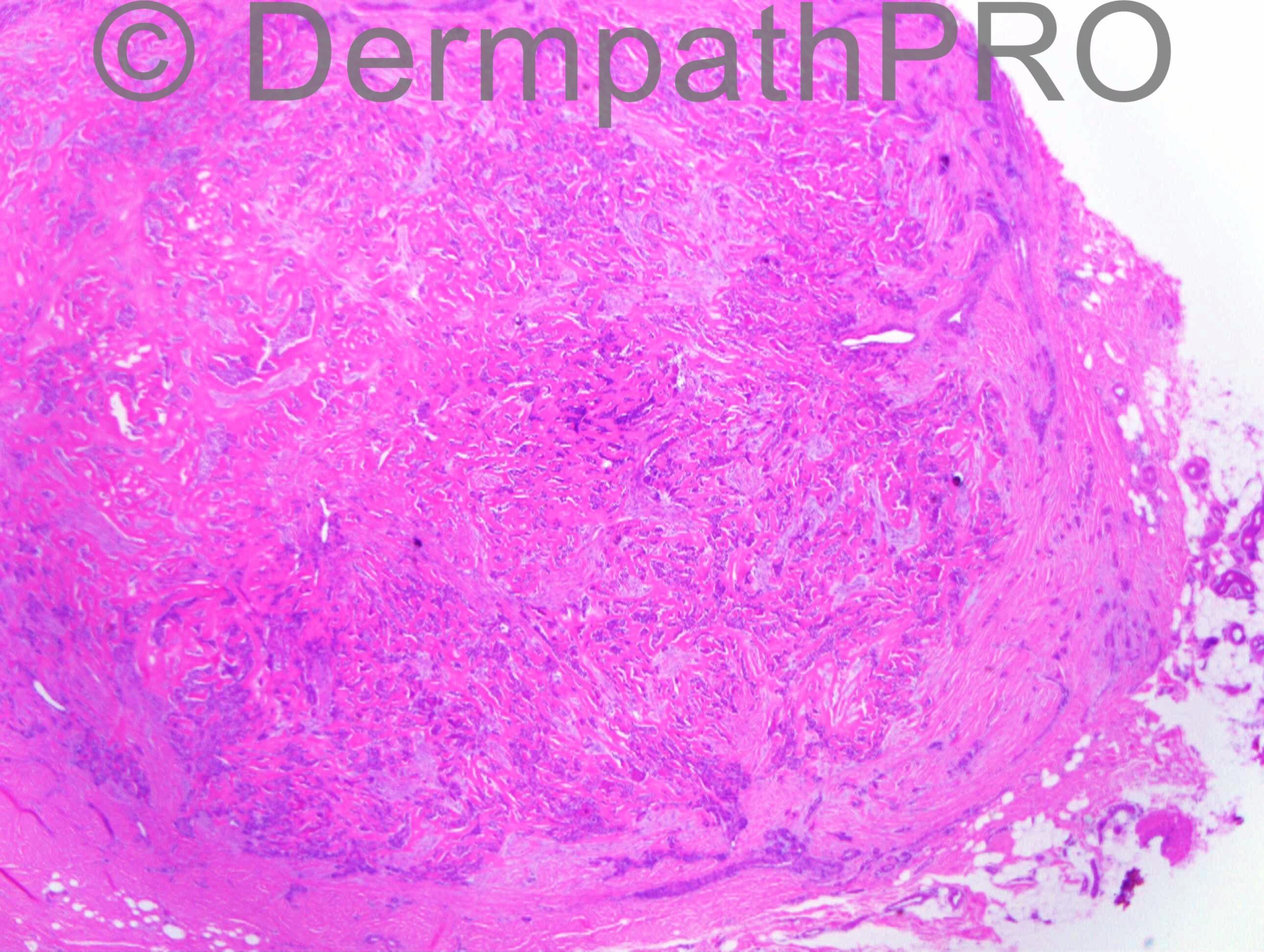
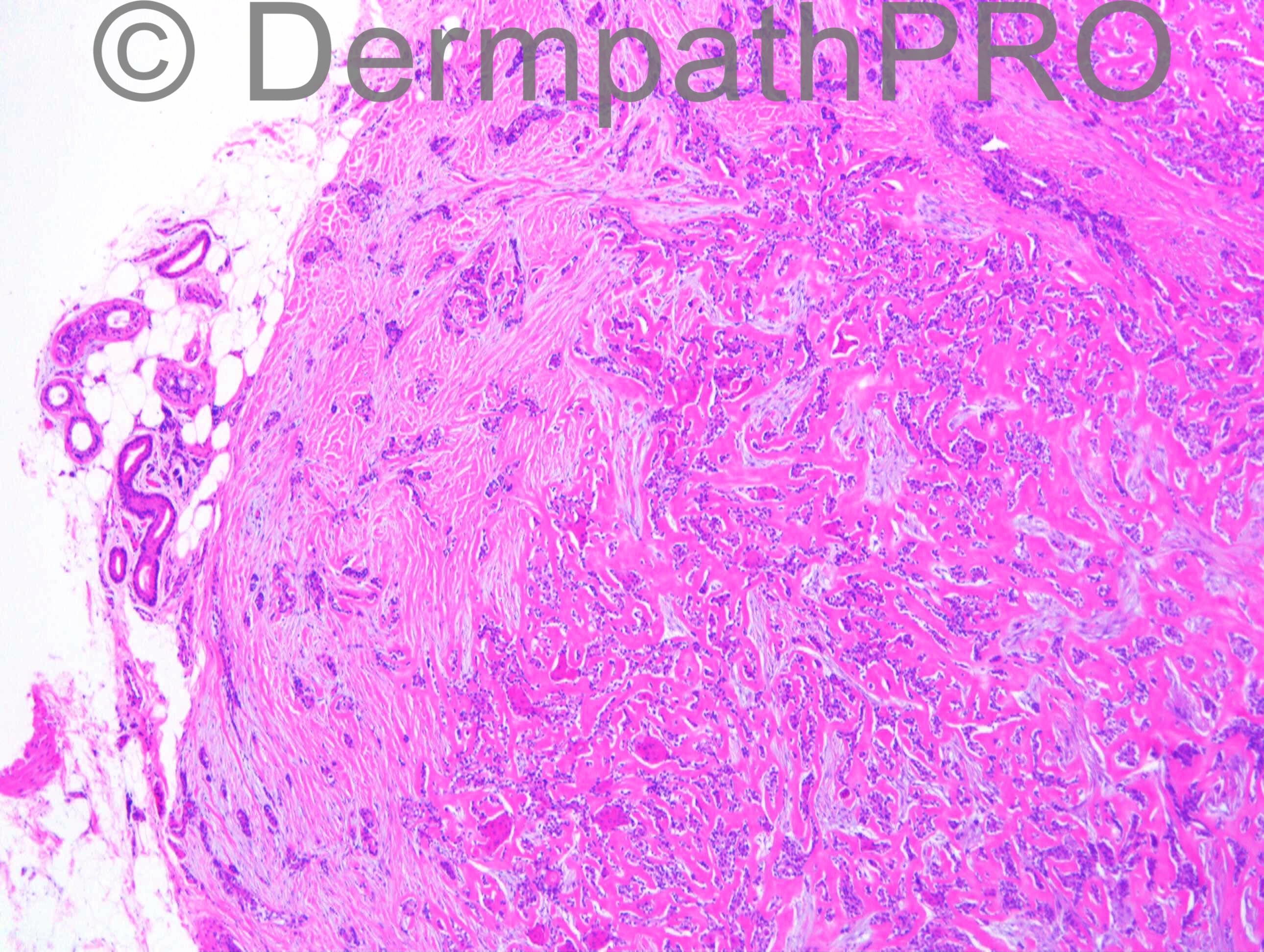
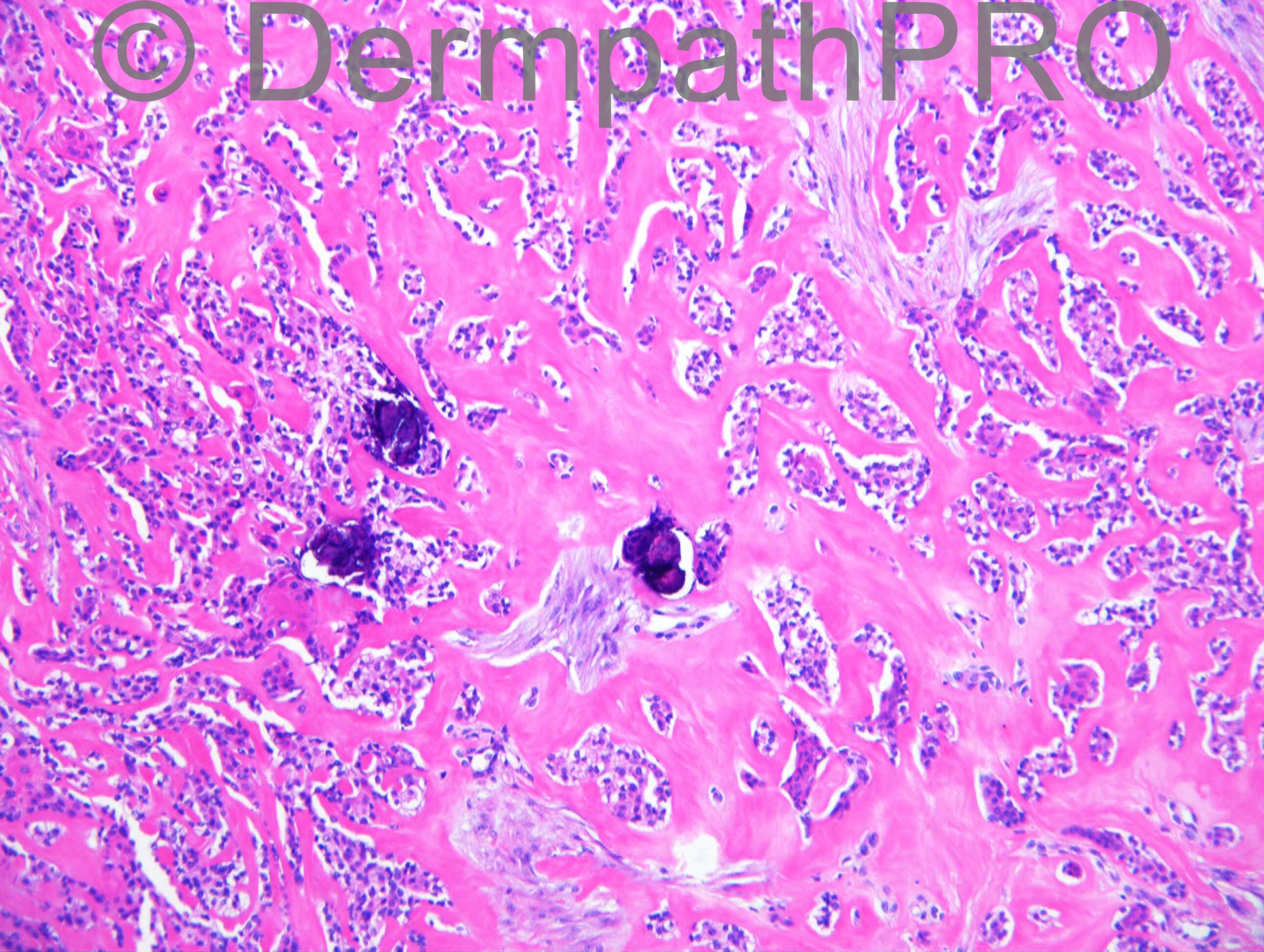
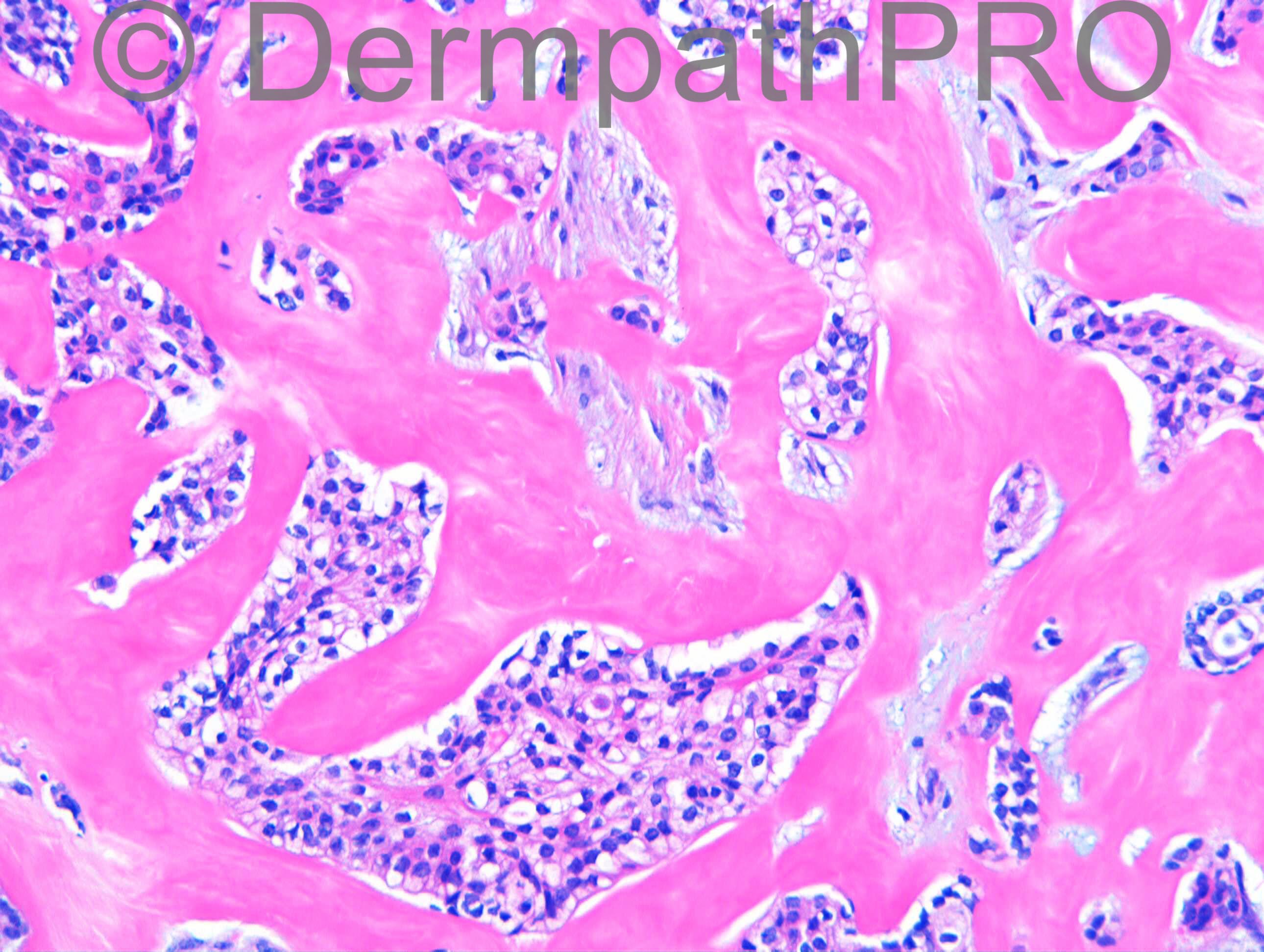
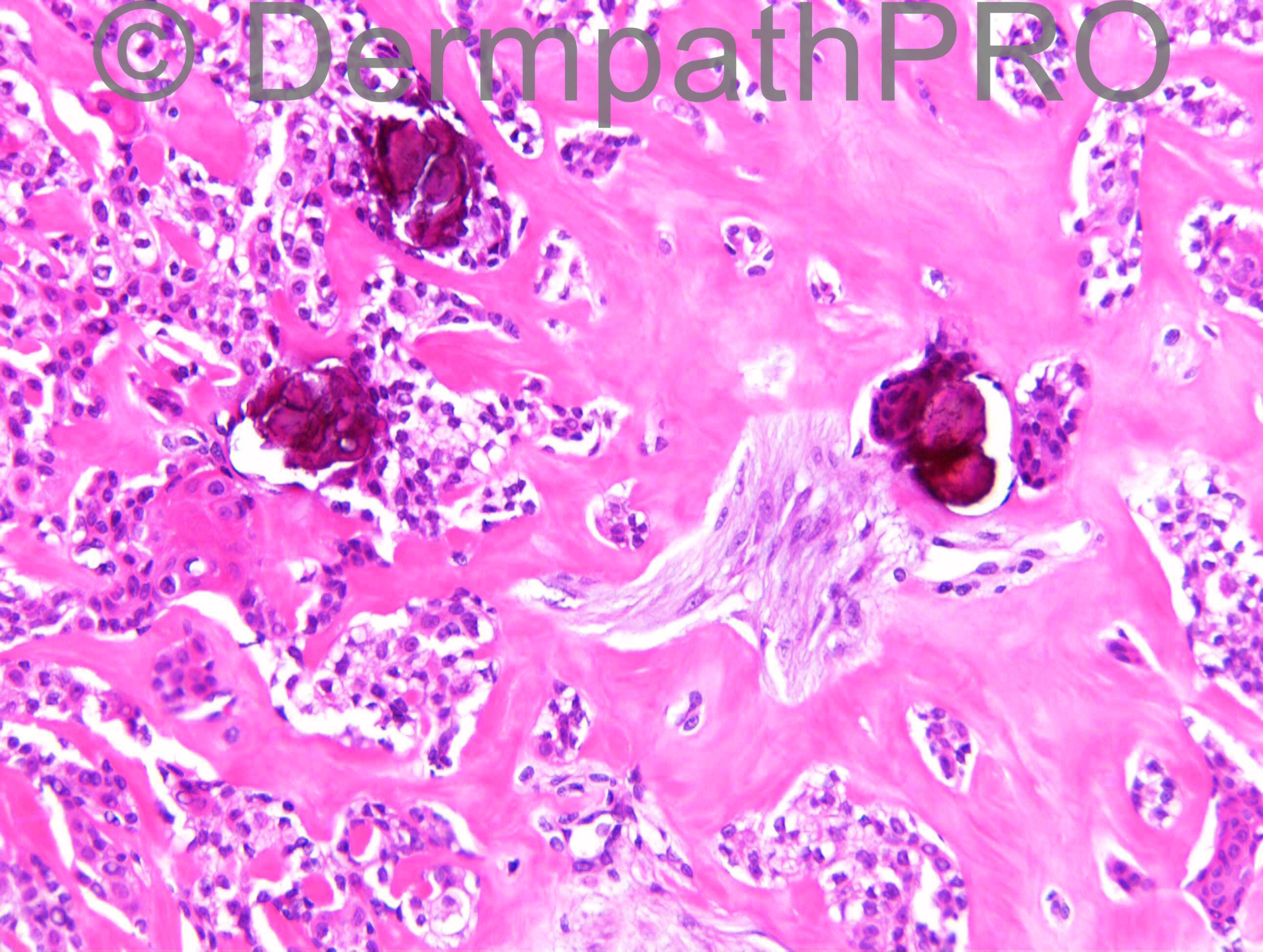
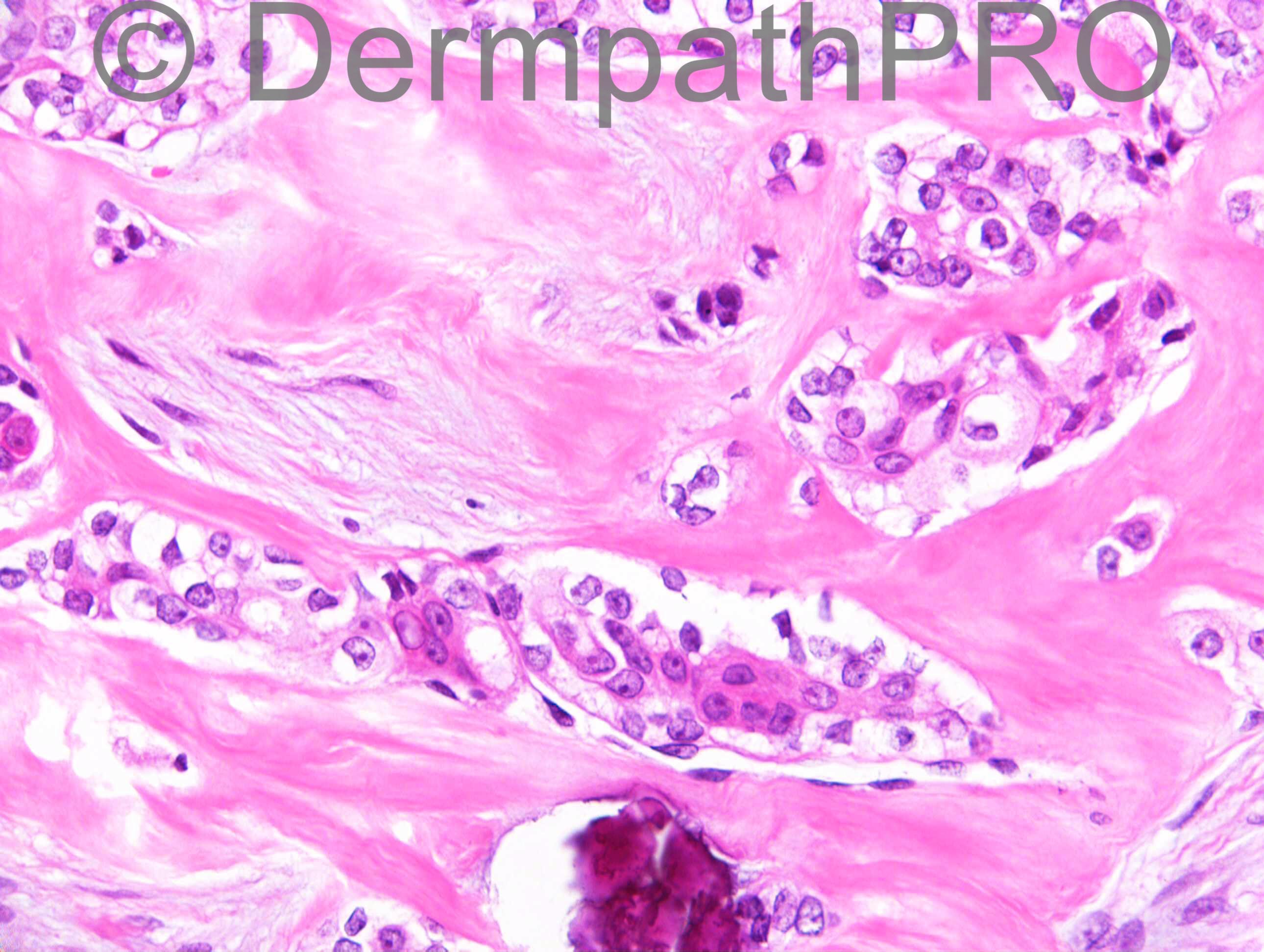
F63. Recurrent swelling right medial aspect of big toe. Previous benign eccrine tumour excised from left foot 2001. Previous cylindroma excised from ear.

Case posted by Dr Richard Carr

  Report Record

User Feedback


Dr. Mona Abdel-Halim

Posted

My hypothesis is: malignant sweat gland tumor, duct differentiation is evident (fig 7). The cells are biphasic, some have esinophilic cytoplasm, some have clear cytoplasm. The stroma is hyalinized with ? mucinous foci. First suggestion: hidradenocarcinoma, second possibility: clear cell porocarcinoma.

Share this comment


Link to comment
share_externally

Sweat gland ductal carcinoma. There is clear cell cytology, ductal differentiation and keratinisation. I think this tumour belongs to the category which in Kazakov's book is under a long heading titled "Eccrine ductal carcinoma, clear cell eccrine carcinoma, clear cell syringoid eccrine carcinoma, syringomatous carcinoma, squamoid eccrine ductal carcinoma and solid carcinoma".

Share this comment


Link to comment
share_externally

Dr. Mona Abdel-Halim

Posted

Agree with Abdul Kadir, may be we just call it eccrine sweat gland ductal carcinoma and according to Kazakov, he says that this group represents a very heterogenous and poorly defined group of lesions representing variations within the spectrum of a single entity...

Share this comment


Link to comment
share_externally

Eman El-Nabarawy

Posted

Agree with Dr. Abdul Kadir. Microcystic adnexal carcinoma top the list of my DD.

Share this comment


Link to comment
share_externally

Dr. Richard Carr

Posted

[img]https://dermpathpro.com/uploads/spot_diagnosis_comment_img/Case_1237_RAC7077x20_CK7_4pm.jpg[/img]

[img]https://dermpathpro.com/uploads/spot_diagnosis_comment_img/Case_1237_RAC7077x20_p63_4pm.jpg[/img]

[img]https://dermpathpro.com/uploads/spot_diagnosis_comment_img/Case_1237_RAC7077x20_BerEP4_4pm.jpg[/img]

[img]https://dermpathpro.com/uploads/spot_diagnosis_comment_img/Case_1237_RAC7077x20_Ki67_4pm.jpg[/img]

[img]https://dermpathpro.com/uploads/spot_diagnosis_comment_img/Case_1237_RAC7077x20_CEA_4pm.jpg[/img]

[img]https://dermpathpro.com/uploads/spot_diagnosis_comment_img/Case_1237_RAC7077x20_CK5_4pm.jpg[/img]

Share this comment


Link to comment
share_externally

Dr. Richard Carr

Posted

Forgot to thank Dr Rand Hawari for kindly sharing this case with me. I will post my opinion on Monday morning but I hope to receive more opinions before then!!!

Share this comment


Link to comment
share_externally

Dr. Richard Carr

Posted

Many thanks for your opinions although I was hoping for a few more - if you are watching cases but not having a go please do as we all learn from the range of opinions especially on difficult cases. My report as follows:

Many thanks for sending this interesting case.
The overall profile is of a relatively rounded tumour which has been
partially shelled-out. The lesion comprises approximately 50 % of the
stroma with distinctive hyaline appearances. The epithelial component
is rather stranded, comprising cohesive pale and clear cells with areas
of central squamous differentiation and a hint of focal lumen
development. Ki67 is exceedingly low (< 5 %).
Immuno markers are supportive of an adnexal tumour with BerEP4 and low
molecular keratins widespread, as well as CK5, p40 and p63. Very sparse
ductal differentiation on CEA. Moderate glycogen content on PAS, but
only very sparse luminal secretions not typical of mucin.

In my opinion, the histological features fit best with a hidradenoma
showing typical amorphous sclerotic stromal change. This stranded
epithelial nature is known to occur in hidradenomas; particularly in the
centre of lesions, although in your case, this feature is particularly
extensive and I think is giving a pseudo-infiltrative appearance of the
lesion. The lesion is incompletely excised and local recurrence would
be a possibility, but I suspect there is an exceedingly low chance for
metastasis from this lesion.

Additional comments: Kazakov's book, in discussing hidradenoma, makes the following statement "The stroma in most cases is sclerosing and hyalinising, even in examples with only minimal amount of the stroma. In areas abundant hyalinisation the stroma has a distinct homogenous glassy appearance. Often fibrotic and desmoplastic foci as well as edema and/or myxoid change are present. In some tumours stromal desmoplasia is prominent and imparts an infiltrative appearance to the neoplasm. Such cases often come from acral regions including the palm, sole, toe, and finger." I think mixed tumour is also a reasonable suggestion but the typical stroma in this case led me to favour [b]hidradenoma (pauciluminal, sclerosing and pseudoinfiltrative variant)[/b].

Share this comment


Link to comment
share_externally

Arif Usmani

Posted

It will be interesting to see if this patient has Brooke-Spiegler syndrome due to previous history of benign eccrine tumor and cylindroma although hidradenoma is not a feature of BSS.

Share this comment


Link to comment
share_externally


Join the conversation

You can post now and register later. If you have an account, sign in now to post with your account.

Guest
Add a comment...

×   Pasted as rich text.   Paste as plain text instead

  Only 75 emoji are allowed.

×   Your link has been automatically embedded.   Display as a link instead

×   Your previous content has been restored.   Clear editor

×   You cannot paste images directly. Upload or insert images from URL.

Loading...

×
×
  • Create New...