Jump to content
In this section we have spot diagnoses posted on a daily basis since June 2010, now over 4000! You can review the archived cases and read the suggested diagnoses by users and the final comment by the contributors.
Case are uploaded each week day by 10 am UK time with the correct diagnosis will generally be posted at 8 pm UK time. Why not view the most recent spot diagnosis and proffer a diagnosis?

Case Number : Case 1239 - 24 March Posted By: Guest

Please read the clinical history and view the images by clicking on them before you proffer your diagnosis.
Submitted Date :
   (0 reviews)

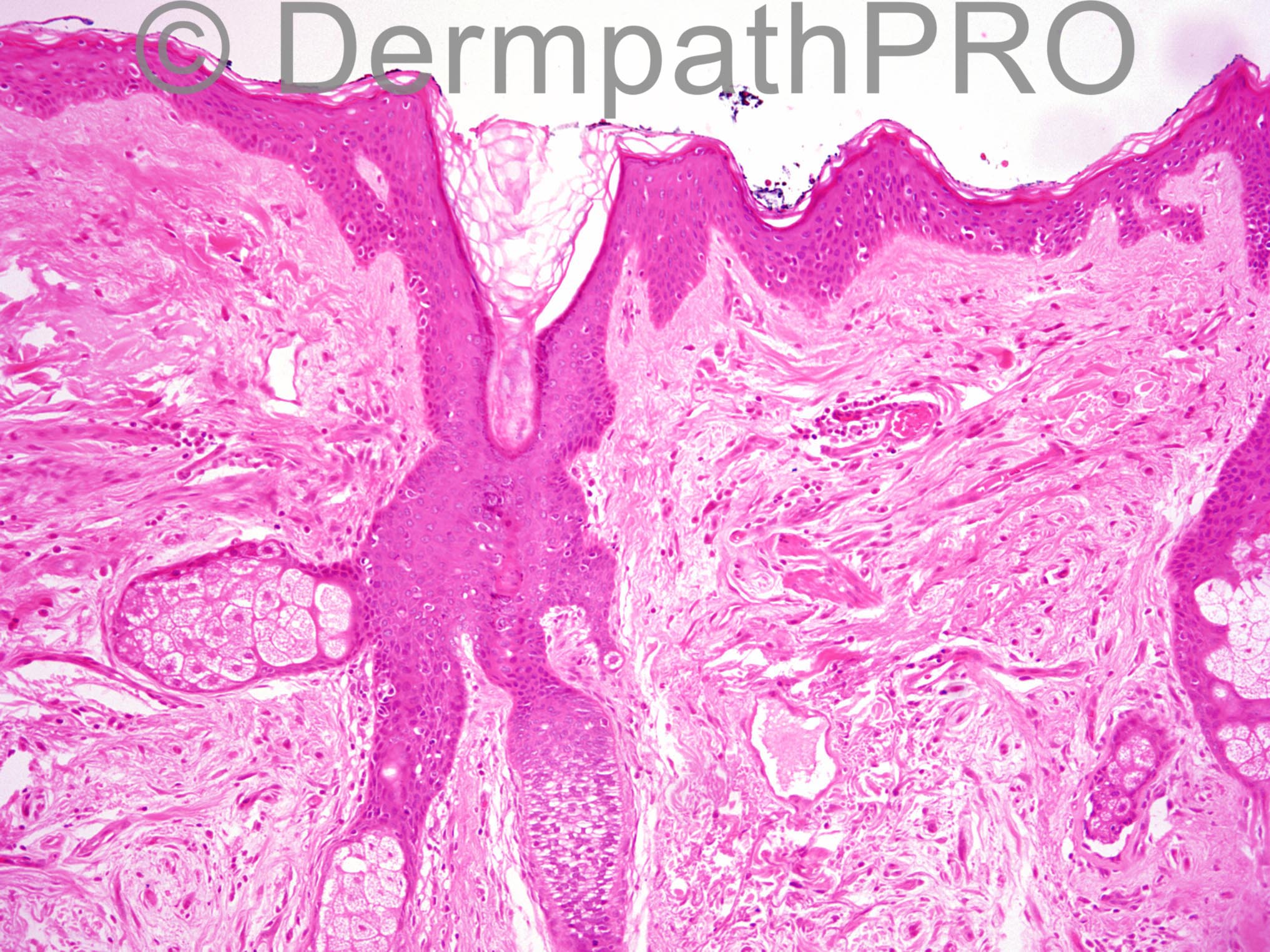
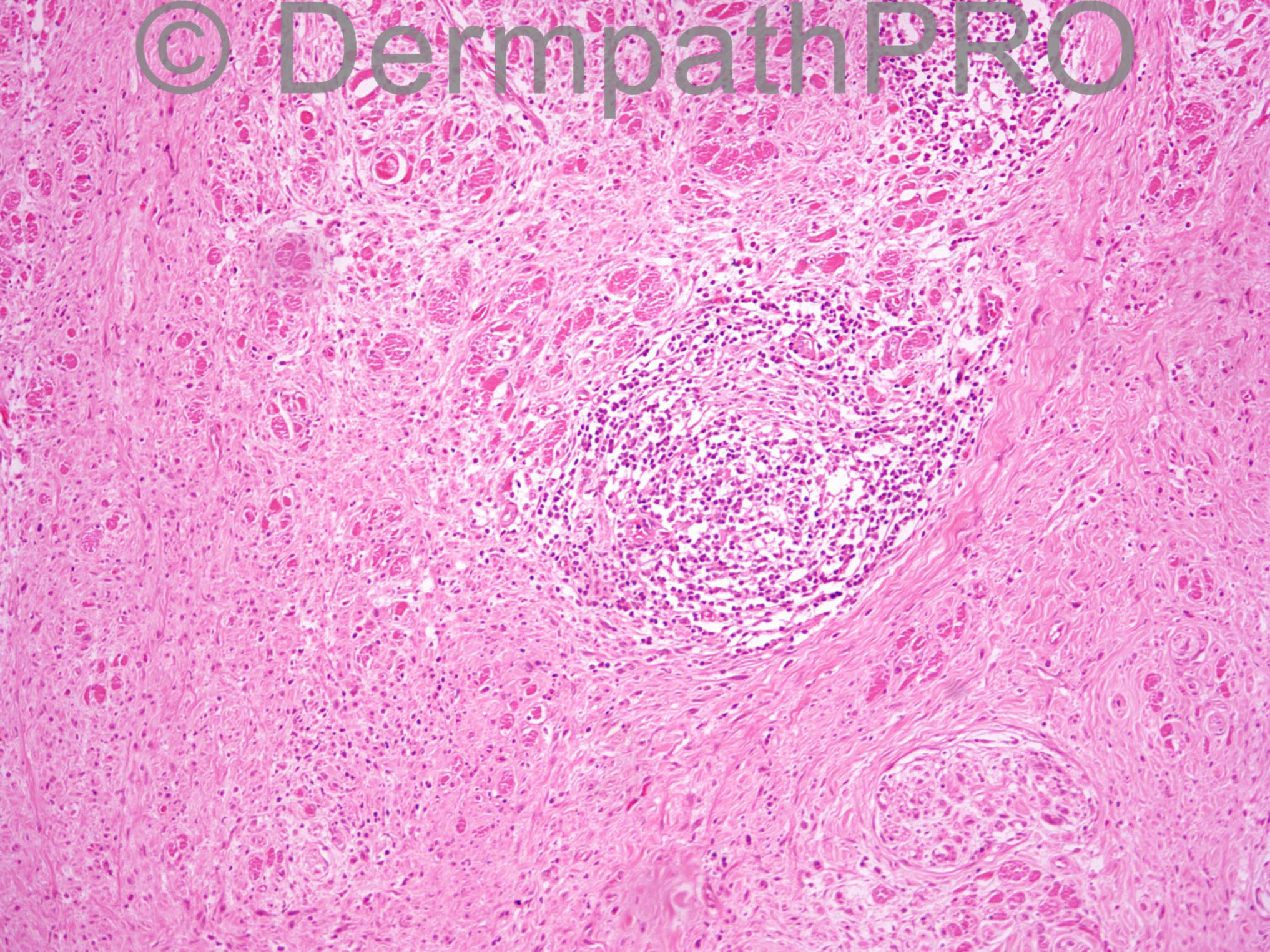
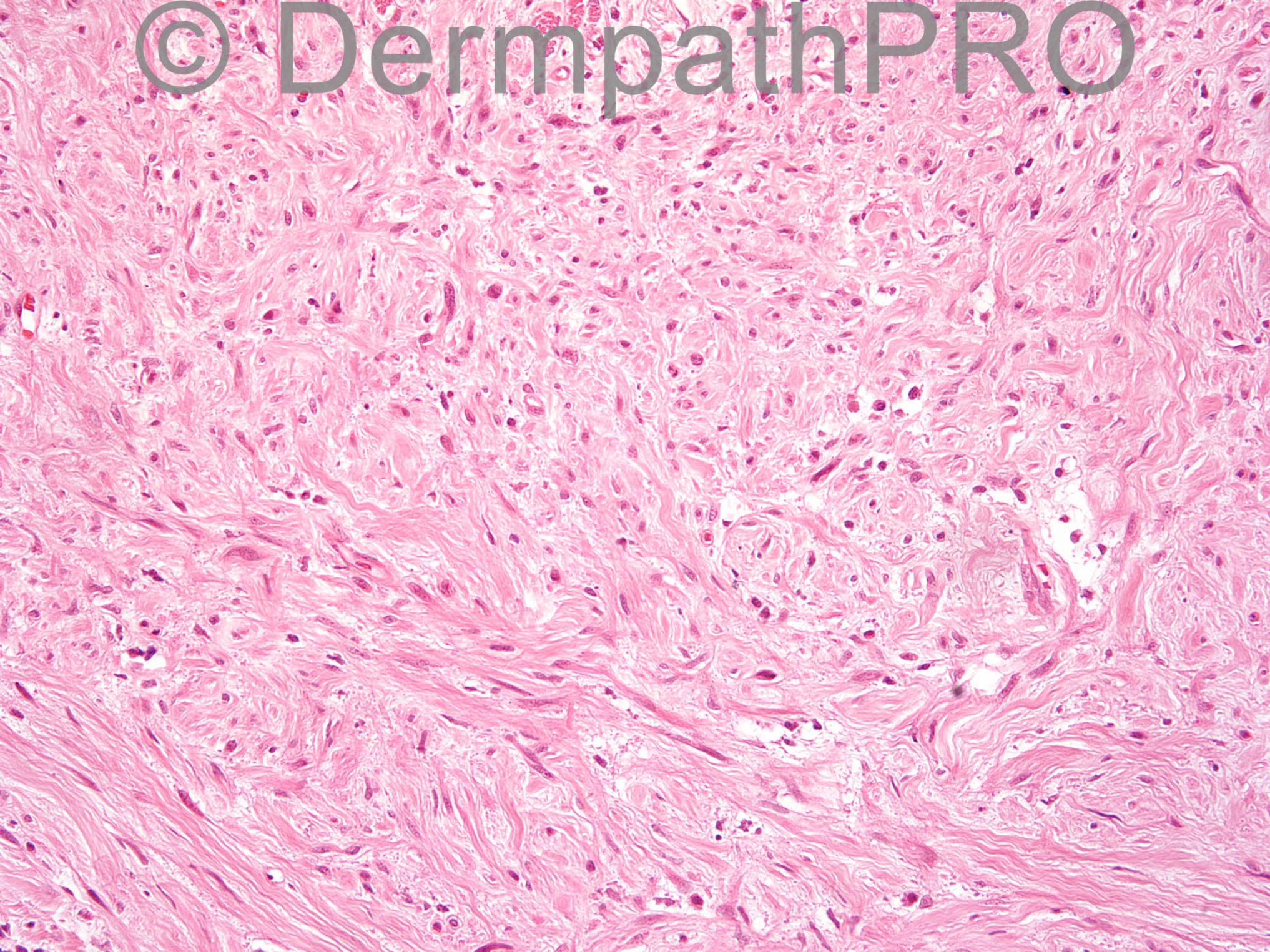
70 year old man with 3 cm plaque on back, previously diagnosed as a scar. This is a deep incisional biopsy; F1 shows the surface of the lesion and F2-F4 shows the deeper aspects.

Case posted by Dr Uma Sundram


  Report Record

User Feedback


Dr. Mona Abdel-Halim

Posted

I will call it desmoplastic melanoma, nodular lymphoid infiltrate is a clue, previous diagnosis as a scar is a common mistake.

Share this comment


Link to comment
share_externally

Dermatomyofibroma just to be different but I appreciate it is usually seen on the back of the neck in young females. Some of the spindle cells are parallel to the skin surface with sparing of the papillary dermis. The overlying epidermis is not showing any features of lentigo maligna. But there is that collection of lymphocytes in the dermis and desmoplastic melanoma presents clinically just like this and in this age group!

Share this comment


Link to comment
share_externally

Guest Romualdo

Posted

Mona is correct! I thought of desmoplastic melanoma but wrote spindle cell melanoma!

Share this comment


Link to comment
share_externally

Arti Bakshi

Posted

Desmoplastic melanoma most likely. Not on a sun exposed site though... would need immunos for confirmation (expect S100 to be positive).

Share this comment


Link to comment
share_externally

Guest Jim Davie MD

Posted

Agree desmoplastic melanoma is most likely (S100 to support). Randomly hyperchromatic atypical spindle cells with aggressive dissection through skeletal muscle, but large nerves appear uninvolved. Desmoplastic stroma has a patchy lymphoplasmacytic infiltrate, and may have some granular mucin.

Would also add spindle-cell SCC to expand the differential (high-molecular weight cytokeratin CK903 may support), although there is no epidermal atypia in these sections.

Share this comment


Link to comment
share_externally

Uma Sundram

Posted

Great comments. This is indeed desmoplastic melanoma, so called pure type--no conventional melanoma component. S100 was positive within the atypical cells and CK 5/6 was negative. A very subtle lentiginous proliferation of melanocytes was present within the epidermis but it was small; no nesting was seen.

Share this comment


Link to comment
share_externally


Join the conversation

You can post now and register later. If you have an account, sign in now to post with your account.

Guest
Add a comment...

×   Pasted as rich text.   Paste as plain text instead

  Only 75 emoji are allowed.

×   Your link has been automatically embedded.   Display as a link instead

×   Your previous content has been restored.   Clear editor

×   You cannot paste images directly. Upload or insert images from URL.

Loading...

×
×
  • Create New...