Jump to content
In this section we have spot diagnoses posted on a daily basis since June 2010, now over 4000! You can review the archived cases and read the suggested diagnoses by users and the final comment by the contributors.
Case are uploaded each week day by 10 am UK time with the correct diagnosis will generally be posted at 8 pm UK time. Why not view the most recent spot diagnosis and proffer a diagnosis?

Case Number : Case 1268 - 04 May Posted By: Guest

Please read the clinical history and view the images by clicking on them before you proffer your diagnosis.
Submitted Date :
   (0 reviews)

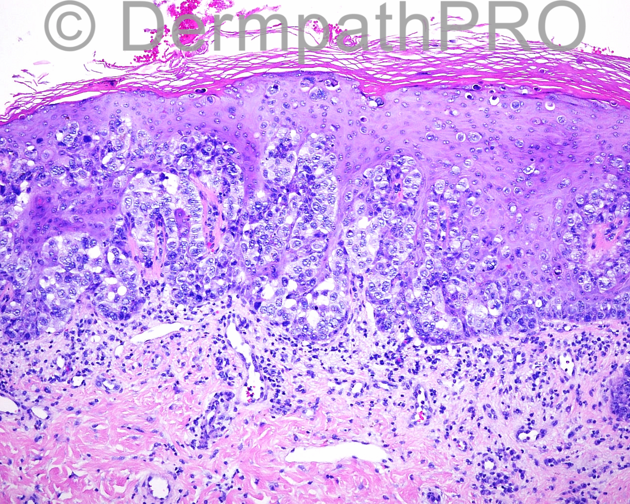
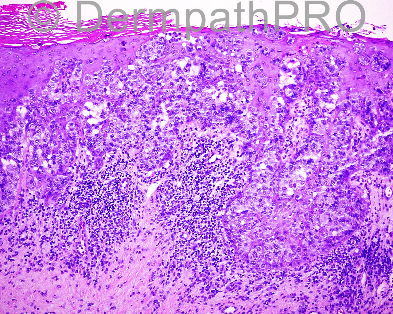
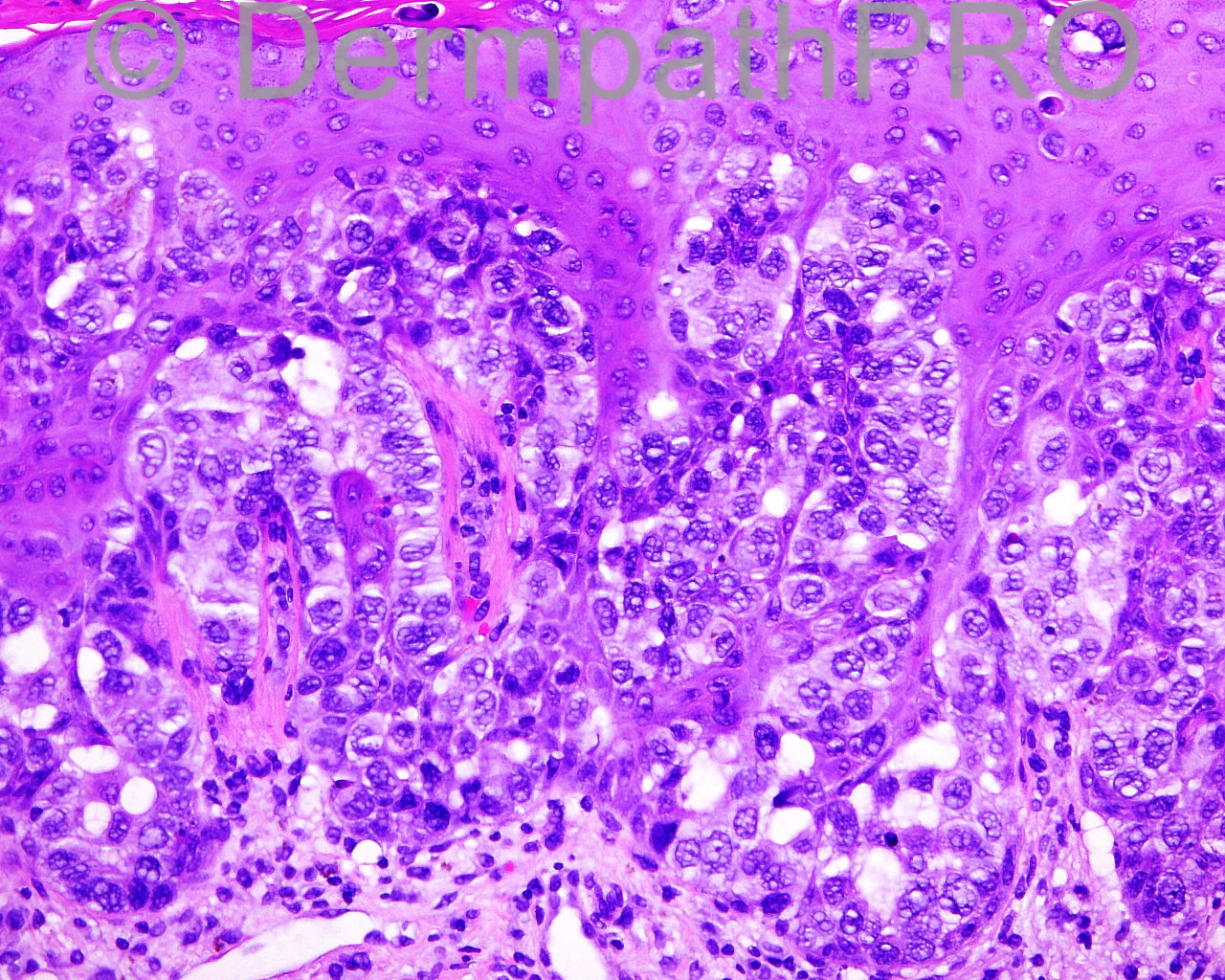
The patient is a 53 year old woman with a punch biopsy of a 13 mm psoriasiform patch taken from the right nipple. Clinical Diagnosis: Basal cell carcinoma vs squamous cell carcinoma vs Paget's.

Case posted by Dr Mark Hurt


  Report Record

User Feedback


Dr. Mona Abdel-Halim

Posted

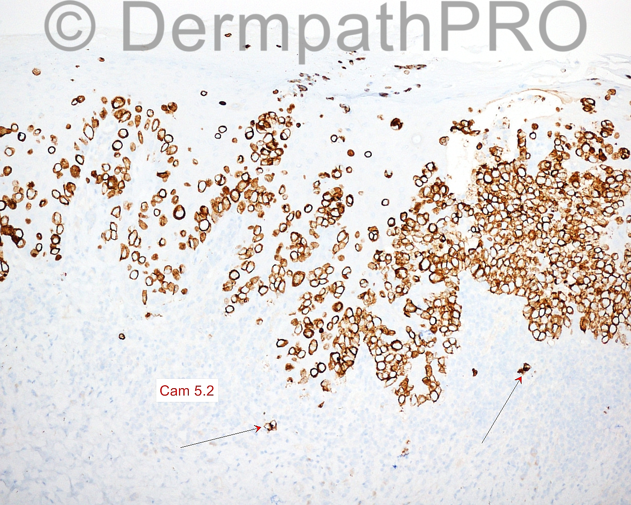
Paget's. CAM5.2 positivity is against pagetoid Bowen's.

Share this comment


Link to comment
share_externally

IgorSC

Posted

I agree with Paget´s disease diagnosis.

Share this comment


Link to comment
share_externally

Guest Romualdo

Posted

Agree, Paget's disease.

Share this comment


Link to comment
share_externally

Mark A. Hurt MD

Posted

I also thought this was Paget's disease. My diagnosis is below. I thought it was curious that there were Cam 5.2 cells present in the dermis, thus my comment:

SKIN, RIGHT NIPPLE , PUNCH BIOPSY :
[b]-- PAGET'S DISEASE[/b]
[indent=1][b]COMMENT: [/b]The presence of Paget's cells within the dermis was a recent subject of study by Korean authors (Lee et al), in 2014, where they found that "the prognosis was similar for invasive mammary Paget's disease and non-invasive mammary Paget's disease, and the mammary Paget's disease must be distinguished from locally advanced breast cancer presenting as satellite skin nodules.” Thus, further correlation for breast disease is warranted at this point.[/indent]


[center]Reference:[/center]


[indent=1]Lee HW, Kim TE, Cho SY, Kim SW, Kil WH, Lee JE, Nam SJ, Cho EY. Invasive Paget disease of the breast: 20 years of experience at a single institution. Hum Pathol. 2014 Dec;45(12):2480-7. doi: 10.1016/j.humpath.2014.08.015. Epub 2014 Sep 7. PubMed PMID: 25288235.[/indent]

Share this comment


Link to comment
share_externally

Dr. Richard Carr

Posted

Mark,
Did you consider these papillary dermal cells may be necrotic neoplastic cells that have dropped out akin to colloid bodies or was there visibile invasion on H&E
Regards
Richard

Share this comment


Link to comment
share_externally

Mark A. Hurt MD

Posted

Richard,

No, I didn't consider it; it didn't come up in my mind. Do you know if that has been studied any any systematic way?

Best,

Mark

Share this comment


Link to comment
share_externally

Dr. Richard Carr

Posted

No I have not but looking at the images my impression was they were limited to the papillary dermis and smaller than the cells in the epidermis - perhaps shrunken - you would be better to judge of course. Dual IHC might be of interest e.g. Ki67/Cam5.2 to see if they are still in cell cycle?
Regards
Richard

Share this comment


Link to comment
share_externally

Mark A. Hurt MD

Posted

Richard, that's a thought. Actually, I posted this case because there are rare examples of extramammary Paget's disease of the nipple, and sometimes in EMPD, there are dermal elements. In my experience, I have sometimes seen tubules in the epidermis of EMPD but I have never seen them in Paget's disease of the nipple. When I saw these dermal Cam 5.2 positive cells in the dermis in the posted case, I was surprised, because I couldn't think of case of Paget's disease with dermal elements (perhaps reflecting the fact that I don't see all that many cases of it anyway), but I didn't see any tubules. My explanation is a bit convoluted, but I wondered whether anyone had ever written on the presence of dermal cells in Paget's disease; it was the reference I posted. If I ever learn any followup on this case, I will post it here.

Mark

Share this comment


Link to comment
share_externally


Join the conversation

You can post now and register later. If you have an account, sign in now to post with your account.

Guest
Add a comment...

×   Pasted as rich text.   Paste as plain text instead

  Only 75 emoji are allowed.

×   Your link has been automatically embedded.   Display as a link instead

×   Your previous content has been restored.   Clear editor

×   You cannot paste images directly. Upload or insert images from URL.

Loading...

×
×
  • Create New...