Jump to content
In this section we have spot diagnoses posted on a daily basis since June 2010, now over 4000! You can review the archived cases and read the suggested diagnoses by users and the final comment by the contributors.
Case are uploaded each week day by 10 am UK time with the correct diagnosis will generally be posted at 8 pm UK time. Why not view the most recent spot diagnosis and proffer a diagnosis?

Case Number : Case 1272 - 08 May Posted By: Guest

Please read the clinical history and view the images by clicking on them before you proffer your diagnosis.
Submitted Date :
   (0 reviews)

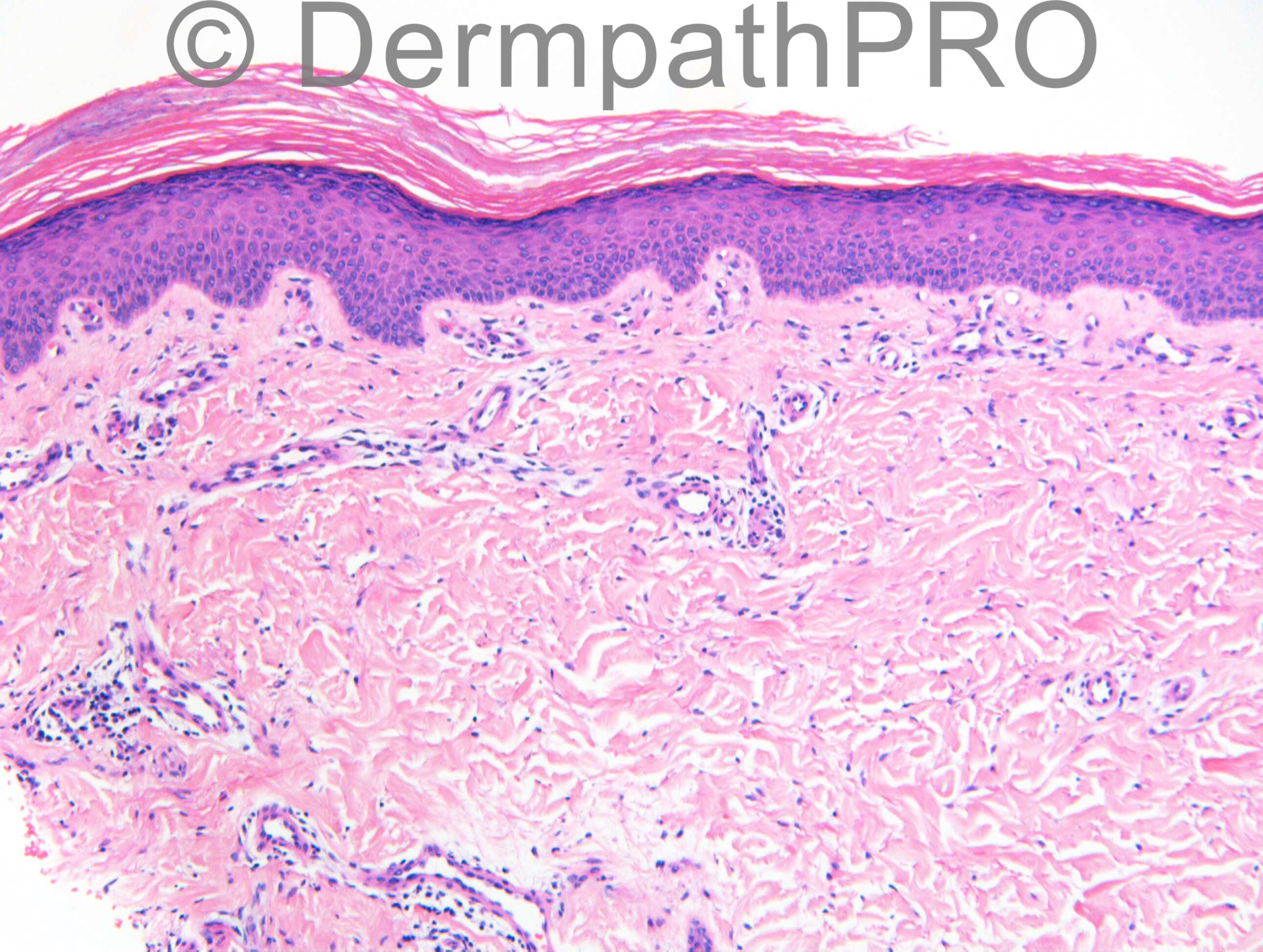
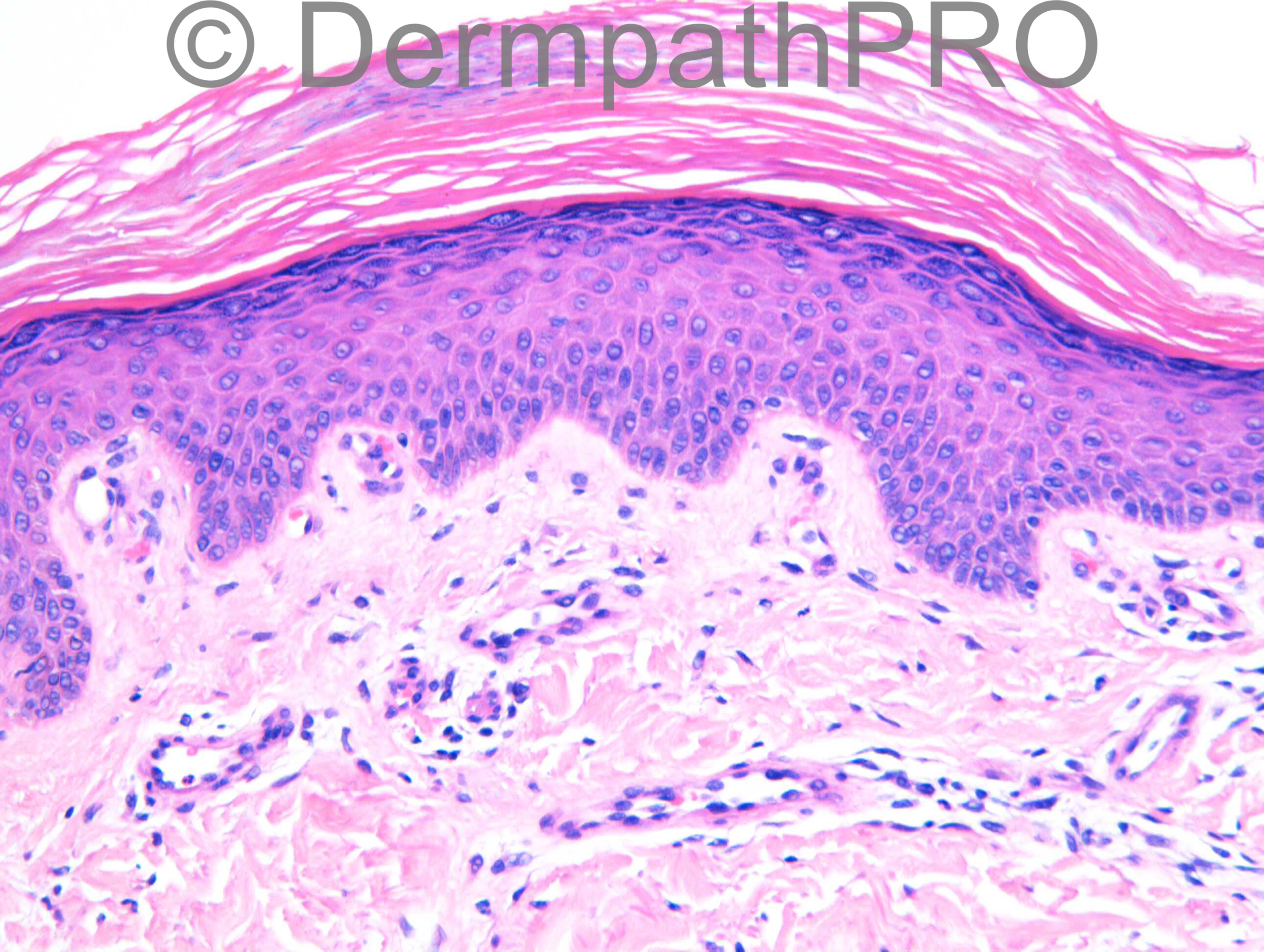
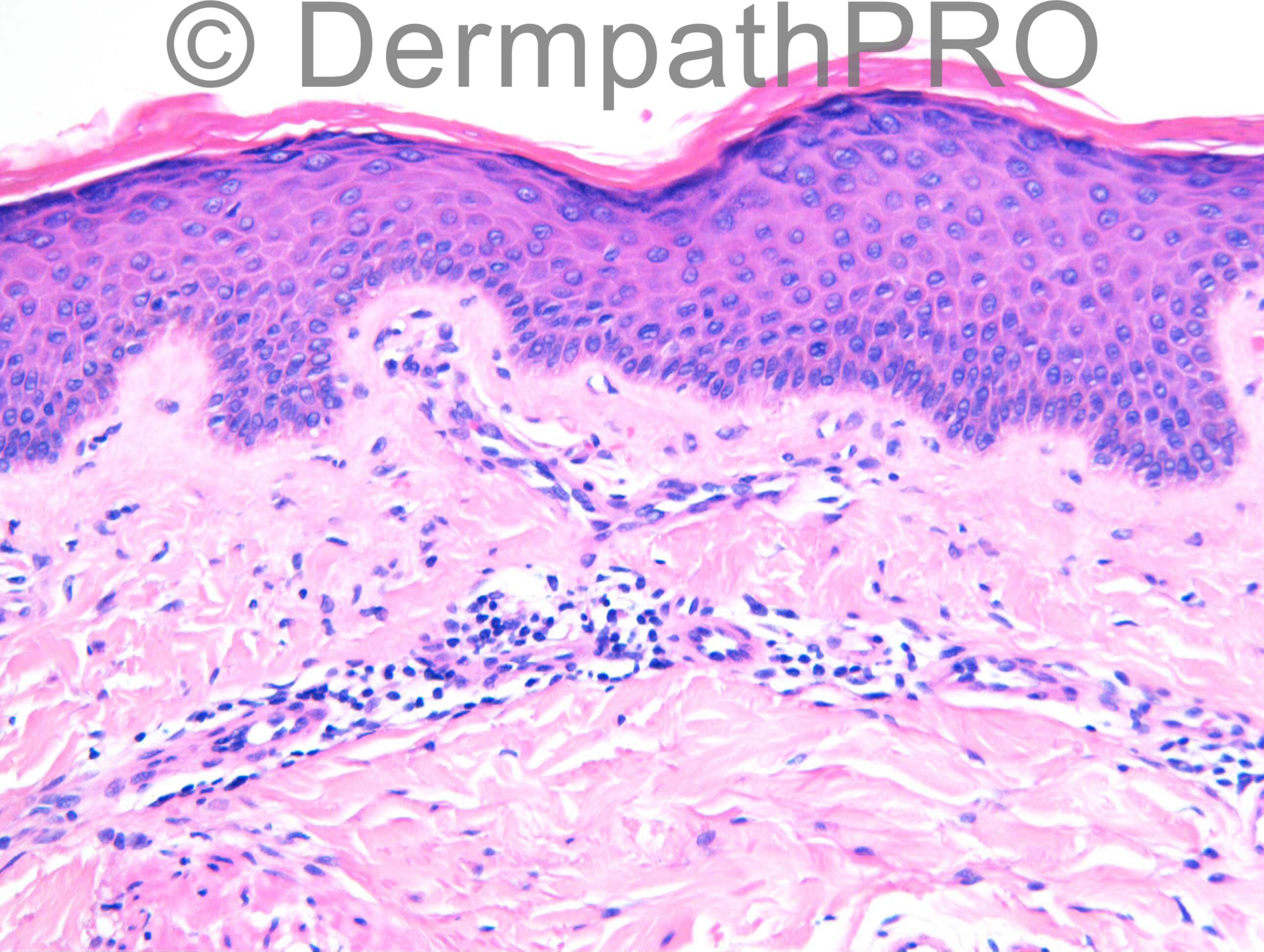
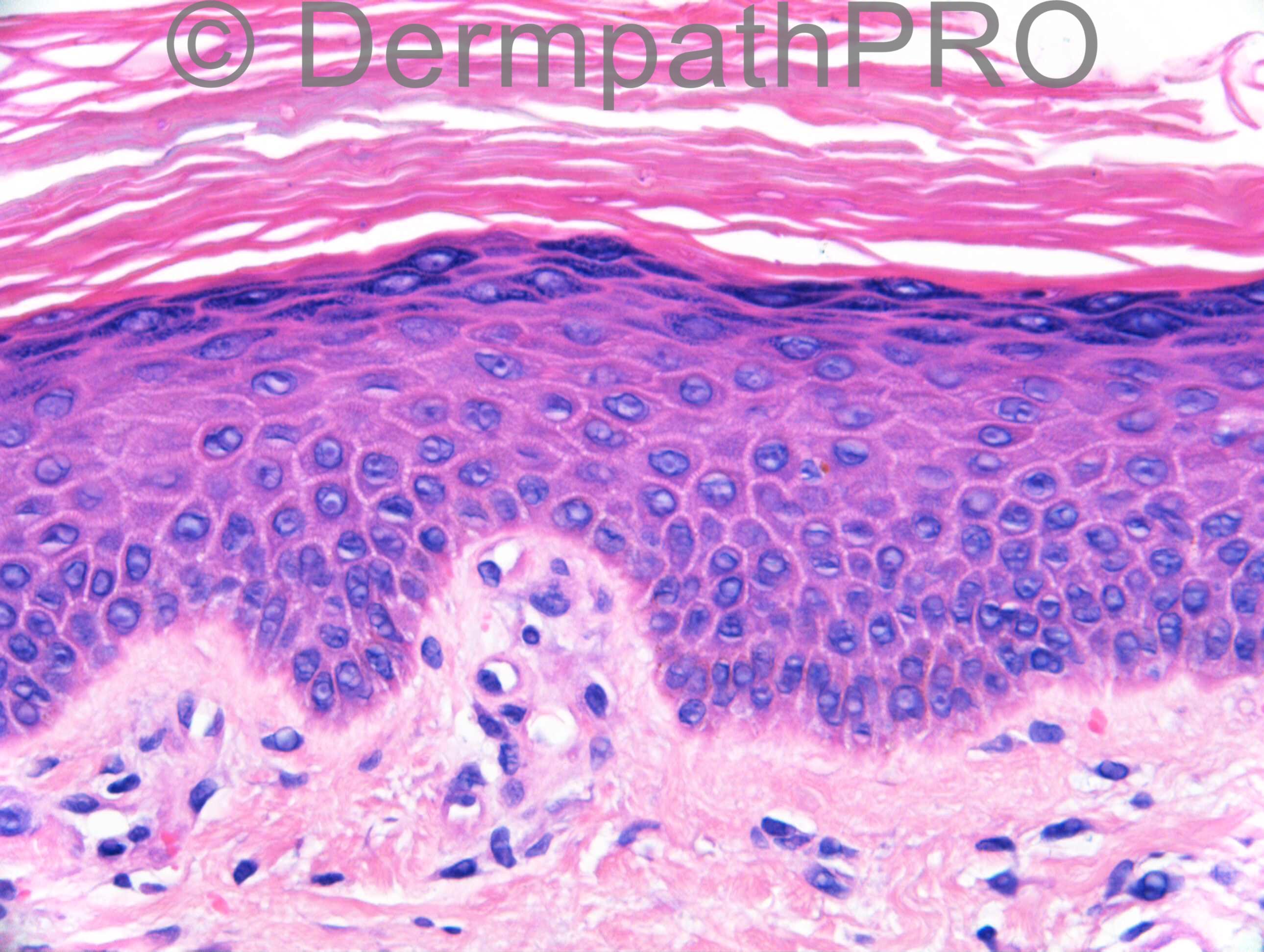
F54. 1 year rash on right thigh not responding to antifungals. Worse with steroids. ?tinea incognito ?what

Case posted by Dr Richard Carr


  Report Record

User Feedback


IgorSC

Posted

Interesting, I see no melanin in the basal layer and almost no melanocytes. Is there any suspicious for Vitiligo in this case? I saw some reports with these two disease in association or infliximab-induced assocition Vitiligo for treatment for PRP.

Share this comment


Link to comment
share_externally

Dr. Richard Carr

Posted

[img]https://dermpathpro.com/uploads/spot_diagnosis_comment_img/Case%201272%20-%2008%20May%20-%20RAC7103x20_PAS_4pm.jpg[/img]

[img]https://dermpathpro.com/uploads/spot_diagnosis_comment_img/Case%201272%20-%2008%20May%20-%20RAC7103x40_PAS_4pm.jpg[/img]

Share this comment


Link to comment
share_externally

Dr. Mona Abdel-Halim

Posted

with the PAS, this is tinea cruris even if he is not responsive to anti-fungals. May be he is not compliant, may be he needs systemic in addition to topical antifungals. May be he has an associated tinea pedis causing recurrence.. Many things can explain... I actually suspected fungal elements from the last image before PAS was presented.

Share this comment


Link to comment
share_externally

Arti Bakshi

Posted

Saw this case only after the PAS, don't think would have suspected tinea before.
Demonstrates the importance of doing fungal stains! Presumably the steroids have knocked off the acute inflammatory cells.

Share this comment


Link to comment
share_externally

Dr. Richard Carr

Posted

Agree Tinea corporis. I missed the fungal elements on my initial screen but given the clinical information doing a PAS is mandatory. Going back to the H&E with the eye of faith there are a few possible ghosted holes in the last image that correlate with the PAS. The lesson is not to dismiss the clinical diagnosis, especially when from an experienced dermatologist as in this case, without due care and attention! I am confident none of you would have signed this out without doing a PAS in real life. This case would have been more difficult had the clinician not handed me the diagnosis. In general I tend to do a PAS on all anatomically localised, spongiotic, psoriasiform, parakeratotic or "subtle" dermatoses (especially if neutrophils are noted - not this case) but rarely pick up tinea incognito (perhaps one in 50 cases for PAS or 2 to 3 cases a year - normally the fungal elements are very obvious on the H&E if you remember to look (no need for PAS in most cases). In the majority of cases without obvious fungal hyphae on H&E I generally request the PAS at 3 additional levels or ask for PAS ribbons x3 and then scrutinise all the sections as the fungal hyphae can be few and far between (often patient already treated for the disease). Note isolated fungal spores are usually non-pathogenic especially if clustered in the follicular orifices or within hyperkeratosis associated with squamoproliferative lesions, do look carefully in the follicles for hyphae - especially on facial biopsies - as often the pick-up in the follicular orifice is higher). I understand the normal rules don't always apply in Spot Diagnosis, especially with my track record of trying to catch you all out, but today a double bluff. Mona next time you think of something do mention it - I remember well, on one of Phillip Mckee's cases, mentioning and dismissing ossifying fibromyxoid tumour (mentioned as an off chance) and it turned out to be the correct diagnosis!

Share this comment


Link to comment
share_externally


Join the conversation

You can post now and register later. If you have an account, sign in now to post with your account.

Guest
Add a comment...

×   Pasted as rich text.   Paste as plain text instead

  Only 75 emoji are allowed.

×   Your link has been automatically embedded.   Display as a link instead

×   Your previous content has been restored.   Clear editor

×   You cannot paste images directly. Upload or insert images from URL.

Loading...

×
×
  • Create New...