Jump to content
In this section we have spot diagnoses posted on a daily basis since June 2010, now over 4000! You can review the archived cases and read the suggested diagnoses by users and the final comment by the contributors.
Case are uploaded each week day by 10 am UK time with the correct diagnosis will generally be posted at 8 pm UK time. Why not view the most recent spot diagnosis and proffer a diagnosis?

Case Number : Case 1278 - 18 May Posted By: Guest

Please read the clinical history and view the images by clicking on them before you proffer your diagnosis.
Submitted Date :
   (0 reviews)

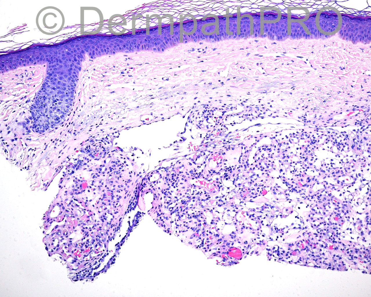
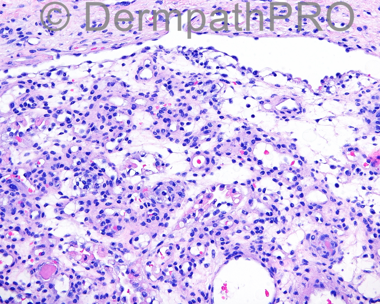
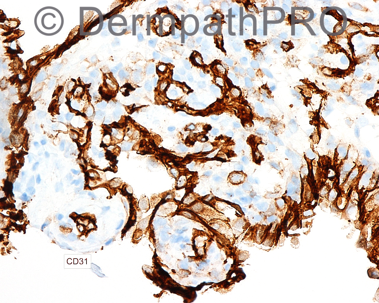
The 60-year-old male with a biopsy of a lesion on the chin.

Case posted by Dr Mark Hurt

  Report Record

User Feedback


Dr. Mona Abdel-Halim

Posted

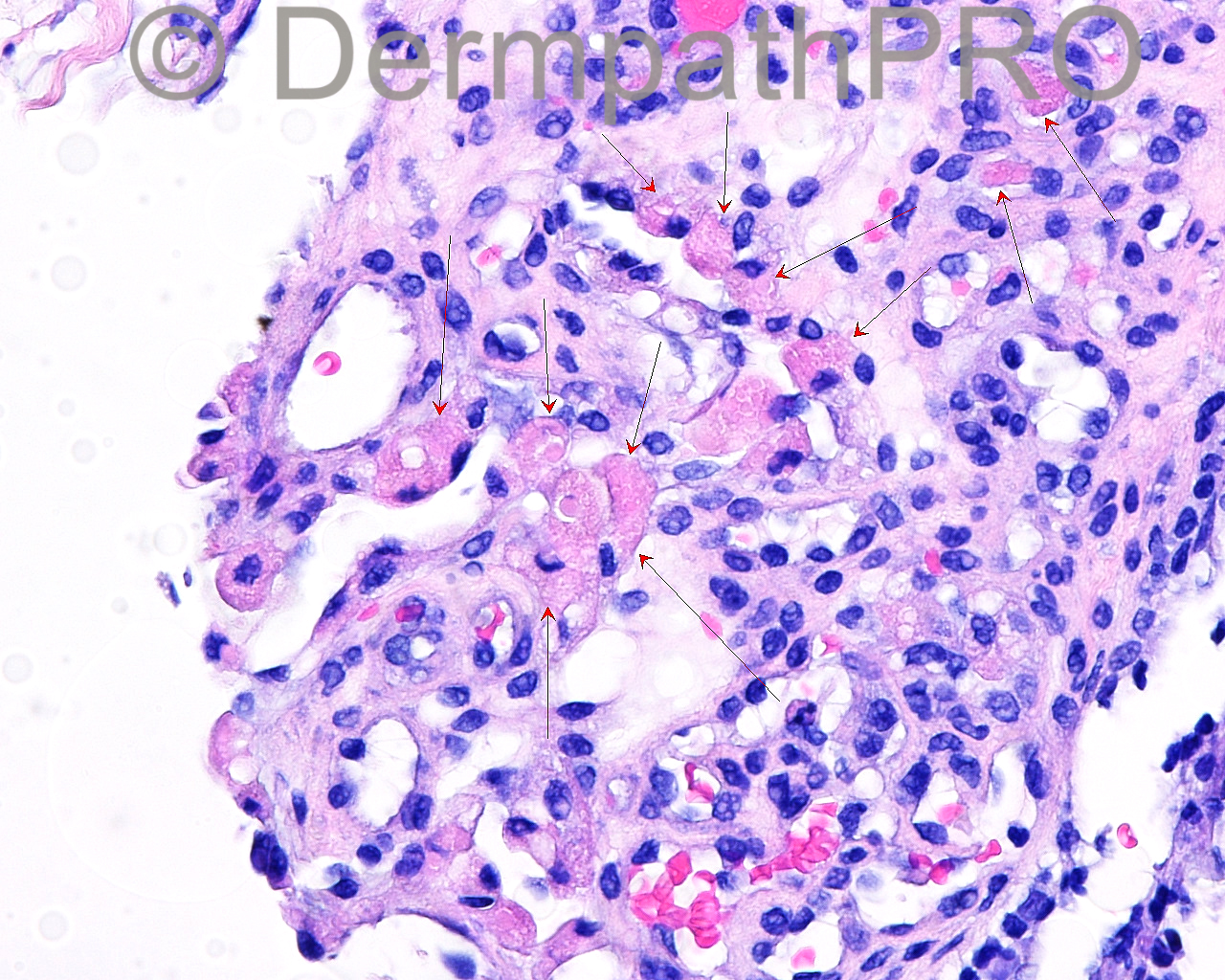
Thought of Glomeruloid Hemangioma ? with lots of esinophilic globules ?

Share this comment


Link to comment
share_externally

Arti Bakshi

Posted

Good possibility, Mona.
Another differential would be Tufted Angioma as there appear to be dilated lymphatic channels at periphery. (and one can get eosinophilic inclusions in this entity too). But the age is unusual, mostly occurs in children.

Share this comment


Link to comment
share_externally

Mark A. Hurt MD

Posted

I thought this was a difficult case, so see my diagnosis note. Thanks for your comments!


[b]-- UNUSUAL HEMANGIOMA[/b]
(With features of glomeruloid hemangioma and papillary hemangioma)
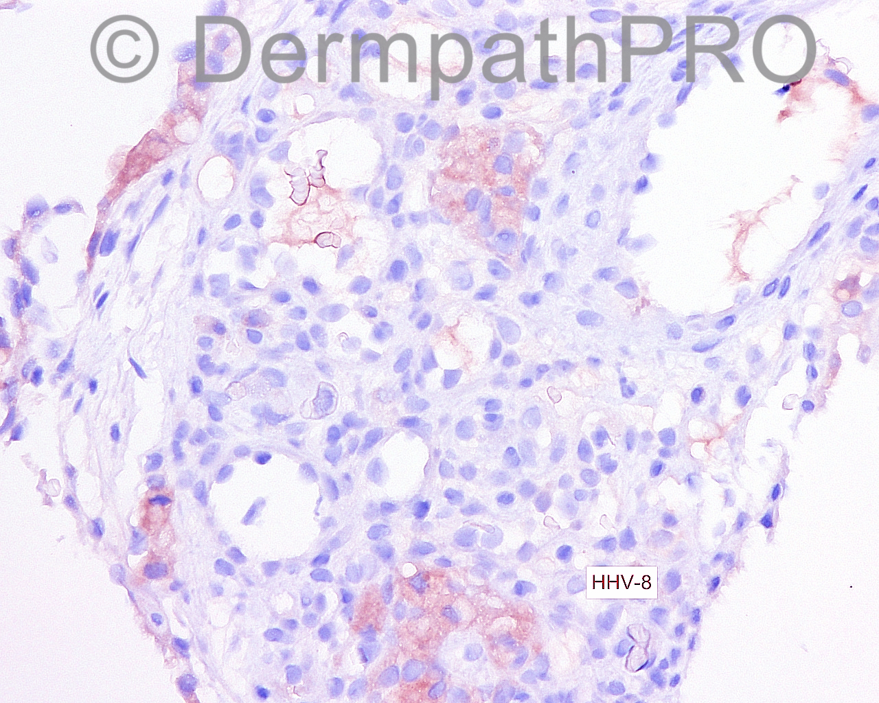
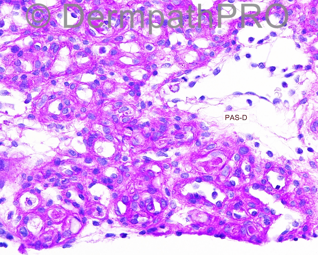
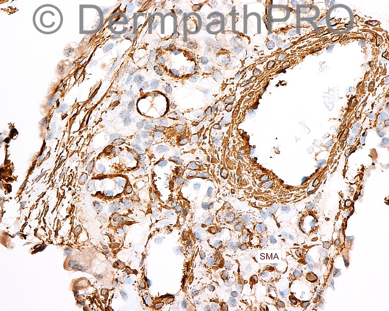
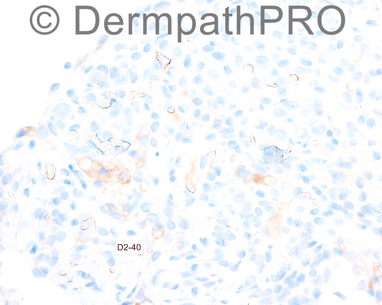
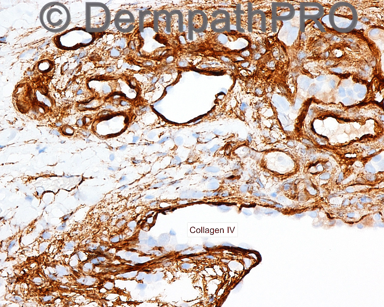
[indent=1][b]COMMENT: [/b]Up until the last few years, the concept of papillary hemangioma had not been put out in the literature, and most of the lesions were classified under glomeruloid hemangioma. Both of the lesions contain PAS-positive hyaline globules in the endothelial cells, and it was thought initially that the lesions of glomeruloid hemangioma were pathognomonic of POEMS syndrome. Today, there is an argument that the papillary hemangiomas, albeit very similar to glomeruloid hemangiomas, are not associated with that syndrome but only the glomeruloid hemangiomas are. In this particular case, the evidence would seem to point toward this being a papillary hemangioma based on the fact that it contains thickened basement membranes with type IV collagen prominence and PAS-positive prominence around the vessels. Nonetheless, the structure of the lesion, to my eye, looks more like a glomeruloid hemangioma. It may just simply be that the small sample precludes the ability to differentiate the two very well. I think at this point, further clinical followup is warranted in order to determine whether the patient has any clinical manifestations of POEMS syndrome. There is some reason to believe that patients without that syndrome can produce papillary hemangiomas that can be very close in structure to the lesion I am seeing here. Usually they are solitary lesions opposed to the multiple ones in glomeruloid hemangiomas.[/indent]

[center]References:[/center]

Suurmeijer AJ, Fletcher CD. Papillary haemangioma. A distinctive cutaneous haemangioma of the head and neck area containing eosinophilic hyaline globules. Histopathology. 2007 Nov;51(5):638-48. PubMed PMID: 17927585.

Suurmeijer AJ. Papillary hemangiomas and glomeruloid hemangiomas are distinct clinicopathological entities. Int J Surg Pathol. 2010 Feb;18(1):48-54. doi:10.1177/1066896908323504. Epub 2008 Sep 19. PubMed PMID: 18805868.

Share this comment


Link to comment
share_externally


Join the conversation

You can post now and register later. If you have an account, sign in now to post with your account.

Guest
Add a comment...

×   Pasted as rich text.   Paste as plain text instead

  Only 75 emoji are allowed.

×   Your link has been automatically embedded.   Display as a link instead

×   Your previous content has been restored.   Clear editor

×   You cannot paste images directly. Upload or insert images from URL.

Loading...

×
×
  • Create New...