Jump to content
In this section we have spot diagnoses posted on a daily basis since June 2010, now over 4000! You can review the archived cases and read the suggested diagnoses by users and the final comment by the contributors.
Case are uploaded each week day by 10 am UK time with the correct diagnosis will generally be posted at 8 pm UK time. Why not view the most recent spot diagnosis and proffer a diagnosis?

Case Number : Case 1405- 11 November Posted By: Guest

Please read the clinical history and view the images by clicking on them before you proffer your diagnosis.
Submitted Date :
   (0 reviews)

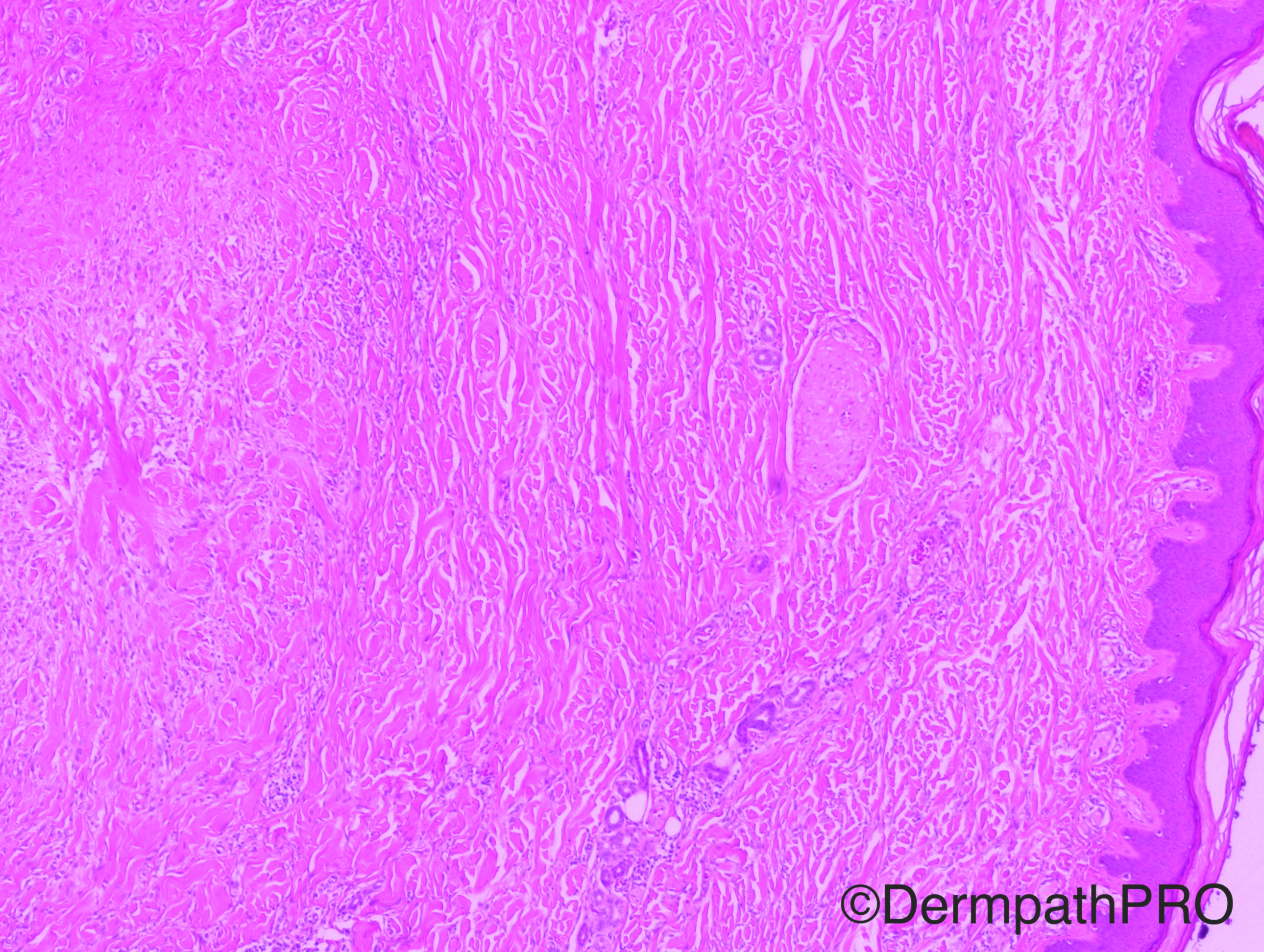
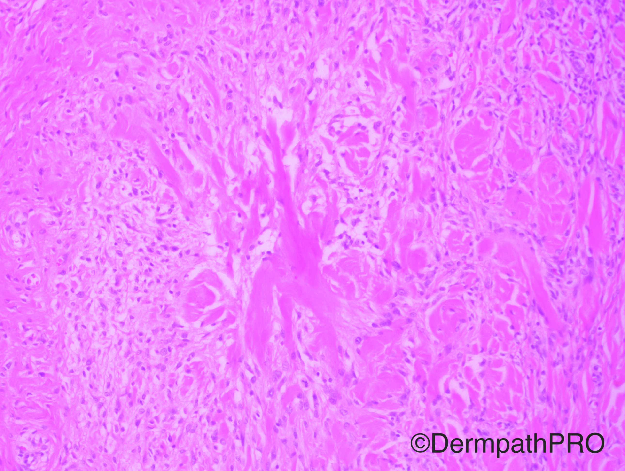
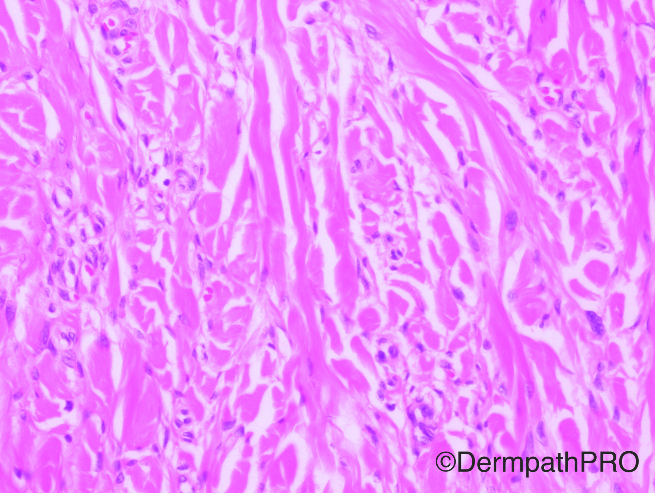
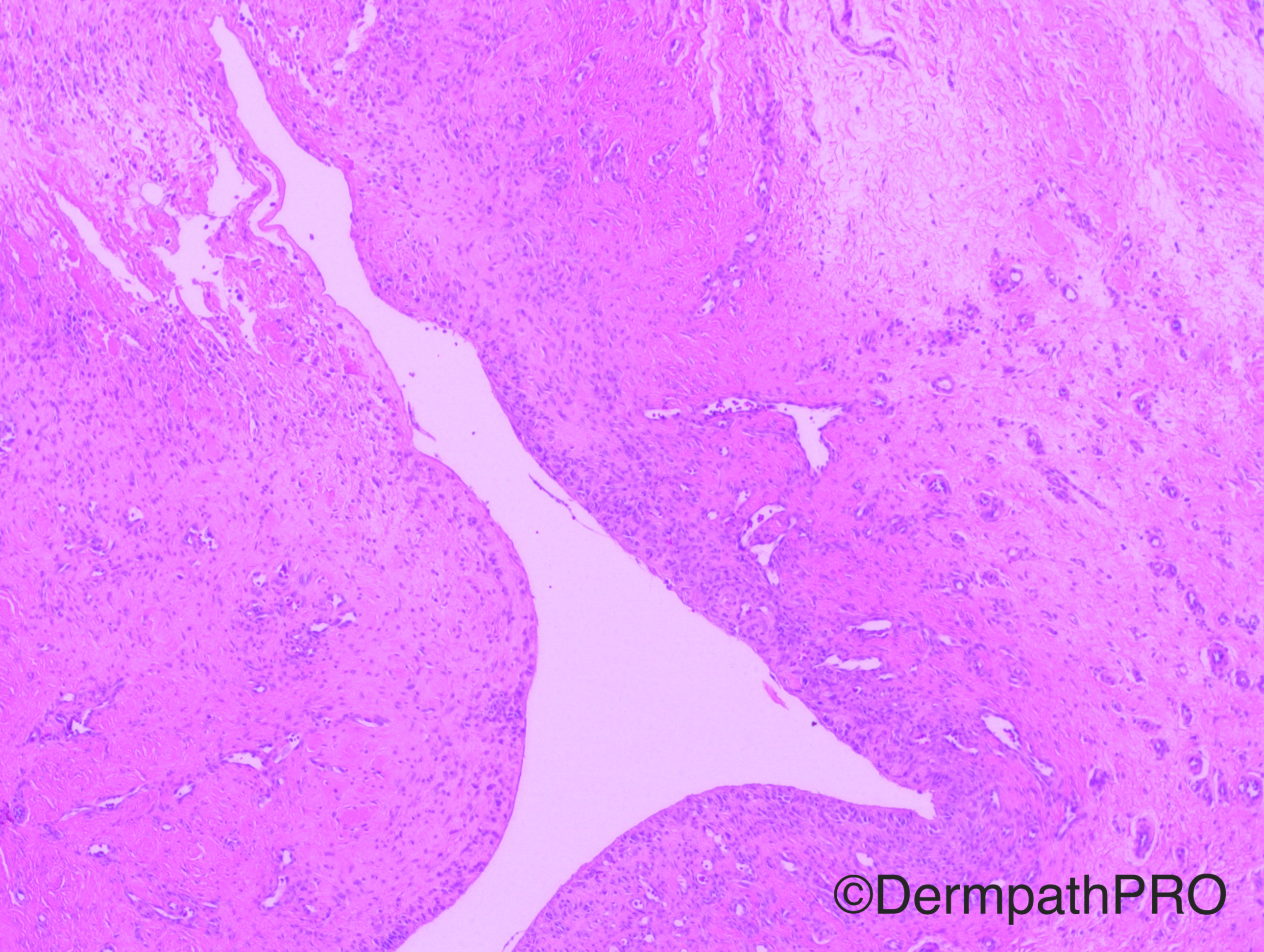
Case History: 66 year-old female with left forearm lesion.

Case posted by Dr Hafeez Diwan


  Report Record

User Feedback


Raul Perret

Posted

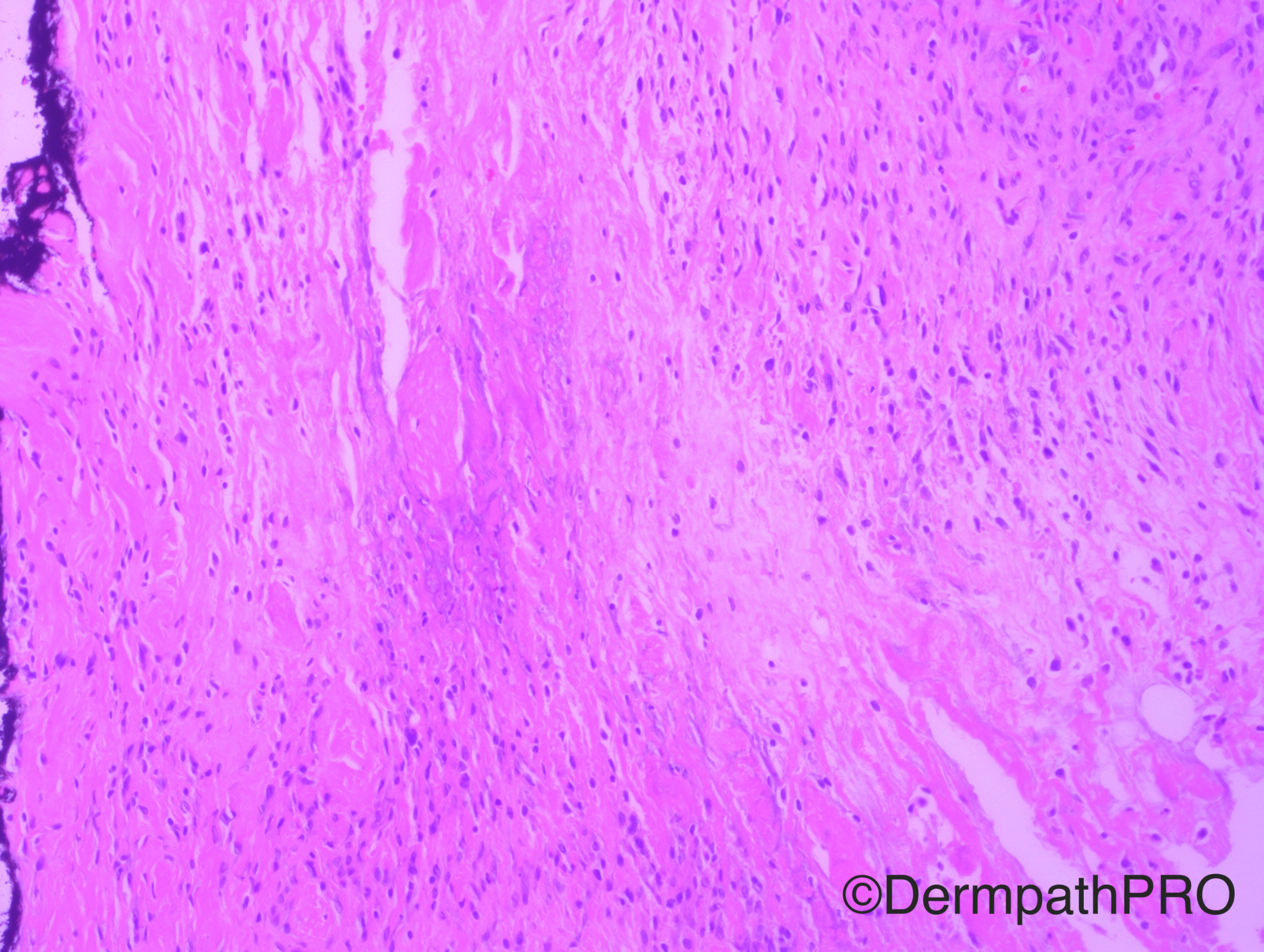
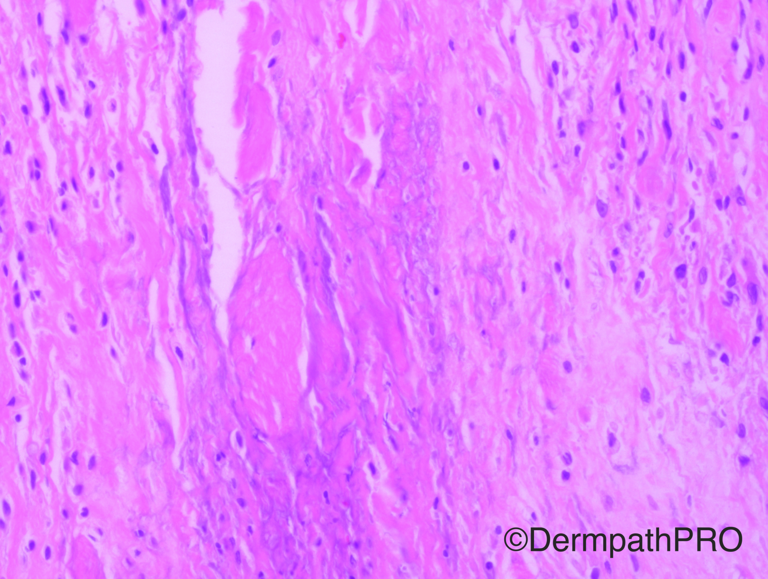
Find it quite hard but i will give it a go. I see a dermal interstitial histiocytic infiltrate with destruction of collagen (collgenolysis) and others with a more ropey collagen (keloid like). Deeper on the dermis there is a vein with histiocytic infiltrate on its Wall surrounded by numerous dilated low caliber vessels. It makes me think of necrobiosis lipoidica vs another granulomatous dermal reaction.

Share this comment


Link to comment
share_externally

Guest Romualdo

Posted

One image shows a cavity without an epithelial lining. Other images show some histiocytes or myofibroblasts maybe secondary to spillage of cystic contents. Ganglion cyst?

Share this comment


Link to comment
share_externally

Deep granulomatous and intersticial dermatitis with areas of collagen degeneration and cystic formation. I think this is a case of an old rheumatoid nodule with cystic degeneration.

Share this comment


Link to comment
share_externally

Arti Bakshi

Posted

Good suggestion by Igor. Either a rheumatoid nodule or a deep granuloma annulare (pseudorheumatoid nodule).

Share this comment


Link to comment
share_externally

Dr. Hafeez Diwan

Posted

I went with deep granuloma annulare. The top two images show what appears to be a necrobiotic focus, and the second row images show an interstitial pattern. The lower three images show some reactive vascular proliferation around fibrinous areas, with a blush of mucin (lowest image).

Share this comment


Link to comment
share_externally

Dr. Richard Carr

Posted

The cavity looks typical of a ganglion cyst or connection with the underlying joint. I think Romauldo is right. Probably pseudo-GA, certainly not typical of deep GA which would be a large well defined necrobiotic nodule.

Share this comment


Link to comment
share_externally


Join the conversation

You can post now and register later. If you have an account, sign in now to post with your account.

Guest
Add a comment...

×   Pasted as rich text.   Paste as plain text instead

  Only 75 emoji are allowed.

×   Your link has been automatically embedded.   Display as a link instead

×   Your previous content has been restored.   Clear editor

×   You cannot paste images directly. Upload or insert images from URL.

Loading...

×
×
  • Create New...