Jump to content
In this section we have spot diagnoses posted on a daily basis since June 2010, now over 4000! You can review the archived cases and read the suggested diagnoses by users and the final comment by the contributors.
Case are uploaded each week day by 10 am UK time with the correct diagnosis will generally be posted at 8 pm UK time. Why not view the most recent spot diagnosis and proffer a diagnosis?

Case Number : Case 1414- 24 November Posted By: Guest

Please read the clinical history and view the images by clicking on them before you proffer your diagnosis.
Submitted Date :
   (0 reviews)

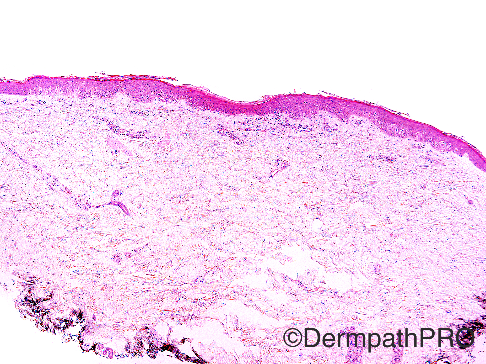
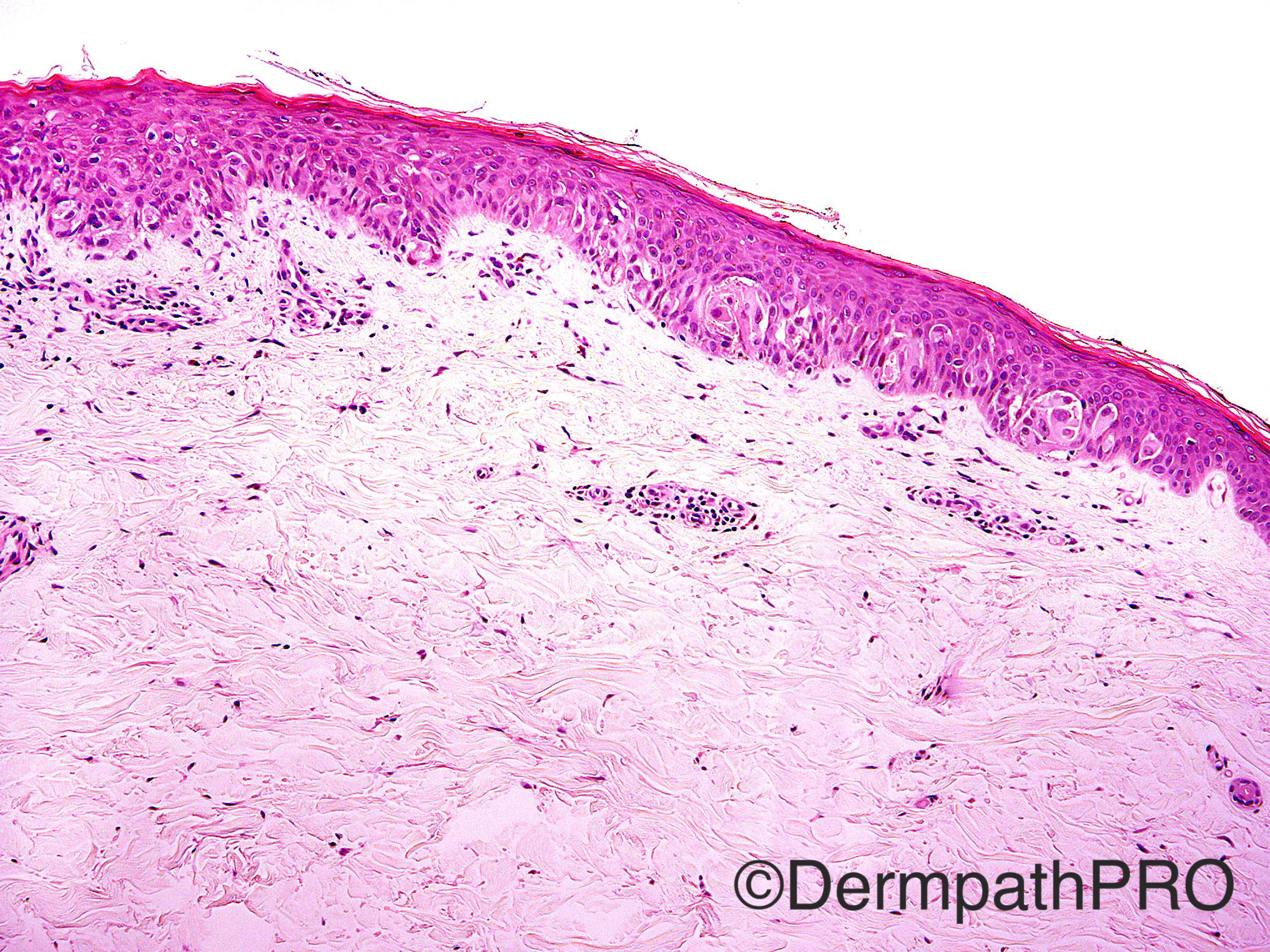
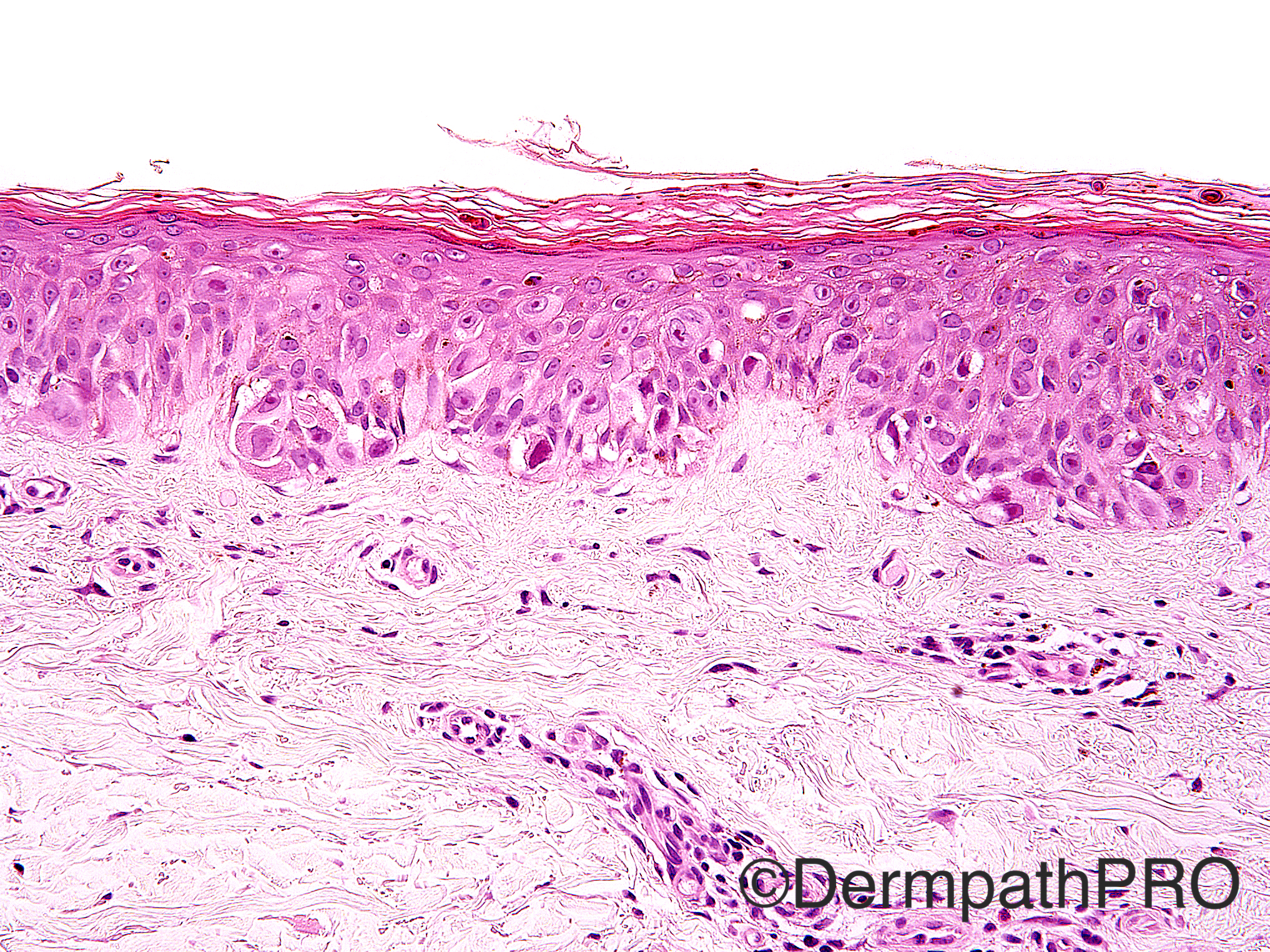
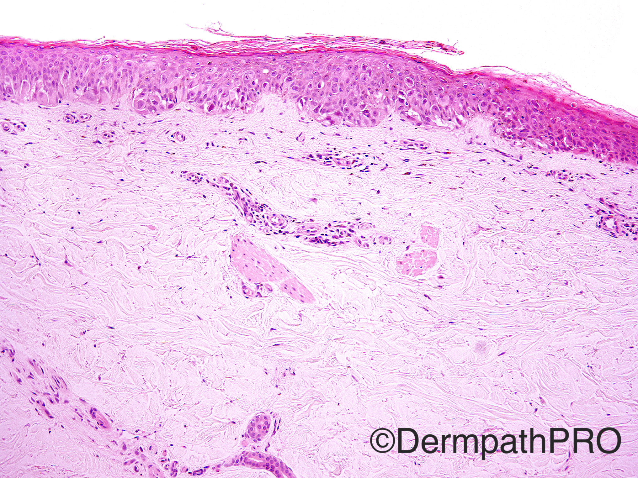
Case History: 29 year old female with nevus on left medial lower leg. Family history of melanoma and multiple atypical nevi. Fifth micrograph-MiTF; Sixth micrograph-p16.

Case posted by Dr Uma Sundram


  Report Record

User Feedback


Raul Perret

Posted

Even if p16 goes against it and the proliferation has clear spitzoid features I would still diagnose this as melanoma in situ.

Share this comment


Link to comment
share_externally

Agree with [color=#1C2837][font=arial, verdana, tahoma, sans-serif][size=4]melanoma-in-situ. [/size][/font][/color]

Share this comment


Link to comment
share_externally

Dr. Mona Abdel-Halim

Posted

Melanoma in situ with spitzoid features, the growth is mainly lentiginous with contiguous melanocytes and there is extensive pagetoid spread.

Share this comment


Link to comment
share_externally

Agree with Melanoma in situ with spitzoid features. This is a fascinating field. Maybe this melanoma is not associated with CDKN2A mutation (considering that this is a case of familial melanoma), but other kind of germline mutation. CDKN2A mutations are now recognised as an important step in the progression of melanoma to the invasive growth. Spitzoid tumors may be associated with HRAS, ALK, BAP-1, BRAF and other oncogenes and tumor supressors, but little is said about in situ lesions. Most reported spitozid tumors we read are polypoid.

Share this comment


Link to comment
share_externally

Arti Bakshi

Posted

Agree, I would report this as malignant melanoma in situ.
The main differential is a pagetoid Spitz, which typically shows single cell pattern of spread with intra-epidermal ascent, as also seen in this case. However, it is not a diagnosis that I am very comfortable making as the distinction from in situ lentiginous melanoma is difficult. According To Mckee's textbook, pagetoid Spitz is 'essentially a lesion seen in children and adolescents and if a similar lesion is seen in adults, a diagnosis of in situ melanoma is more appropriate'. Interested to know what others think about this entity.

Share this comment


Link to comment
share_externally

Uma Sundram

Posted

Agreed and GREAT discussion. I called this melanoma in situ w spitzoid features.

Share this comment


Link to comment
share_externally

Dr. Richard Carr

Posted

This is a [b]small, atypical, intra-epidermal spitzoid lesion[/b] could be an evolving, or pagetoid Spitz, naevus but one cannot exclude MIS (I don't think one can make a diagnosis with confidence either way) - important point is completeness of excision and given history keep an eye on patient. I don't waste a lot of time worrying about the diagnosis of "SMALL DARK ATYPICAL INTRA-EPIDERMAL LESIONS" when the management / treatment is more important. Try to train your clinical colleagues never to take very narrow margins on small recently grown dark lesions (if they are not worried watch and wait, if they are concernced excision with 3mm of normal skin all the way round) - they are always close to impossible to classify microscopically and this becomes academic if margins are satisfactory!

Share this comment


Link to comment
share_externally

Arti Bakshi

Posted

Thanks for your comment, Richard.
But I guess, the problem is what defines a satisfactory margin! In our practice, if a lesion like this was excised by say a margin of 1-2mm, our clinicians would go back and do a further 5 mm excision if we were to call it melanoma in situ. If labelled pagetoid Spitz, they wouldn't do anything further. If the lesion is on a cosmetically sensitive site, I suppose the distinction does become important. Agree completely that histological distinction in such lesions is virtually impossible, so which way should one err towards??

Share this comment


Link to comment
share_externally

Dr. Richard Carr

Posted

Our clinicians won't go back to an in situ melanoma if it is excised histologically (after MDM discussion and aggreement of the team). Obviously what constitues a complete excision is down to discussion but certainly if there was a nice 2mm (on small atypical intra-epidermal lesions) I would be happy for watchful waiting in most instances (depending on lesion size degree of atypia etc. etc.). MelanA can be helpful to define the borders and you can also do levels. Far too many patient's get pointless re-excisions in my opinion. It emphasises my point to give it a slightly generous margin ab initio (i.e. train your team and don't spare their blushes for the patient's ultimate benefit).

Share this comment


Link to comment
share_externally


Join the conversation

You can post now and register later. If you have an account, sign in now to post with your account.

Guest
Add a comment...

×   Pasted as rich text.   Paste as plain text instead

  Only 75 emoji are allowed.

×   Your link has been automatically embedded.   Display as a link instead

×   Your previous content has been restored.   Clear editor

×   You cannot paste images directly. Upload or insert images from URL.

Loading...

×
×
  • Create New...