Jump to content
In this section we have spot diagnoses posted on a daily basis since June 2010, now over 4000! You can review the archived cases and read the suggested diagnoses by users and the final comment by the contributors.
Case are uploaded each week day by 10 am UK time with the correct diagnosis will generally be posted at 8 pm UK time. Why not view the most recent spot diagnosis and proffer a diagnosis?

Case Number : Case 1415- 25 November Posted By: Guest

Please read the clinical history and view the images by clicking on them before you proffer your diagnosis.
Submitted Date :
   (0 reviews)

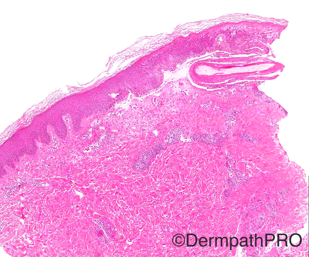
Case History: 19/M tense blisters on legs

Case posted by Dr Arti Bakshi


  Report Record

User Feedback


Raul Perret

Posted

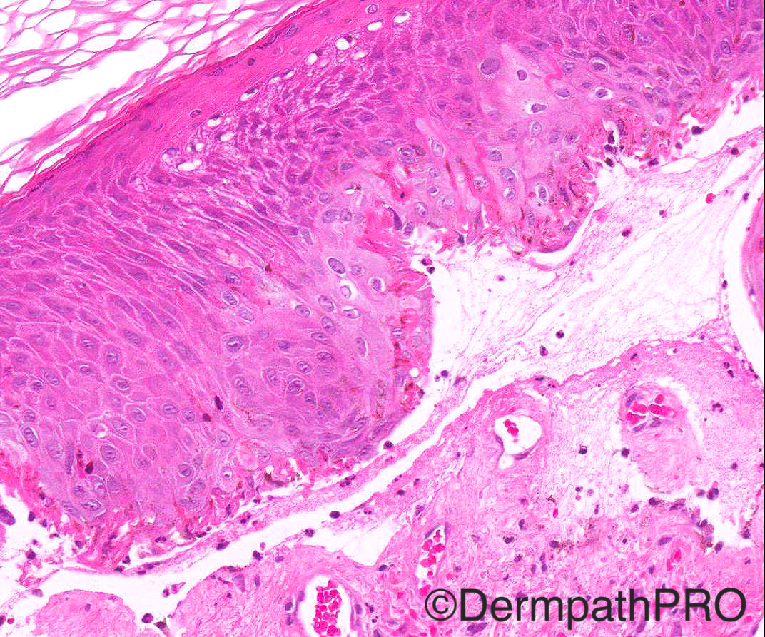
The images correspond to a cell poor subepidermal blíster with areas of spongiosis. Immunofluorescence is of course primary and the differential is wide but would have to think about epidermolysis bullosa, bullous LE (that can be cell poor), etc. Still the clinical setting in this case as well as vertical elongation of keratinocytes makes me consider also bullae developing after physical trauma like cryotherapy or burns

Share this comment


Link to comment
share_externally

Difficult case. I see a subepidermal blister with scattered inflammatory cells and my ddx is mostly EB and Porphyria cutenea with the eosinophilic bodies seen representing "caterpillar" bodies. DIF is necessary.

Share this comment


Link to comment
share_externally

Dr. Mona Abdel-Halim

Posted

Agree with all above suggestions, also will add diabetic bullae

Share this comment


Link to comment
share_externally

Arti Bakshi

Posted

Will add more clinical information: 4 day history of acute onset of blisters, not on any medications, otherwise fit and well. Not diabetic. Porphyrin screen negative. Direct IMF negative.
Hope that narrows the differential...

Share this comment


Link to comment
share_externally

Raul Perret

Posted

I dont favor bullous insect bite because in the clinical description is written legs (bilateral). I still think it could be a physical damage of skin like cryotherapy, burns, suction, etc (even if usually the epidermis is necrotic in these cases). There is also bullous amyloidosis which I dont favor. Any history of physical injury or therapy Arti?

Share this comment


Link to comment
share_externally

I once saw a case like this with necrotic keratinocytes caused by Munchausen syndrome. Another ddx.

Share this comment


Link to comment
share_externally

Arti Bakshi

Posted

This is a case of Bullous Dermatitis Artefacta, most likely due to thermal/cryo injury. Many of you were on the right track, so well done for that! Extra kudos to Raul for nailing the diagnosis even before the full clinical history and rightly observing the characteristic 'vertical elongation of keratinocytes' that I was trying to show in images 3 and 4. There is also superficial epidermal necrosis with loss of cytoplasmic detail in superficial keratinocytes. These features are indicative of thermal/cryo injury. (see reference below which describes remarkable similar histology in 2 patients with bullous DA). The patient had not had any medical procedure and the patient denied any trauma. When I discussed the case with the dermatologist, she felt the clinical presentation (large bizarre shaped blisters, acute onset, patient demeanour etc) were consistent with DA. Ofcourse, other differentials incl porphyria, pseudoporphyria and autoimmune bullous disorders needed to be excluded, which was done by appropriate tests.
[url="http://www.ncbi.nlm.nih.gov/pubmed/22771895"]http://www.ncbi.nlm.nih.gov/pubmed/22771895[/url]

Share this comment


Link to comment
share_externally

Dr. Richard Carr

Posted

Shame I did not have a go yesterday. The age and rather abrupt necrosis of the epidermis (upper left image), and lack of any bone fide pathology underneath shouts external factors. Good that the clinical colleagues were thinking likewise.

Share this comment


Link to comment
share_externally


Join the conversation

You can post now and register later. If you have an account, sign in now to post with your account.

Guest
Add a comment...

×   Pasted as rich text.   Paste as plain text instead

  Only 75 emoji are allowed.

×   Your link has been automatically embedded.   Display as a link instead

×   Your previous content has been restored.   Clear editor

×   You cannot paste images directly. Upload or insert images from URL.

Loading...

×
×
  • Create New...