Jump to content
In this section we have spot diagnoses posted on a daily basis since June 2010, now over 4000! You can review the archived cases and read the suggested diagnoses by users and the final comment by the contributors.
Case are uploaded each week day by 10 am UK time with the correct diagnosis will generally be posted at 8 pm UK time. Why not view the most recent spot diagnosis and proffer a diagnosis?

Case Number : Case 1382 - 09 October Posted By: Guest

Please read the clinical history and view the images by clicking on them before you proffer your diagnosis.
Submitted Date :
   (0 reviews)

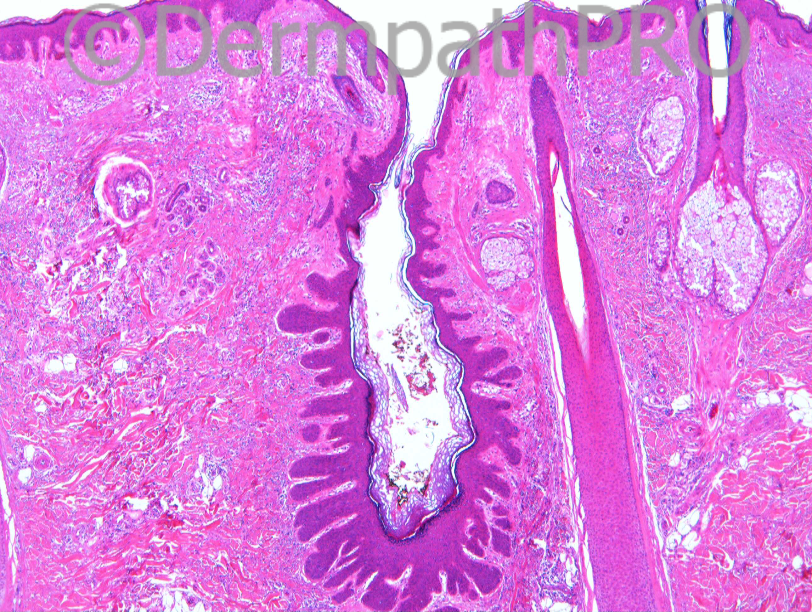
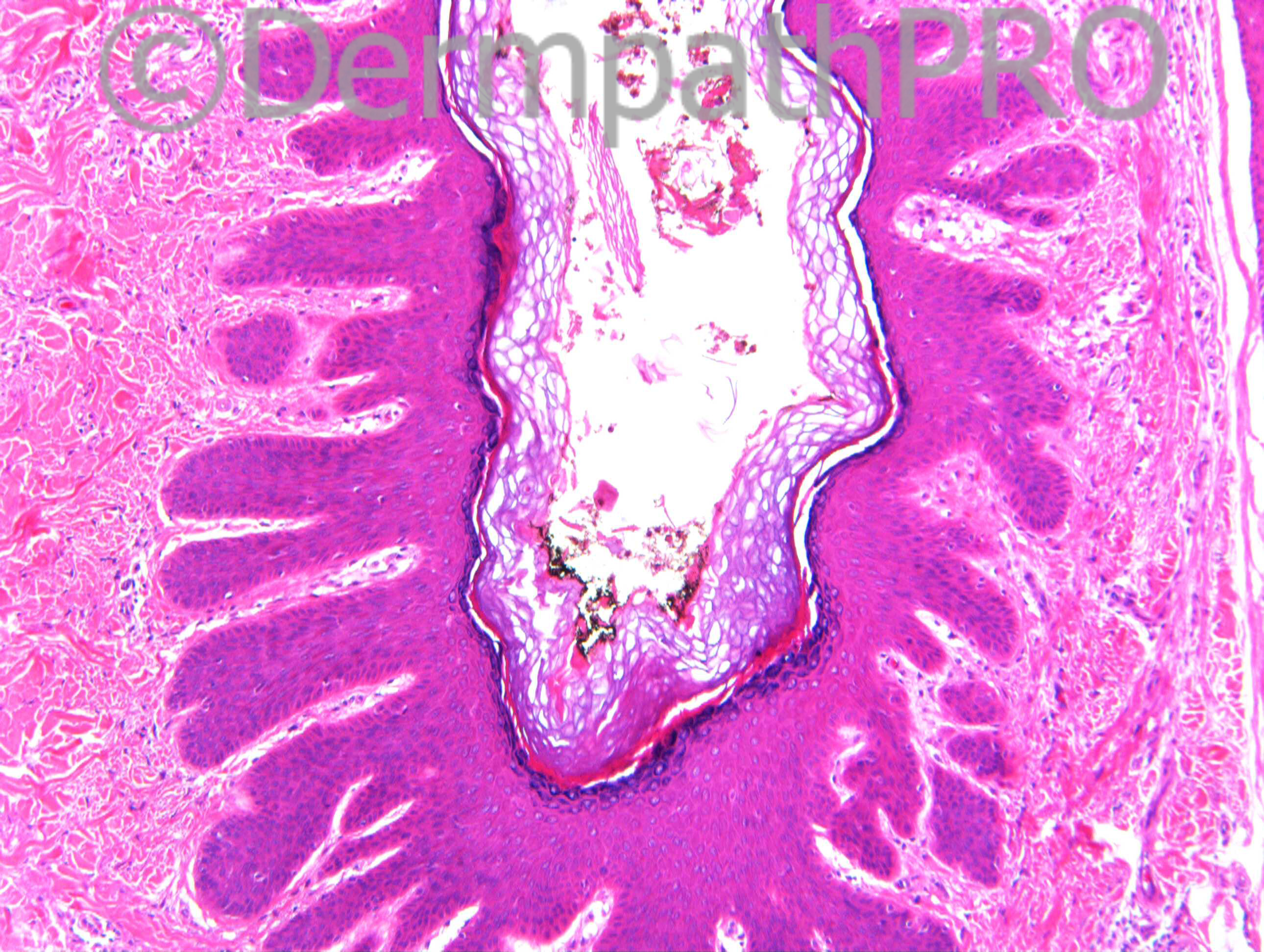
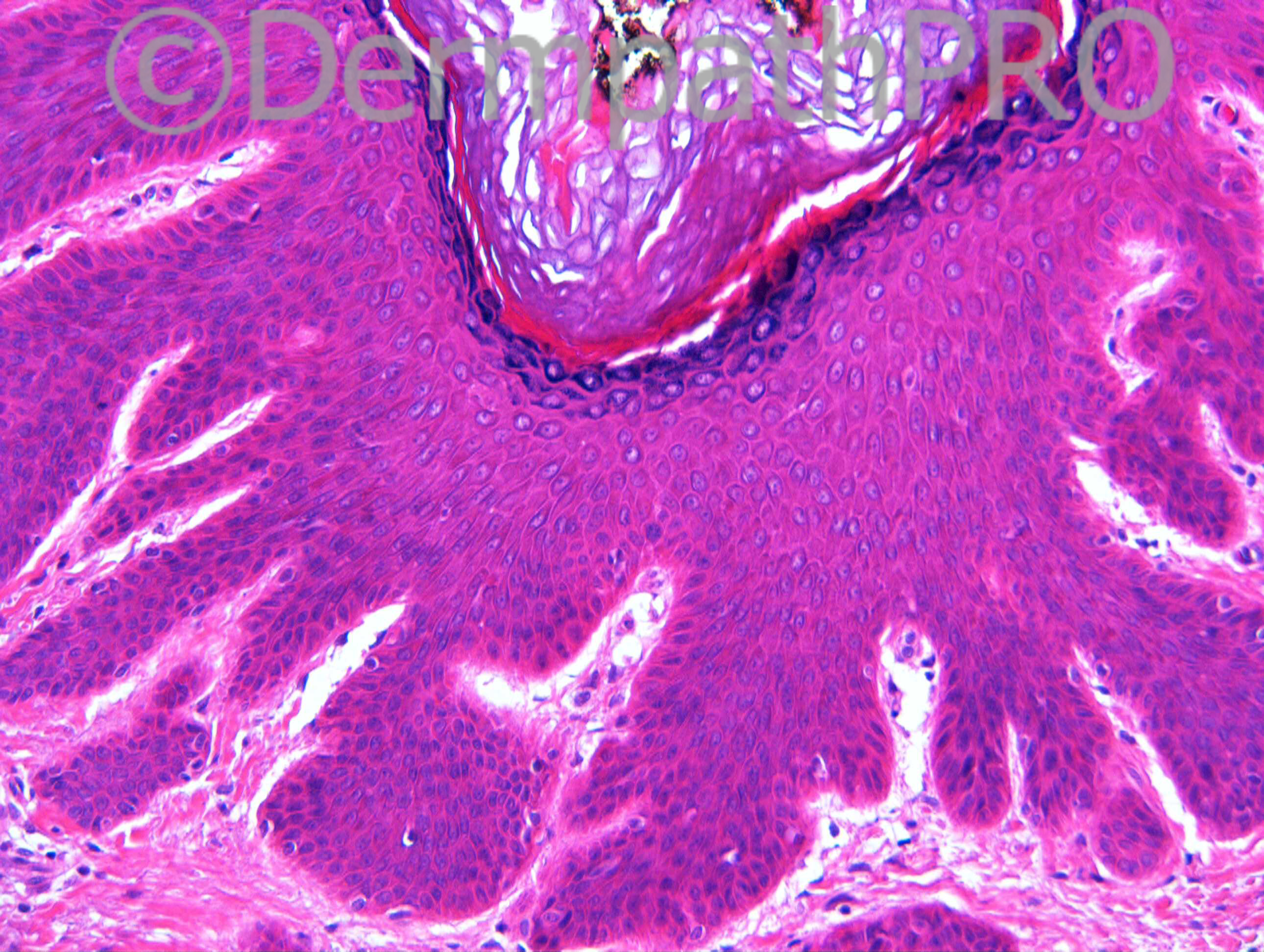
M50. Cyst on cheek

Case posted by Dr Richard Carr


  Report Record

User Feedback


Dr. Richard Carr

Posted

We will put up a few more images at 4pm today.

Share this comment


Link to comment
share_externally

Dr. Mona Abdel-Halim

Posted

Then I will wait for the rest of the images :-))

Share this comment


Link to comment
share_externally

Arti Bakshi

Posted

I thought there was a catch!!….lets see what turns up at 4.00pm!

Share this comment


Link to comment
share_externally

Dr. Richard Carr

Posted

[img]https://dermpathpro.com/uploads/spot_diagnosis_comment_img/Case%201382%20-%20RAC7276x10b_4pm.jpg[/img]

[img]https://dermpathpro.com/uploads/spot_diagnosis_comment_img/Case%201382%20-%20RAC7276x10c4pm.jpg[/img]

[img]https://dermpathpro.com/uploads/spot_diagnosis_comment_img/Case%201382%20-%20RAC7276x20b_4pm.jpg[/img]

[img]https://dermpathpro.com/uploads/spot_diagnosis_comment_img/Case%201382%20-%20RAC7276x2_4pm.jpg[/img]

[img]https://dermpathpro.com/uploads/spot_diagnosis_comment_img/Case%201382%20-%20RAC7276x2b_4pm.jpg[/img]

[img]https://dermpathpro.com/uploads/spot_diagnosis_comment_img/Case%201382%20-%20RAC7276x5b_4pm.jpg[/img]

Share this comment


Link to comment
share_externally

Arti Bakshi

Posted

Several hair follicular infundibula are involved with one area of rupture and granulomatous inflammation.
? Nevus comedonicus ( althoug clinical does not fit). This can show dilate pore type epithelial proliferation.

Share this comment


Link to comment
share_externally

Dr. Mona Abdel-Halim

Posted

I think the main pathology is that of a dilated pore of Winer, adjacent (to the left), yet away from the pore (unrelated to it), there is a focus of granulomatous inflammation that may be due to unrelated folliculitis and ruptured follicle.

Share this comment


Link to comment
share_externally

Arti Bakshi

Posted

Another possibility (to tie everything together) would be inflammatory cystic acne!
This would explain the inflammation, fibrosis and comedones (the latter showing Dilated pore of Winer type changes).

Share this comment


Link to comment
share_externally

Dr. Richard Carr

Posted

In Akerman's tome on follicular neoplasms I seem to remember him regarding dilated pore as a reaction within ruptured infundibular cysts and this opinion is also mentions in Kazakov. Some believe dilated pore of Winer is a neoplasm. I don't know the answer. Kazakov mentions a variant of naevus comedonicus with circumferential bulbous projections "dilated pore naevus". So is it an acneiform lesion with secondary changes (dilated pore) or is it dilated pore with secondary acneiform process? I don't know but thought you might like to see this one! I suppose if nearly every dilated pore has either a scar, redisual infundibular cyst or acneiform process (on adequate examination) then we might find the answer.

Share this comment


Link to comment
share_externally


Join the conversation

You can post now and register later. If you have an account, sign in now to post with your account.

Guest
Add a comment...

×   Pasted as rich text.   Paste as plain text instead

  Only 75 emoji are allowed.

×   Your link has been automatically embedded.   Display as a link instead

×   Your previous content has been restored.   Clear editor

×   You cannot paste images directly. Upload or insert images from URL.

Loading...

×
×
  • Create New...