Jump to content
In this section we have spot diagnoses posted on a daily basis since June 2010, now over 4000! You can review the archived cases and read the suggested diagnoses by users and the final comment by the contributors.
Case are uploaded each week day by 10 am UK time with the correct diagnosis will generally be posted at 8 pm UK time. Why not view the most recent spot diagnosis and proffer a diagnosis?

Case Number : Case 1389 - 20 October Posted By: Guest

Please read the clinical history and view the images by clicking on them before you proffer your diagnosis.
Submitted Date :
   (0 reviews)

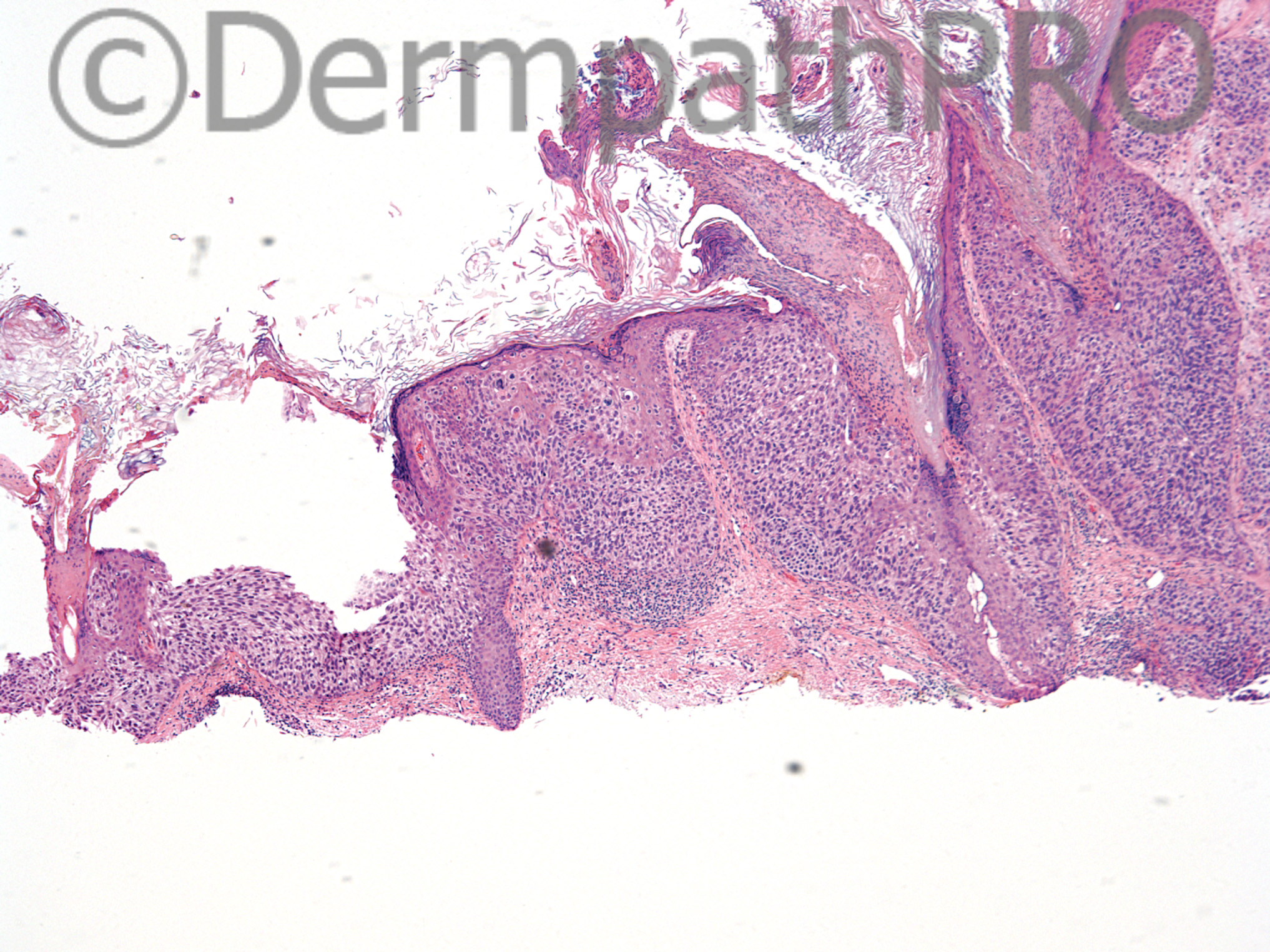
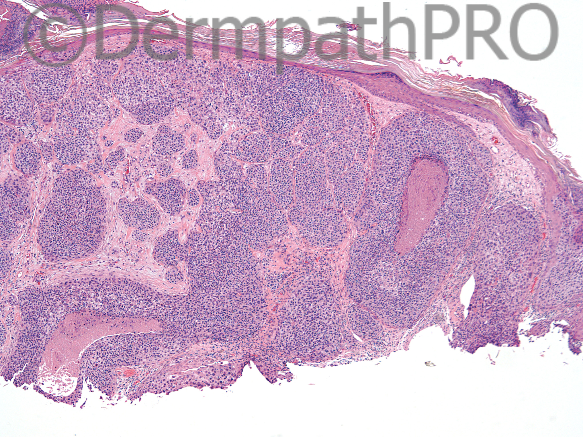
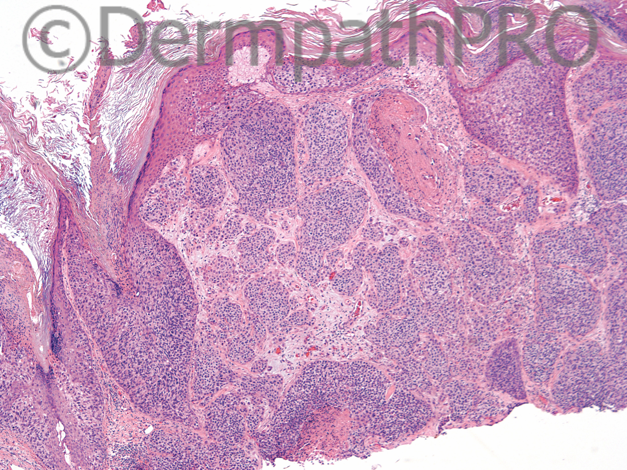
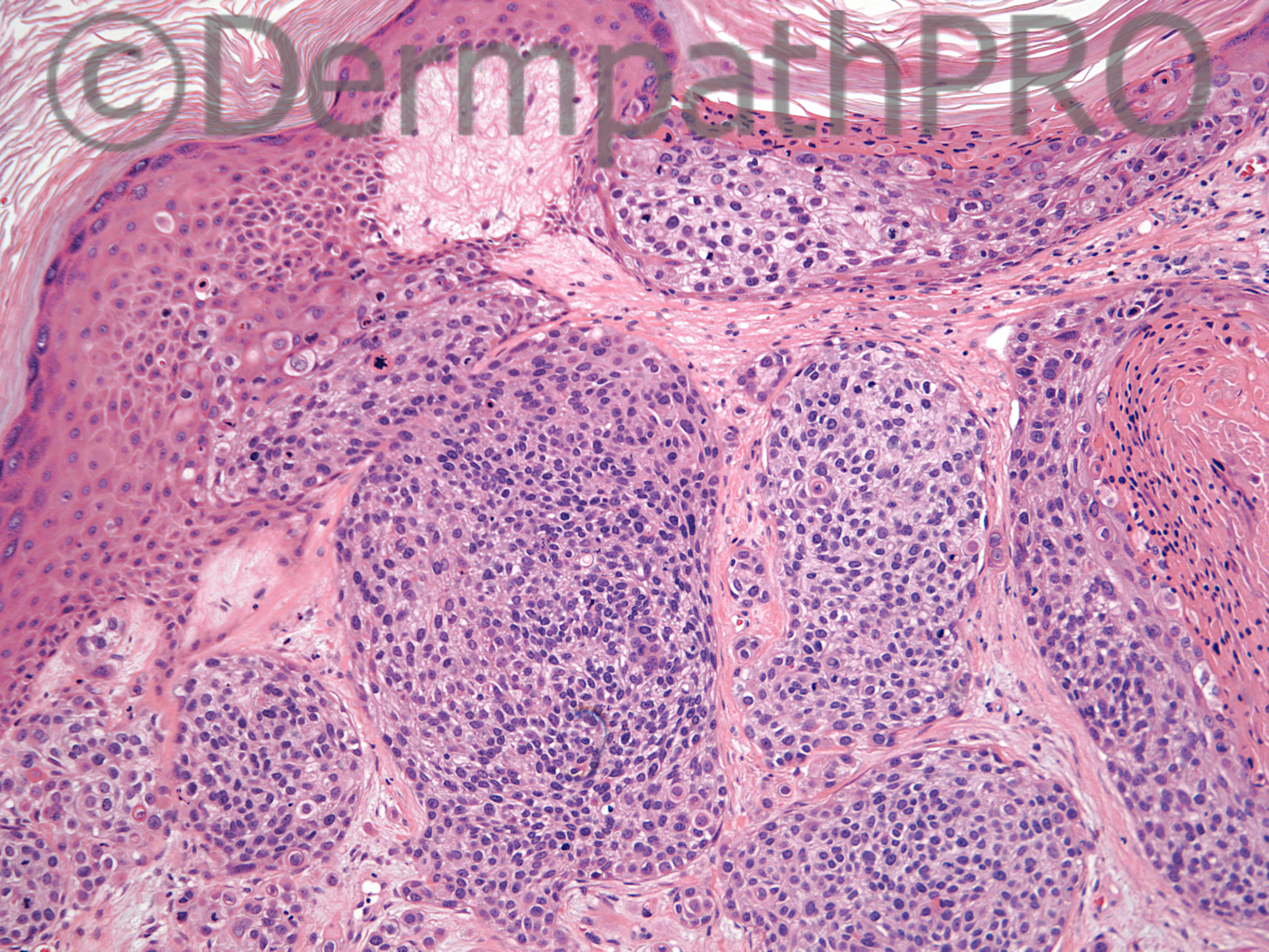
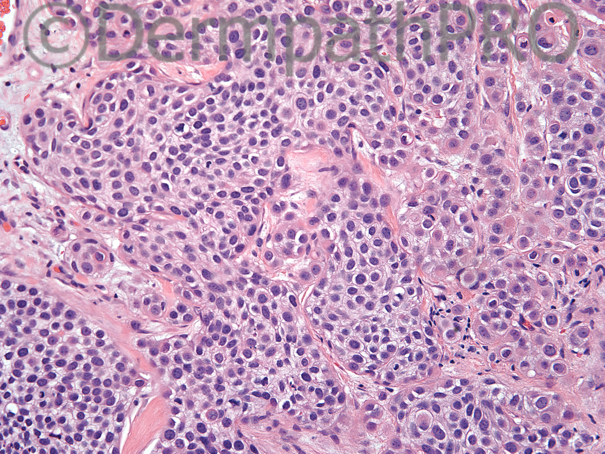
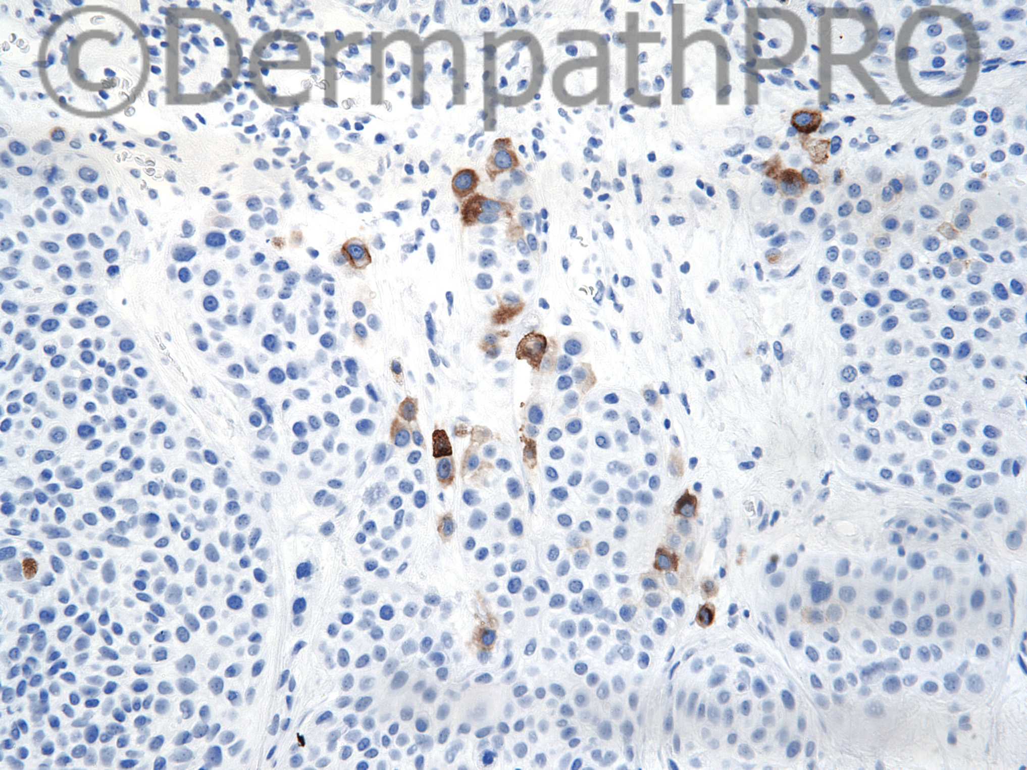
99 year old female with erythematous crusted plaque on left cheek. Rule out SCC vs amelanotic melanoma. Figure 6=monoclonal CEA.

Case posted by Dr Uma Sundram


  Report Record

User Feedback


Guest Romualdo

Posted

CEA is not highlighting ducts or intracytoplasmic lumens. I think positive cells are plasma cells. My suggestion is epidermotropic metastatic carcinoma, possibly from the breast.

Share this comment


Link to comment
share_externally

Dr. Mona Abdel-Halim

Posted

Agree with Romualdo that CEA here is not showing typical pattern of highlighting ducts/epithelial linings. I consider it irrelevant. I believe a wide panel has been done. Waiting for more information from Uma !! Actually, based on morphology, I am highly suspecting melanoma (amelanotic). I have read about CEA positivity in metastatic melanoma but all mentioned polyclonal CEA positivity not monoclonal !! Will love to know the correct diagnosis.

Share this comment


Link to comment
share_externally

Arti Bakshi

Posted

Interesting range of diagnoses!..so will add a few more for variety's sake.
An invasive bowenoid porocarcinoma certainly crossed my mind, but agree that there is no clear ductal differentiation and CEA is not typical.
To my eye, the tumour appears to be arising from the infundibular epithelium (image 1) and shows trichilemmal keratinisation. The epidermal involvement seems too widespread for an epidermotropic mets (but good thought!). Not convinced of melanocytic.
I would favour an invasive poorly differemtiated folllicular scc/trichilemmal carcinoma.

Share this comment


Link to comment
share_externally

Dr. Mona Abdel-Halim

Posted

Looking every now and then on the images, just got this thought: what about undifferentiated extraoccular sebaceous carcinoma with epidermotropic spread??

Share this comment


Link to comment
share_externally

Guest Romualdo

Posted

I think it is a good idea, although I cannot see nuclear identation by cytoplasmic lipid vacuoles. If this is really an undifferentiated sebaceous carcinoma, maybe this undifferentiation is the reason nuclear identation is not seen.

Share this comment


Link to comment
share_externally

Uma Sundram

Posted

My apologies to everyone, I just moved from california to michigan and had nothing else on my mind for a few days! Just realized I never posted the answer to this case. I went with trichilemmal carcinoma but all of these crossed my mind as well. EMA and adipophilin negative and all melanocytic markers are negative. pan cytokeratin positive. Patient had a full work up with no involvement elsewhere. Great ddx!

Share this comment


Link to comment
share_externally


Join the conversation

You can post now and register later. If you have an account, sign in now to post with your account.

Guest
Add a comment...

×   Pasted as rich text.   Paste as plain text instead

  Only 75 emoji are allowed.

×   Your link has been automatically embedded.   Display as a link instead

×   Your previous content has been restored.   Clear editor

×   You cannot paste images directly. Upload or insert images from URL.

Loading...

×
×
  • Create New...