Jump to content
In this section we have spot diagnoses posted on a daily basis since June 2010, now over 4000! You can review the archived cases and read the suggested diagnoses by users and the final comment by the contributors.
Case are uploaded each week day by 10 am UK time with the correct diagnosis will generally be posted at 8 pm UK time. Why not view the most recent spot diagnosis and proffer a diagnosis?

Case Number : Case 1392- 23 October Posted By: Guest

Please read the clinical history and view the images by clicking on them before you proffer your diagnosis.
Submitted Date :
   (0 reviews)

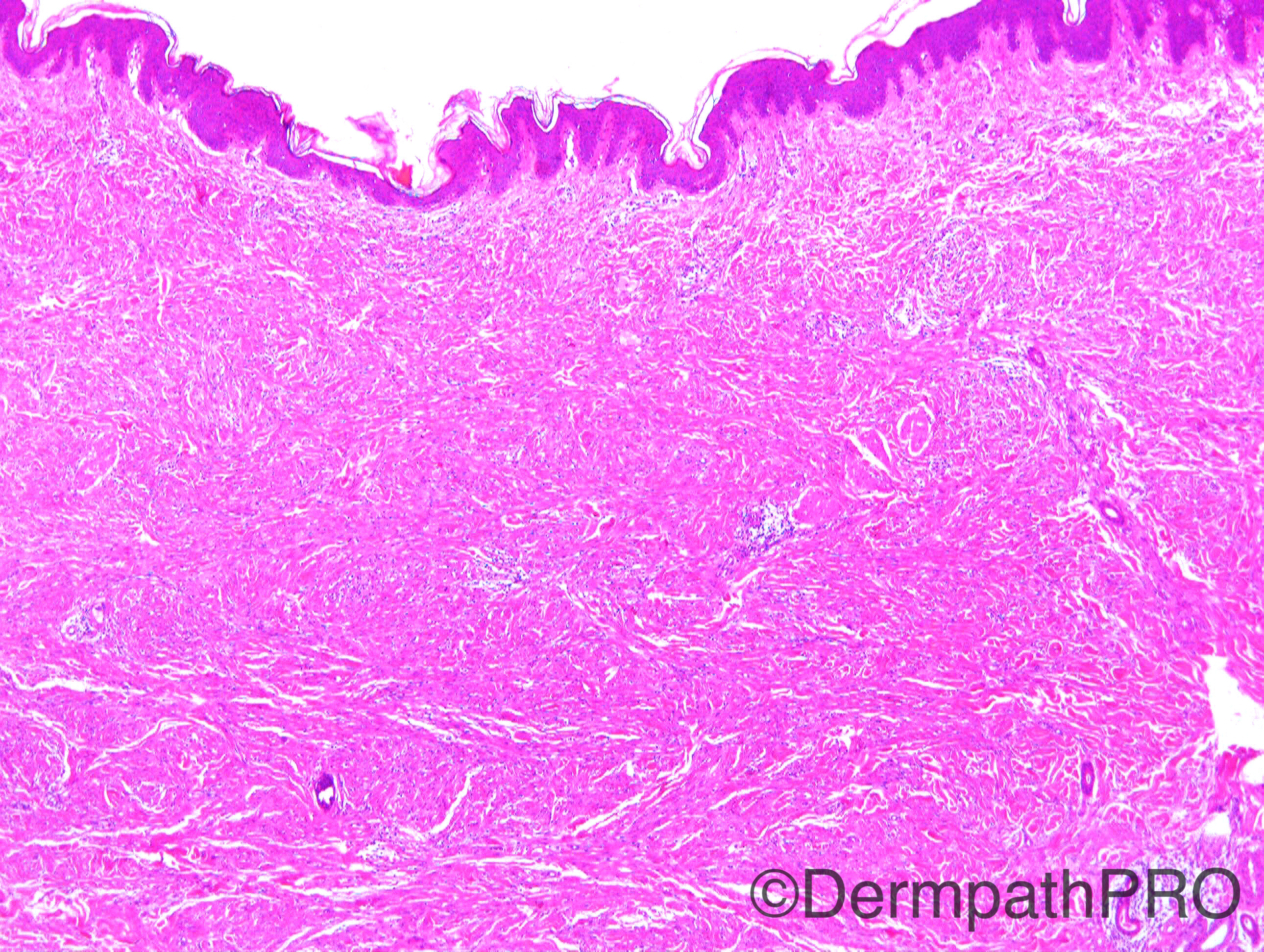
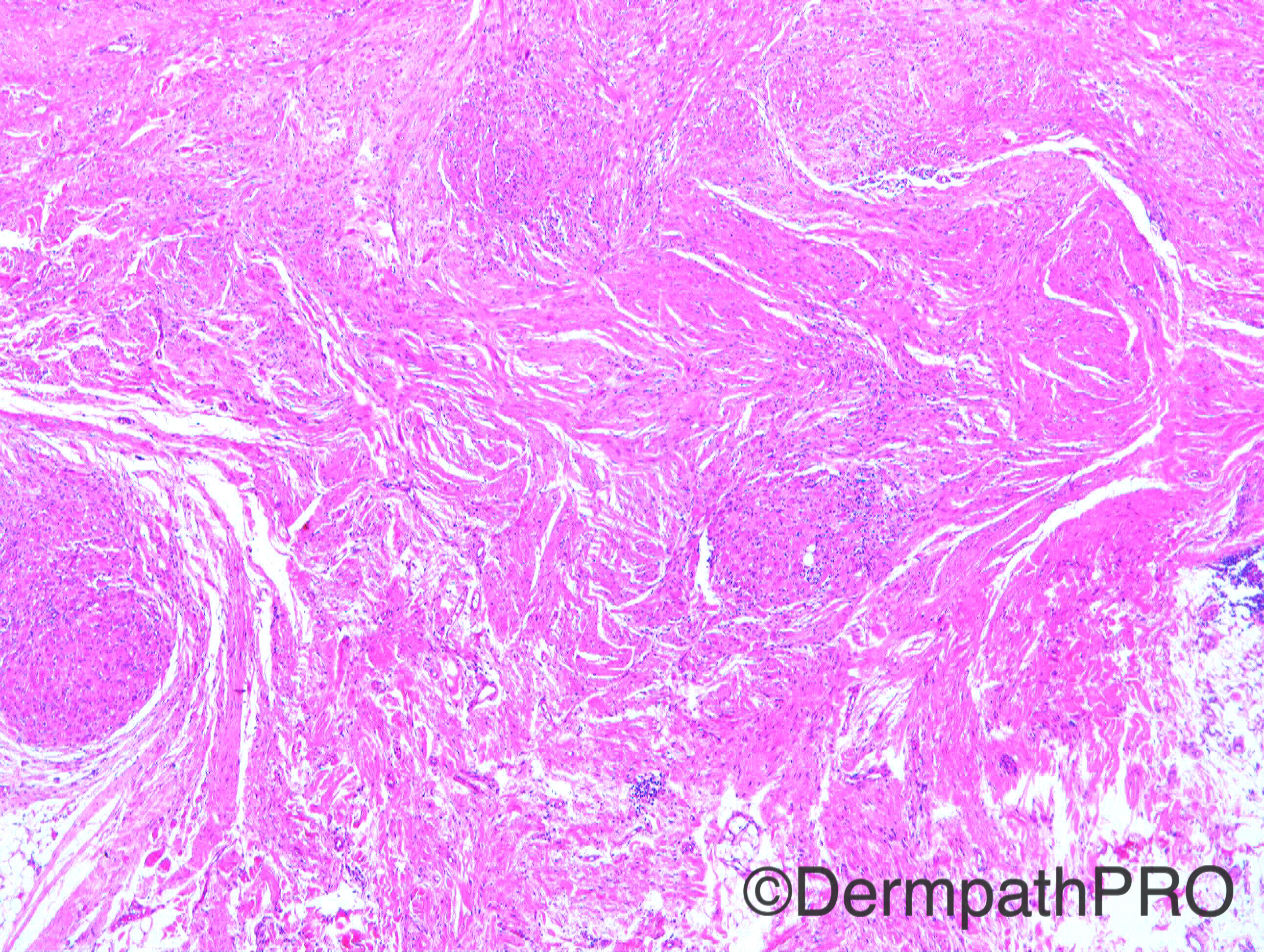
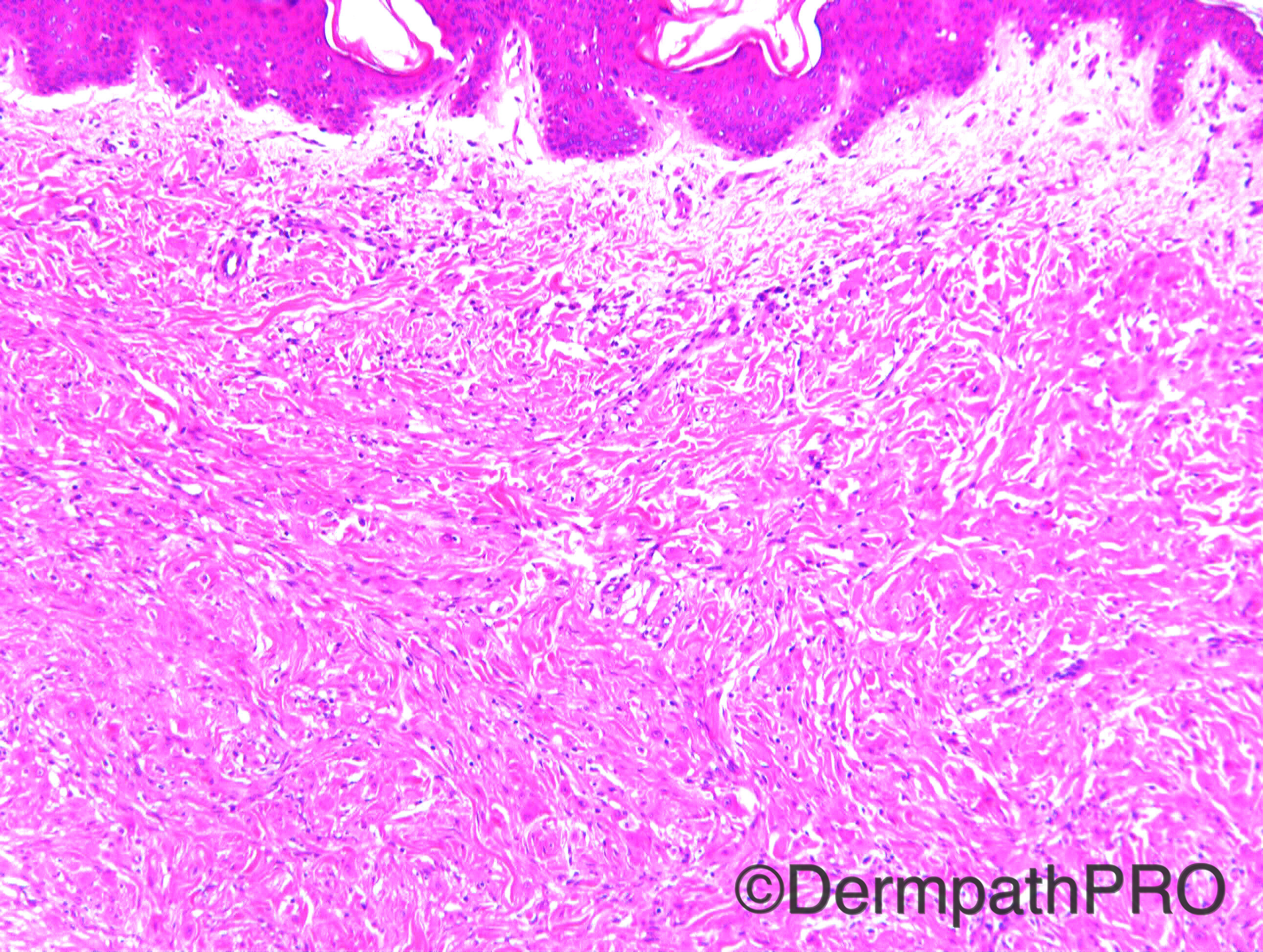
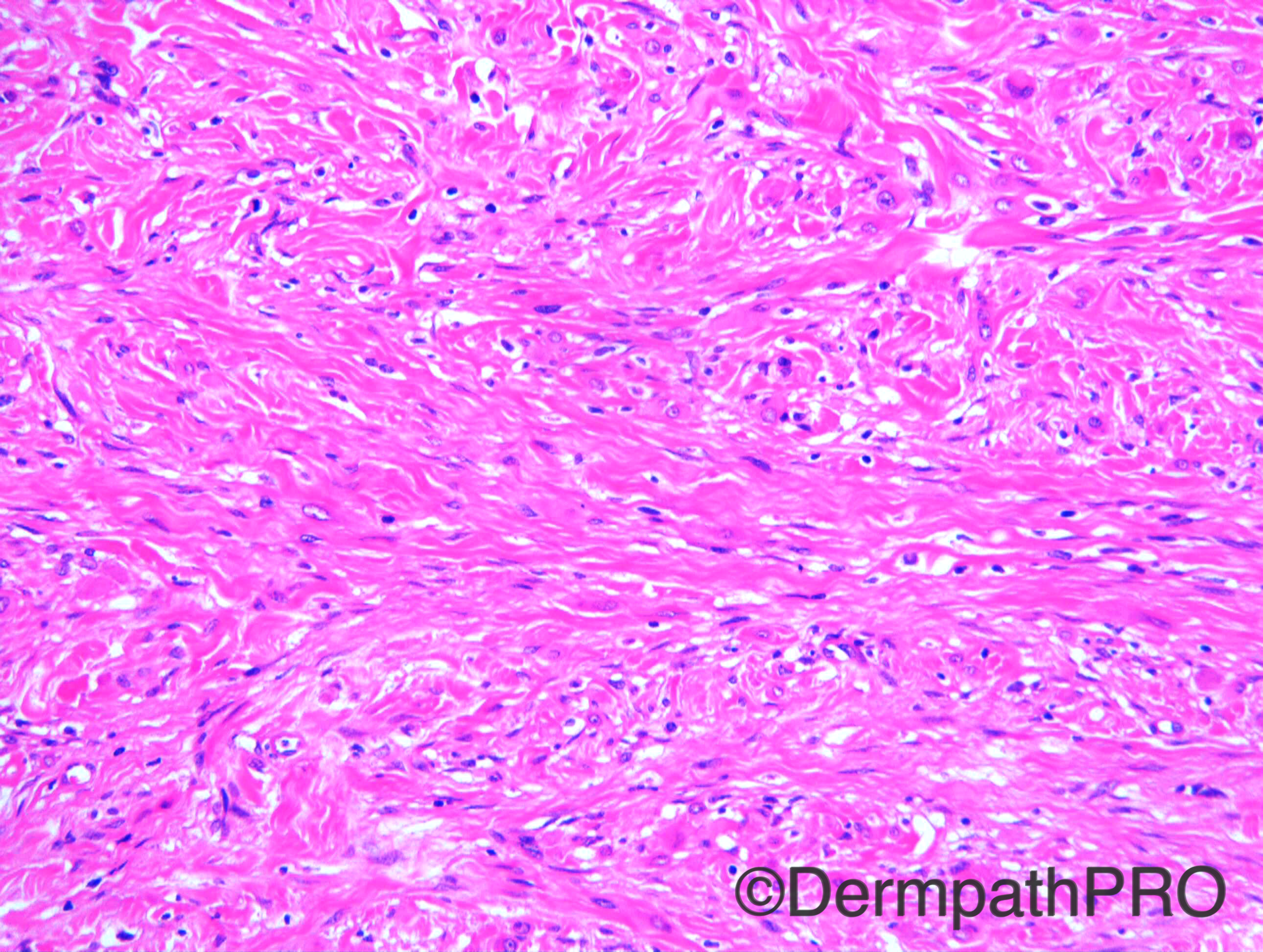
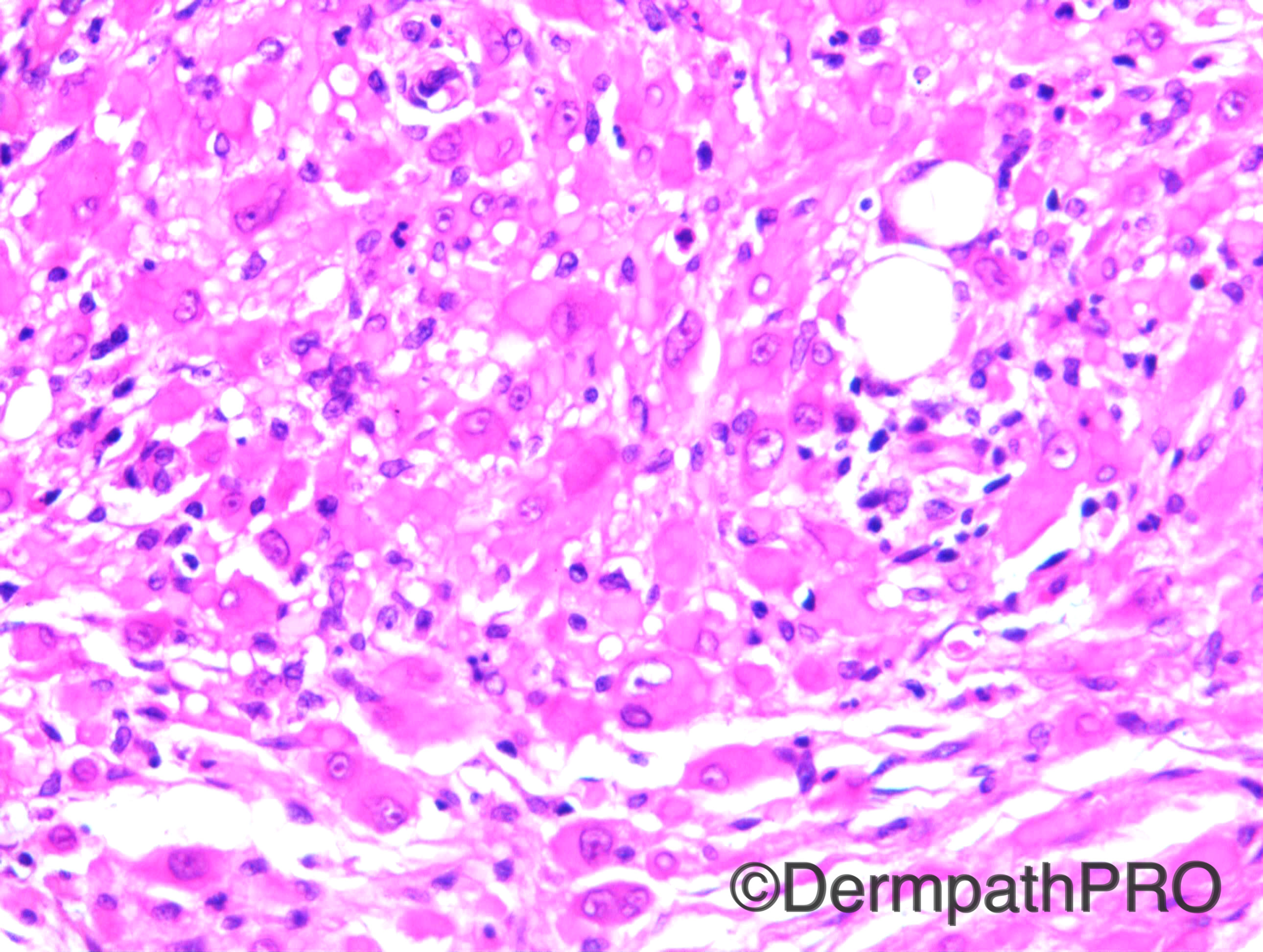
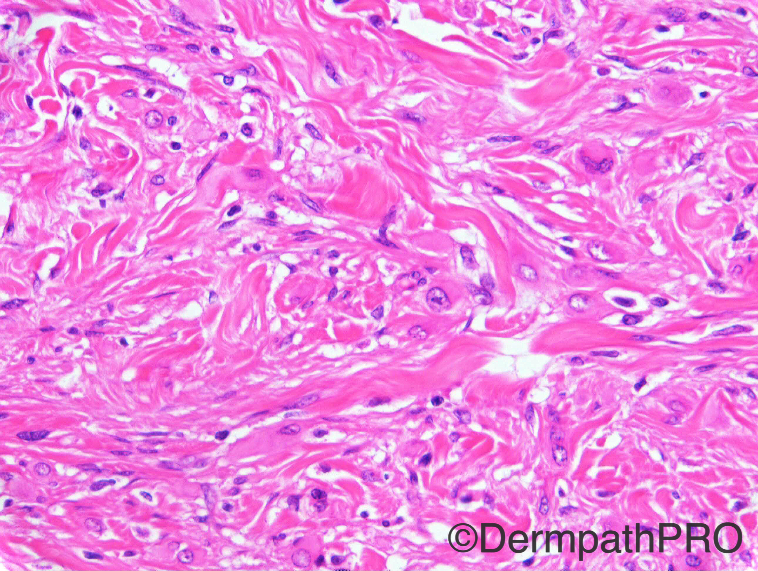
Case History: M20. Multiple subcutaneous lumps, on flank. Case c/o Dr Arti Baskhi

Case posted by Dr Richard Carr

  Report Record

User Feedback


Dr. Mona Abdel-Halim

Posted

Thinking of ? Plexiform fibrohistiocytic tumor

Share this comment


Link to comment
share_externally

Rhabdomyoma ?? rhabdomyomatous mesenchymal hamartoma ??

Share this comment


Link to comment
share_externally

Guest Romualdo

Posted

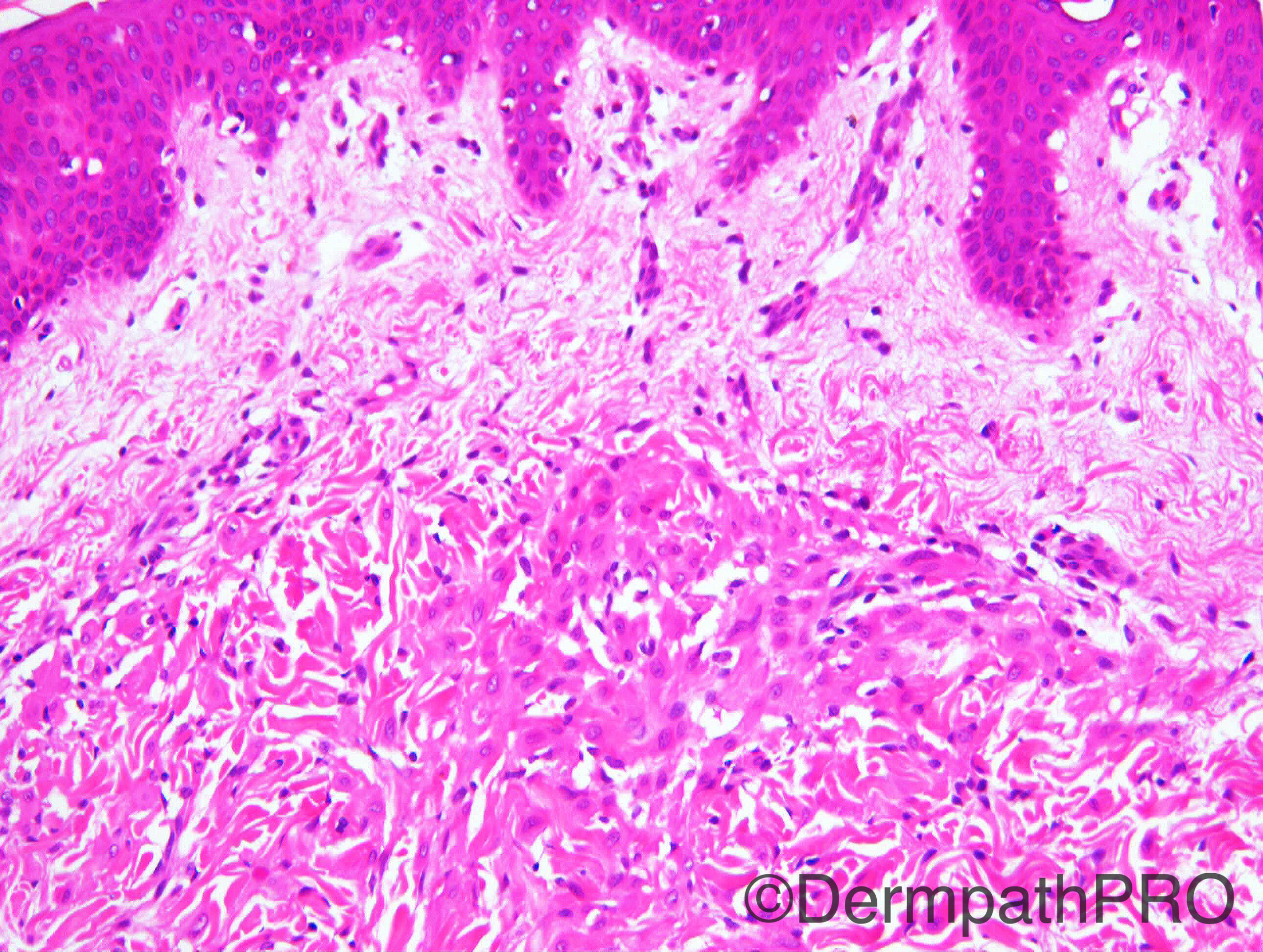
Infiltrative neoplasm with predominant fascicular pattern. Large round/ oval/ spindle cells with abundant brightly eosinophilic cytoplasm resembling rhabdomyoblasts with vesicular nuclei without significant atypia and mitotic figures. Sparse neutrophils are also seen. Young age and multiple lesions concentrated at one anatomic region are also typical of pseudomyogenic hemangioendothelioma, to IHC confirmation.

Share this comment


Link to comment
share_externally

Dr. Mona Abdel-Halim

Posted

Good suggestion Romualdo :-)
Today I am learning from you :-)

Share this comment


Link to comment
share_externally

Dr. Richard Carr

Posted

Another great case from Arti who I am sure can give the answer in due course.

Share this comment


Link to comment
share_externally

Arti Bakshi

Posted

Romualdo's description and diagnosis is spot on! Pretty impressive that you can diagnose this (rather rare) entity on a few images, when it took me quite some time (and many immunos) to crack it, along with the help of my soft tissue colleagues.
Plexiform fibrohistiocytic tumour is very good differential, which we considered as well, however multinucleate cells were lacking and the immunos did not fit.
The clue to the diagnosis of pseudomyogenic haemangioendothlioma lies in the 'rhabdomyoblast like cells' (which also give its name), well demonstrated in the last 2 images.
Immunos confirmed the endothelial nature of the tumour (CD 31 and ERG positive). Desmin, myoD1, myogenin were negative. The tumor is expected to show diffuse cytokeratin positivity but this was disappointing in this case. We tried quite a few cytokeratins and there was only patchy positivity with AE1/3. There was no loss of INI1. The clinical presentation is typical with multicentric spread.
Thanks to Richard for sharing this case on dermpathpro and thank you all for you comments!

Share this comment


Link to comment
share_externally


Join the conversation

You can post now and register later. If you have an account, sign in now to post with your account.

Guest
Add a comment...

×   Pasted as rich text.   Paste as plain text instead

  Only 75 emoji are allowed.

×   Your link has been automatically embedded.   Display as a link instead

×   Your previous content has been restored.   Clear editor

×   You cannot paste images directly. Upload or insert images from URL.

Loading...

×
×
  • Create New...