Jump to content
In this section we have spot diagnoses posted on a daily basis since June 2010, now over 4000! You can review the archived cases and read the suggested diagnoses by users and the final comment by the contributors.
Case are uploaded each week day by 10 am UK time with the correct diagnosis will generally be posted at 8 pm UK time. Why not view the most recent spot diagnosis and proffer a diagnosis?

Case Number : Case 1359 - 08 September Posted By: Guest

Please read the clinical history and view the images by clicking on them before you proffer your diagnosis.
Submitted Date :
   (0 reviews)

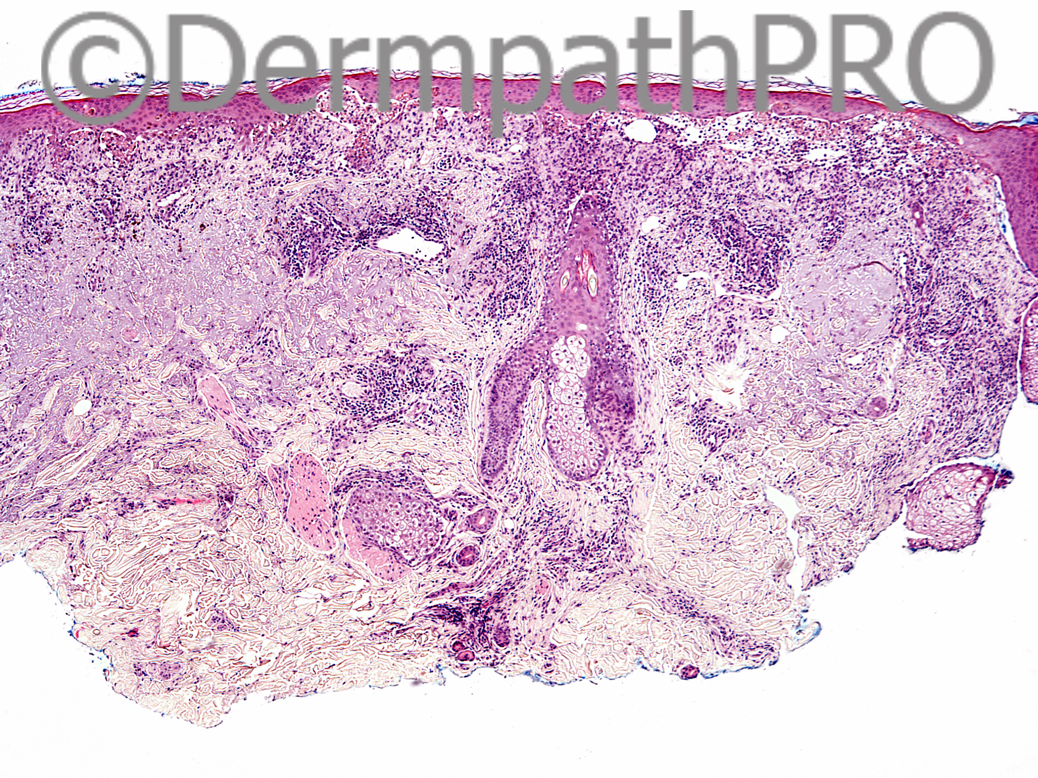
63 year old male with 4 blisters that show up at the same spot 1-2 times a year. Itchy. No changes in medications. Biopsy of left temple.

Case posted by Dr Uma Sundram


  Report Record

User Feedback


Dr. Mona Abdel-Halim

Posted

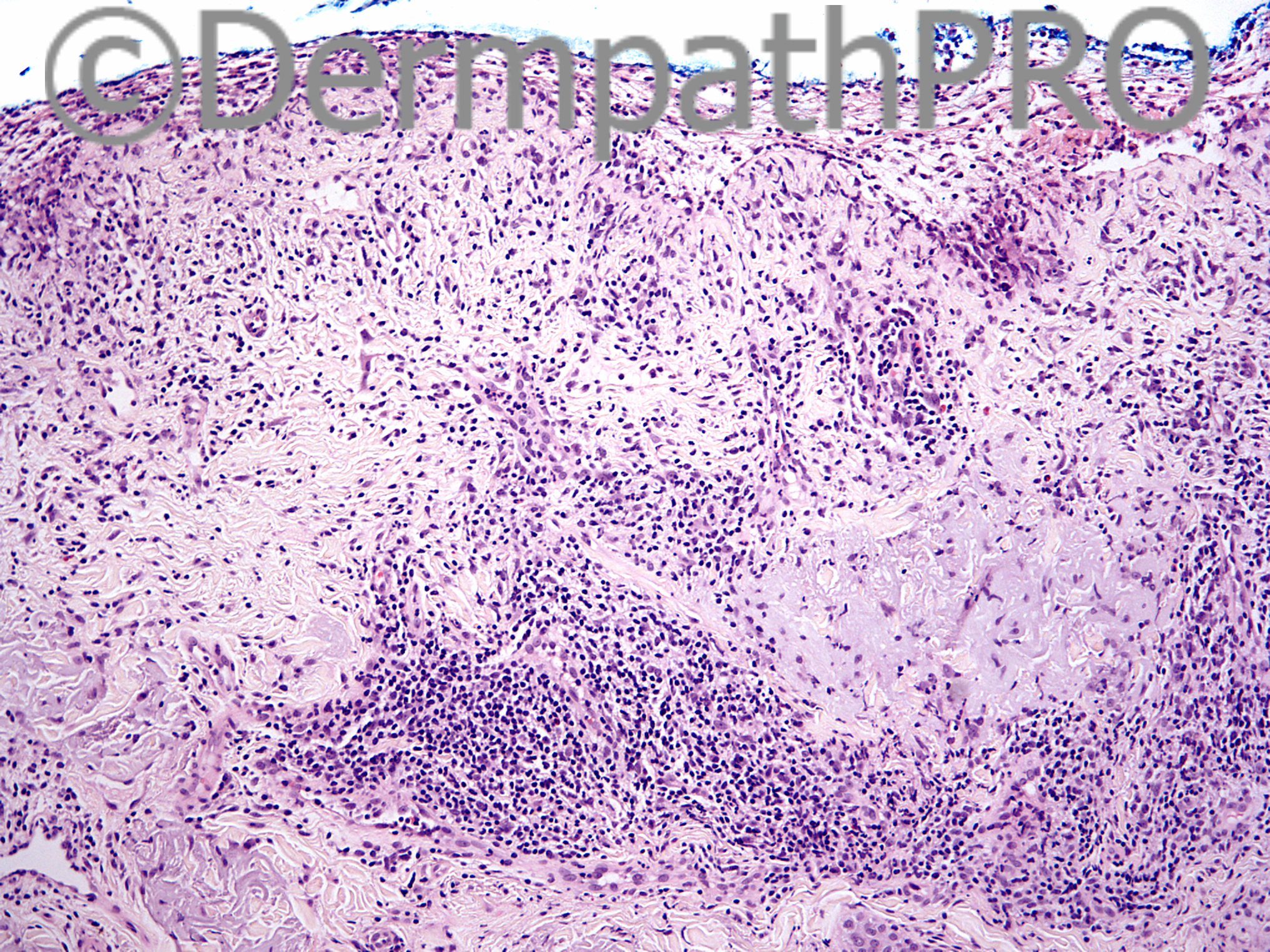
The pathological picture including necrotic KCs along DEJ as well as through all epidermal levels (up to complete necrosis), together with a mixed lymphohistiocytic infiltrate with few esinophils and neutrophils and few melanophages in addition to the deep extension of the infiltrate together with the clinical description of recurring blisters in sun exposed sites once or twice a year with no change in medications make me favour a form of phototoxic reaction. Detailed history can help. May be he is exposed to some plant materials on seasonal backgrounds (once or twice a year!!!!)... May be !!!
A bullous fixed drug reaction can mimic this pathologically, but would have expected more melanophages as pigmentation starts to develop early in these lesions, and with no changes in medications and with the pattern described for the recurrence once or twice yearly, I think this is not the case.

Share this comment


Link to comment
share_externally

Arti Bakshi

Posted

Good possibility, Mona.
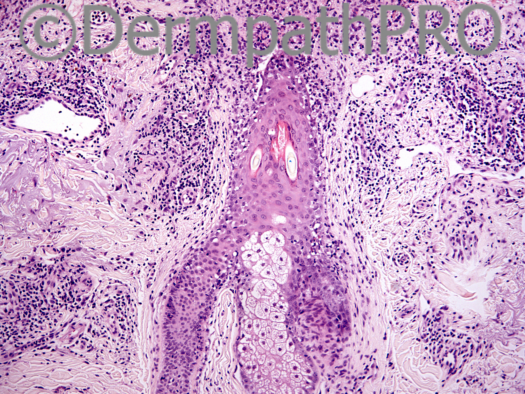
There are certainly necrotic keratinocytes all throughout, but most prominent at the DE junction. This suggests a predominant interface/lichenoid tissue reaction pattern. This is also seen around hair follicular epithelium. Would be unusual to get such an interface pattern with phototoxic dermatitis, but possible!
What about a recurrent erythema multiforme (possibly to herpes), although inflammation is quite deep.
Or a lichenoid contact dermatitis to something else??

Share this comment


Link to comment
share_externally

Guest Romualdo

Posted

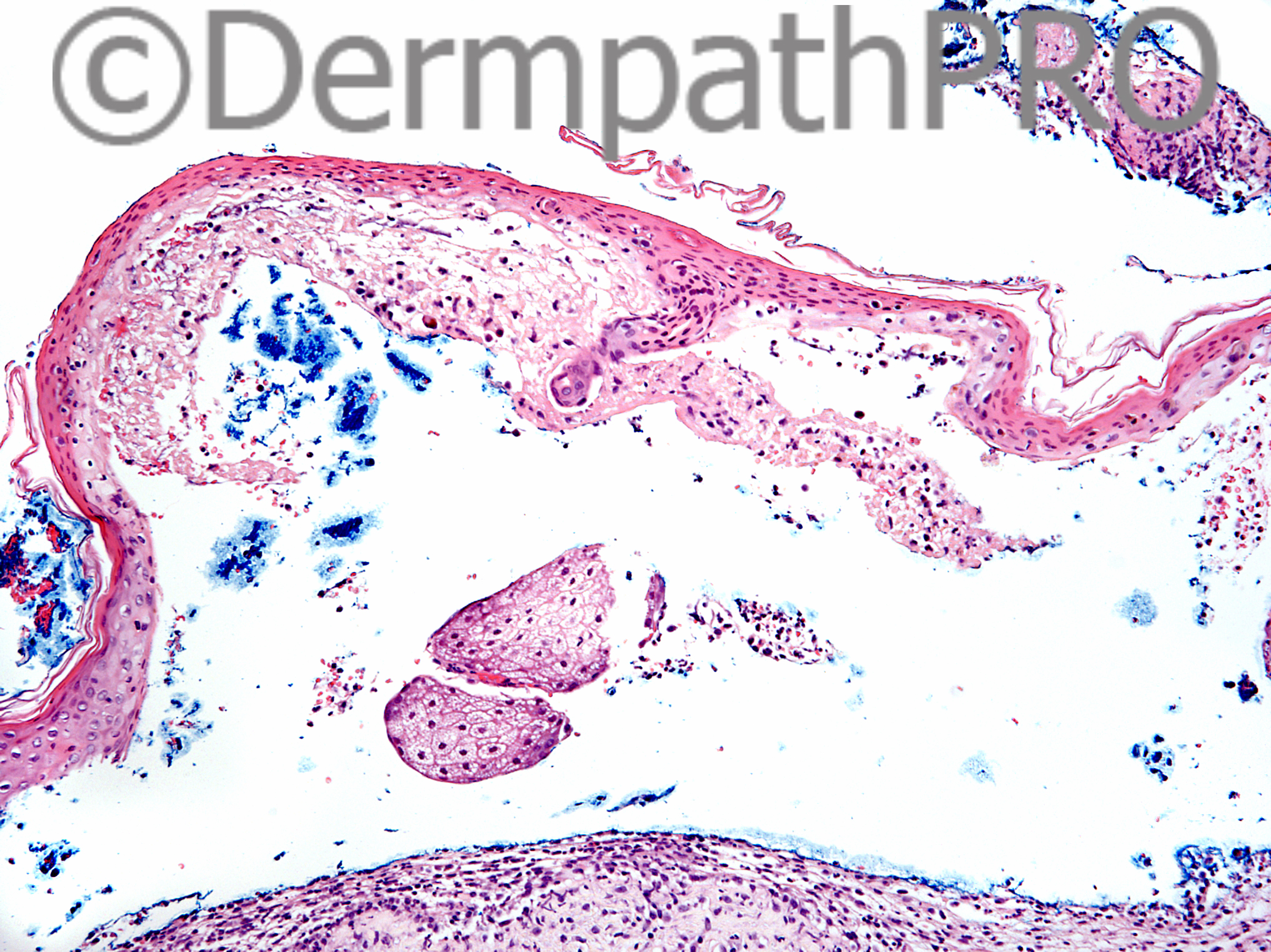
I don't know how to explain the presence of necrotic keratinocytes but I see upper dermal scarring and, most inportantly, sebaceous lobules apparently "floating" in a subepidermal blister, a typical finding of cicatricial pemphigoid.

Share this comment


Link to comment
share_externally

Dr. Richard Carr

Posted

I like the idea of an erythema multiforme-like fixed drug eruption. There are some case reports.

Share this comment


Link to comment
share_externally

Uma Sundram

Posted

Difficult case but this is indeed an example of erythema multiforme like fixed drug eruption. Patient has a known defined reaction to acetaminophen. DIF is negative and the reaction is very localized. No known photo-related activity or known contactants.

Share this comment


Link to comment
share_externally


Join the conversation

You can post now and register later. If you have an account, sign in now to post with your account.

Guest
Add a comment...

×   Pasted as rich text.   Paste as plain text instead

  Only 75 emoji are allowed.

×   Your link has been automatically embedded.   Display as a link instead

×   Your previous content has been restored.   Clear editor

×   You cannot paste images directly. Upload or insert images from URL.

Loading...

×
×
  • Create New...