Jump to content
In this section we have spot diagnoses posted on a daily basis since June 2010, now over 4000! You can review the archived cases and read the suggested diagnoses by users and the final comment by the contributors.
Case are uploaded each week day by 10 am UK time with the correct diagnosis will generally be posted at 8 pm UK time. Why not view the most recent spot diagnosis and proffer a diagnosis?

Case Number : Case 1362 - 11 September Posted By: Guest

Please read the clinical history and view the images by clicking on them before you proffer your diagnosis.
Submitted Date :
   (0 reviews)

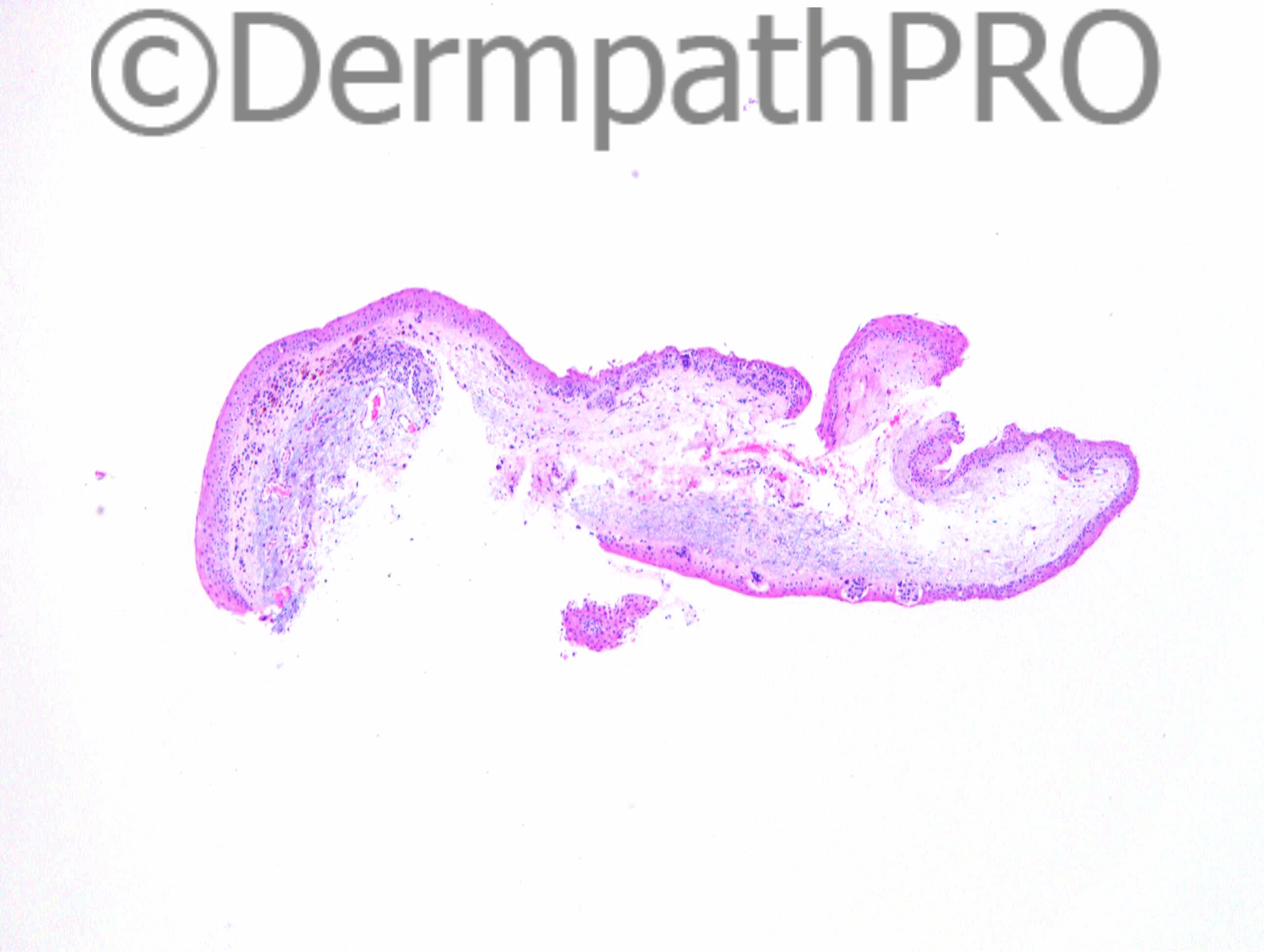
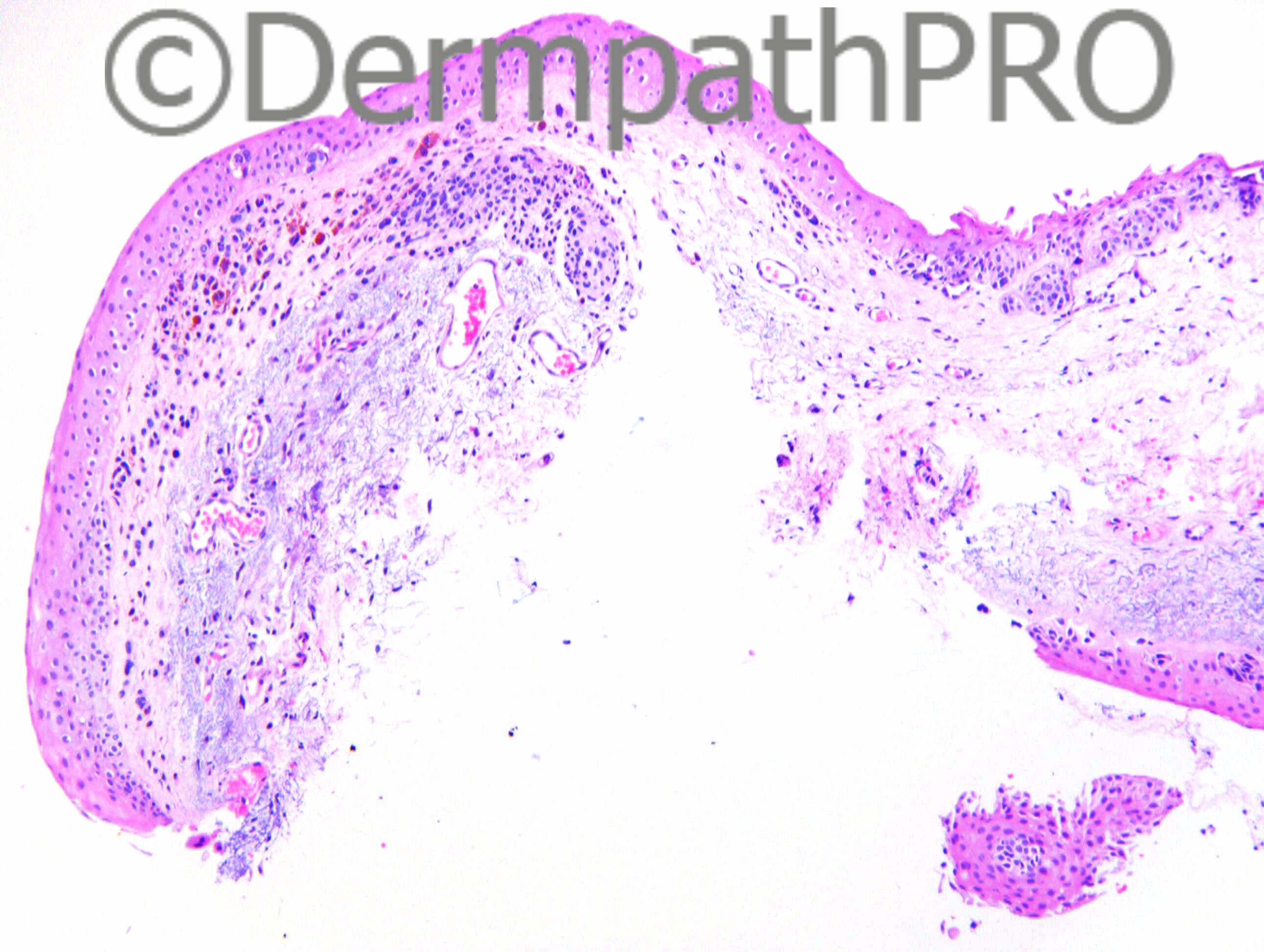
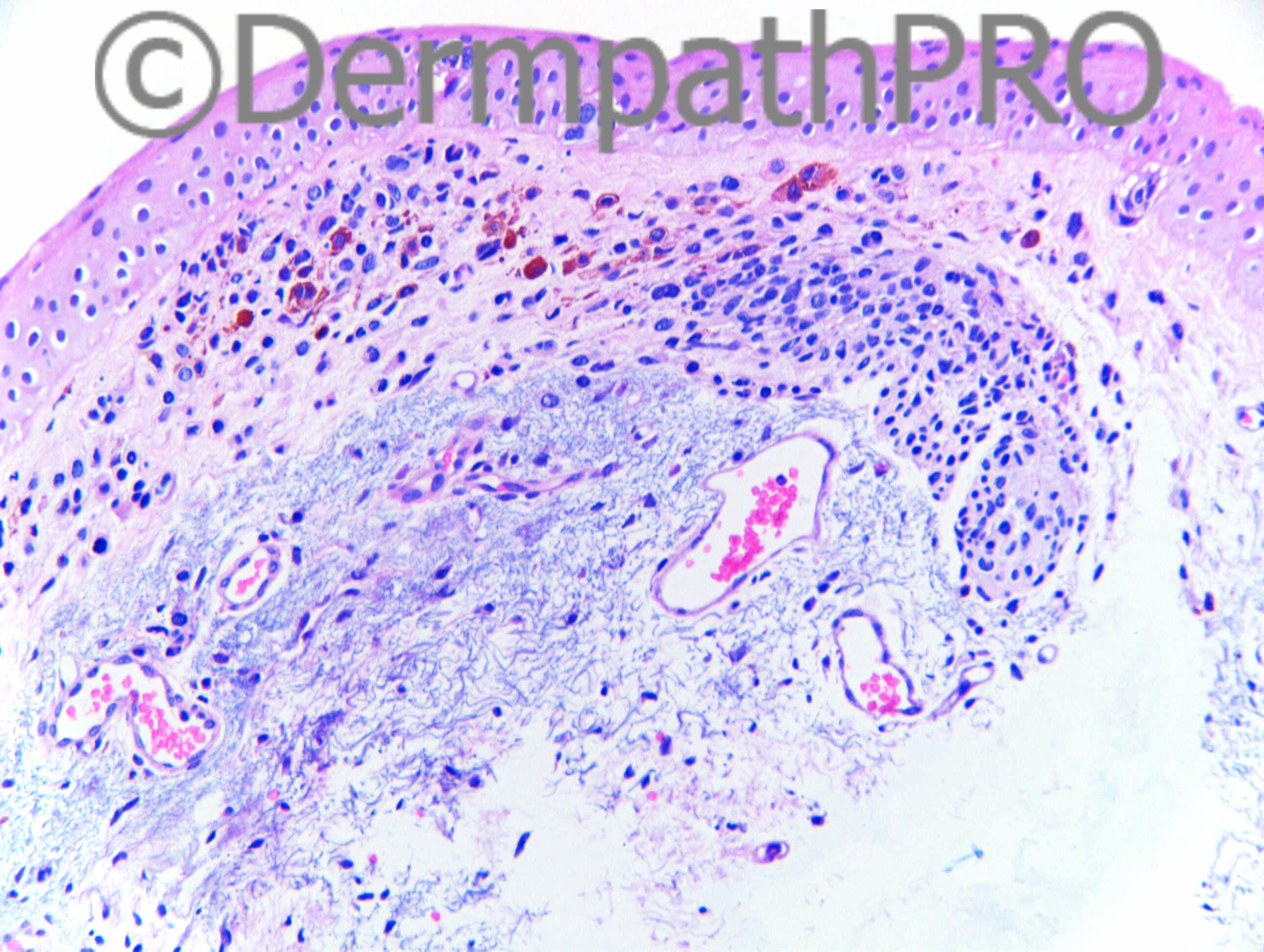
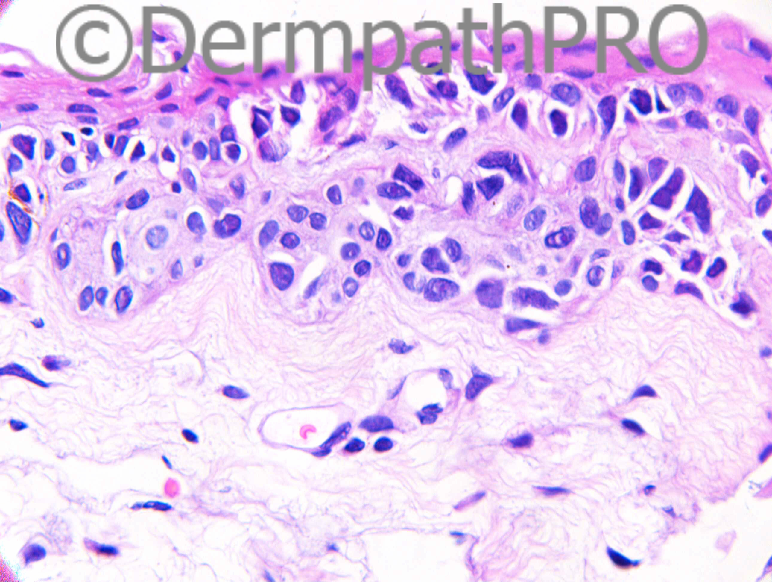
F25. Right sided primary acquired melanosis / pigmentation at 7 o'clock on bulbar conjunctiva.

Case posted by Dr Richard Carr


  Report Record

User Feedback


Dr. Mona Abdel-Halim

Posted

Very difficult with no experience in conjunctival melanocytic lesions !!!!!!
But will try..

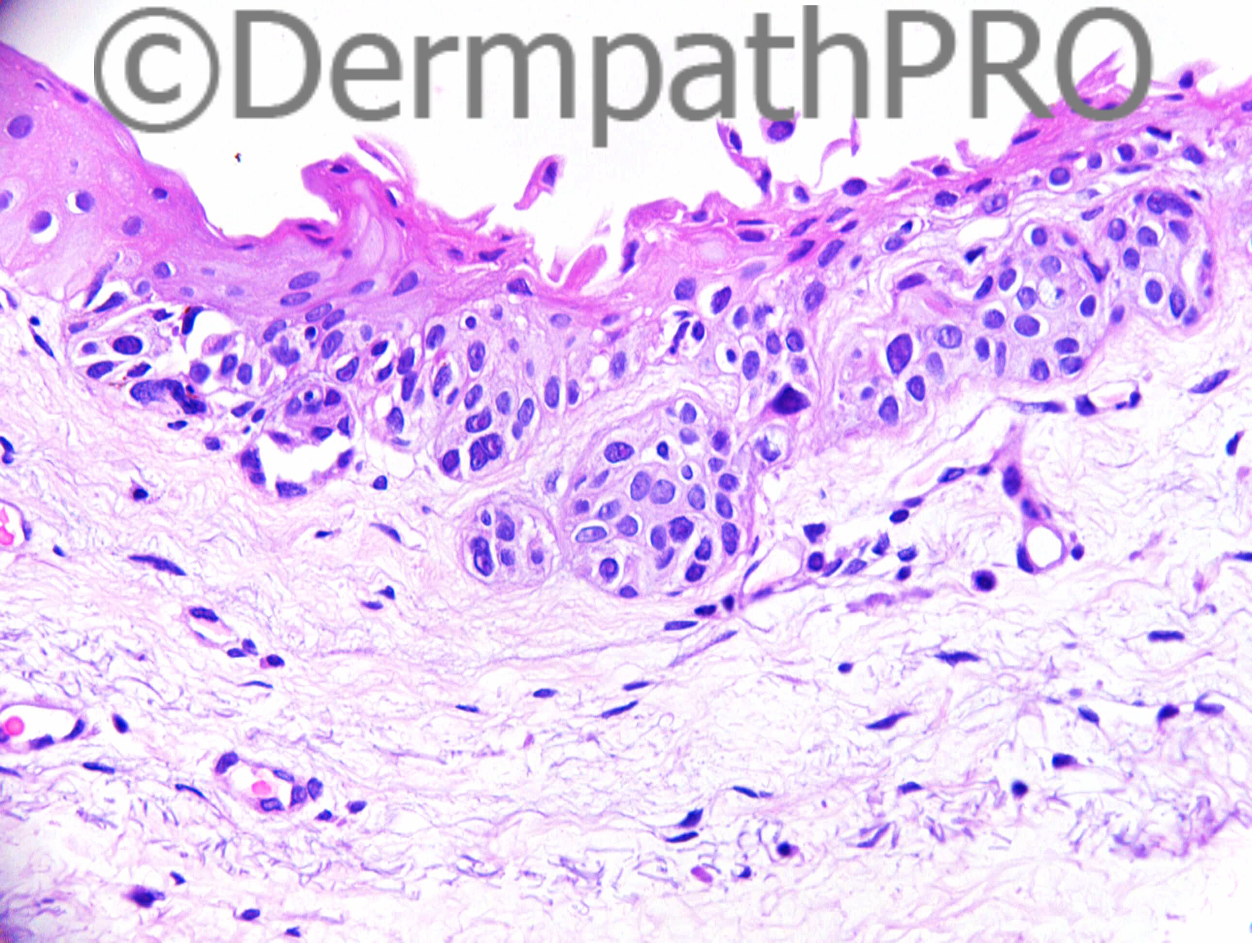
This definitely can not be termed PAM, because PAM should be only an intraepithelial lesion with or without melanocytic proliferation (with or without atypia). We have here both junctional and dermal components. The junctional component is both nested and lentiginous. The last image is a somehow worrying with the junctional cells being mainly lentiginous, pleomorphic, hyperchromatic and contiguous but overall it appears to be focal looking at the lesion as a whole. The dermal component although showing random cytological atypia, I still c the cells singly dispersed with some spindly forms in the lower part, this is to me reassuring especially with the lack of mitoses and the lack of associated inflammatory infiltrate.
I am between conjunctival nevus with atypical (dysplastic) features or less likely early melanoma in situ developing in a conjunctival nevus (considering the last image).... If my case, I would have seeked the opinion of an ocular pathologist specialized in melanocytic lesions.. Would have done proliferation markers also. Would love to read other opinions.

Share this comment


Link to comment
share_externally

Arti Bakshi

Posted

Minimal experience in conjunctival lesions here too, so have had to hit the books!!
I get the impression that there are 2 components to this biopsy. On low power, the lesion to the left is clearly a benign conjunctival compound naevus. However, to the right, there is a predominant junctional proliferation (high power on image 5 and 7). Its unlikely that this is part of the 1st lesion. I think this may represent PAM with atypia (enlarged hyperchromatic cells).

Share this comment


Link to comment
share_externally

Guest Romualdo

Posted

I think this is melanoma in situ developing in a conjunctival nevus.

Share this comment


Link to comment
share_externally

Dr. Richard Carr

Posted

This is a difficult case! Well done those who had a go.
I shared this with my more expert regional (ocular) colleague who commented as follows:

"This is conjunctiva showing an atypical melanocytic lesion with solar elastosis. In my opinion this is at least acquired melanosis with atypia (melanoma in situ) and very suspicious of invasive melanoma. To some extent the clinical history is important here since there is just an outside possibility that the dermal component represents a pre-existing nevus. However, given the fact there is subtle cytologic atypia and active pigment production, and no obvious maturation, I believe this is invasive malignant melanoma."

The case was then reviewed by a National Ophthalmic/ocular pathologist who commented as follows:
"The junctional melanocytes are atypical and are focally confluent although individual nests are also present within the epithelium. The second component is a "naevoid" component that is composed of mostly maturing melanocytes except that superficially there is some focal random cytological atypia. FISH analysis of the junctional component of this lesion, in comparison with the naevus cells have increased copy number of all probes suggesting polysomy for chromosomes 6 and 11. Whilst the Vysis guidelines would class this as positive, the polysomic nature of these cells would suggest this is negative.
Furthermore, FISH of both cell types has shown no evidence of loss of CDKN2A, relative to 9 centromere and no evidence of MYC relative to 8 centromere, however the naevus cells also show additional copies of these probe sets suggestive of polysomy 8 and 9.
Despite the FISH being negative (and a false positive with polysomy) the junctional proliferation is judged to be that of in-situ melanoma given the degree of cytological atypia and the unusual architectural configuration.
The question arises whether there is evidence of early invasive melanoma. In my view there are some cytologically atypical melanocytes in the stroma below the epithelium although these are few in number and therefore the stromal component is judged to be a melanocytic proliferation of uncertain biological behaviour. The excision of the in-situ component is incomplete. The excision of the stromal component is uncertain."

Share this comment


Link to comment
share_externally


Join the conversation

You can post now and register later. If you have an account, sign in now to post with your account.

Guest
Add a comment...

×   Pasted as rich text.   Paste as plain text instead

  Only 75 emoji are allowed.

×   Your link has been automatically embedded.   Display as a link instead

×   Your previous content has been restored.   Clear editor

×   You cannot paste images directly. Upload or insert images from URL.

Loading...

×
×
  • Create New...