Jump to content
In this section we have spot diagnoses posted on a daily basis since June 2010, now over 4000! You can review the archived cases and read the suggested diagnoses by users and the final comment by the contributors.
Case are uploaded each week day by 10 am UK time with the correct diagnosis will generally be posted at 8 pm UK time. Why not view the most recent spot diagnosis and proffer a diagnosis?

Case Number : Case 1369 - 22 September Posted By: Guest

Please read the clinical history and view the images by clicking on them before you proffer your diagnosis.
Submitted Date :
   (0 reviews)

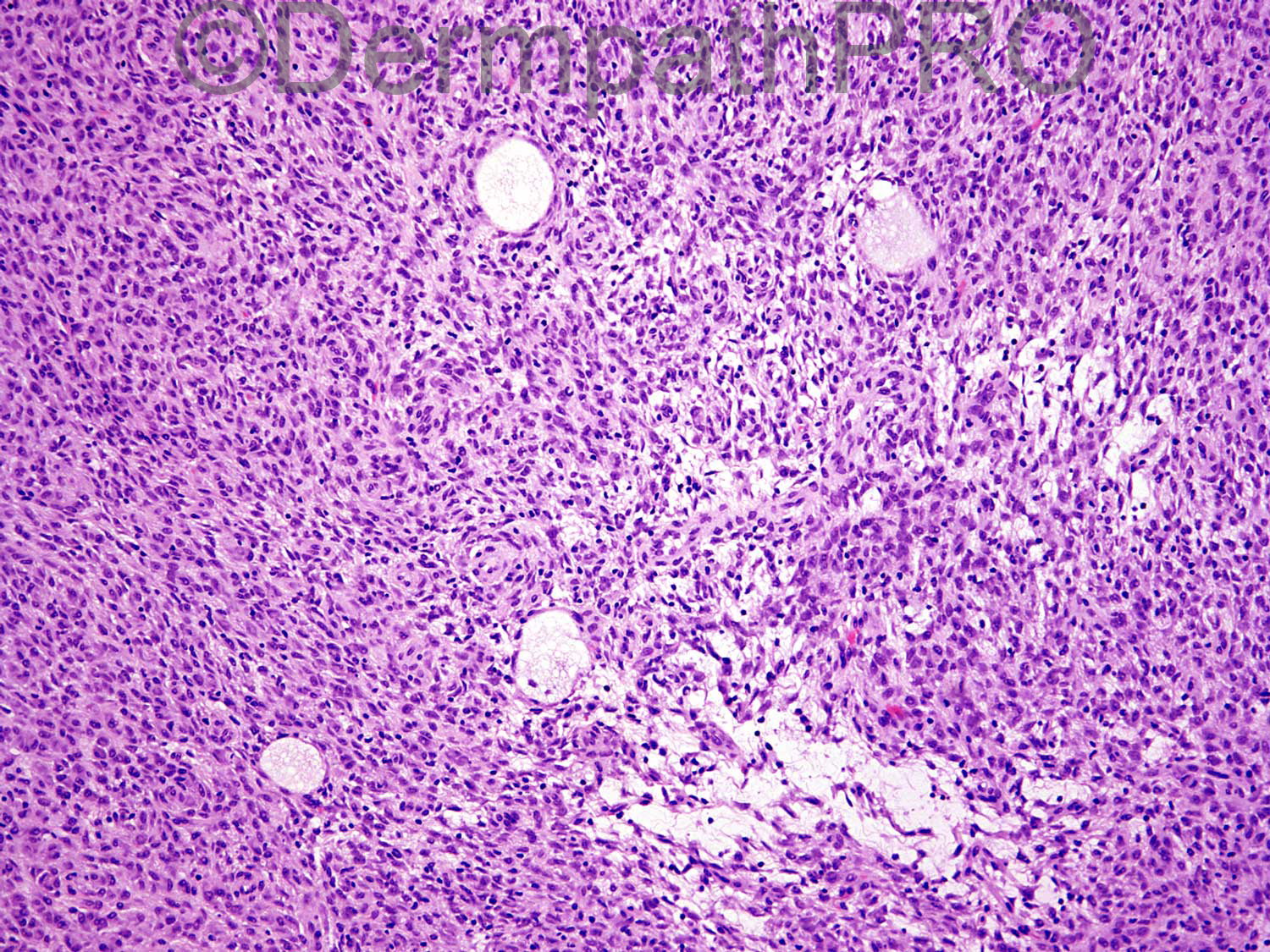
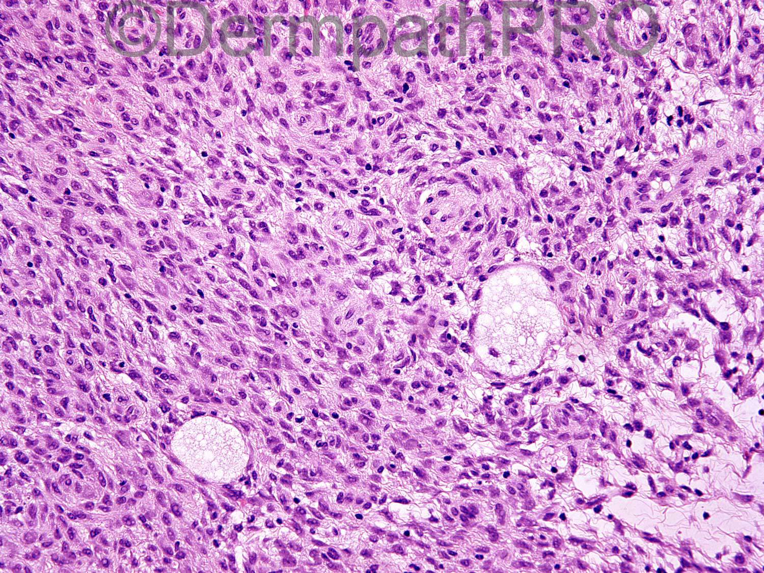
51 year old man with 7 mm erythematous nodule on right upper arm.

Case posted by Dr Uma Sundram


  Report Record

User Feedback


Dr. Mona Abdel-Halim

Posted

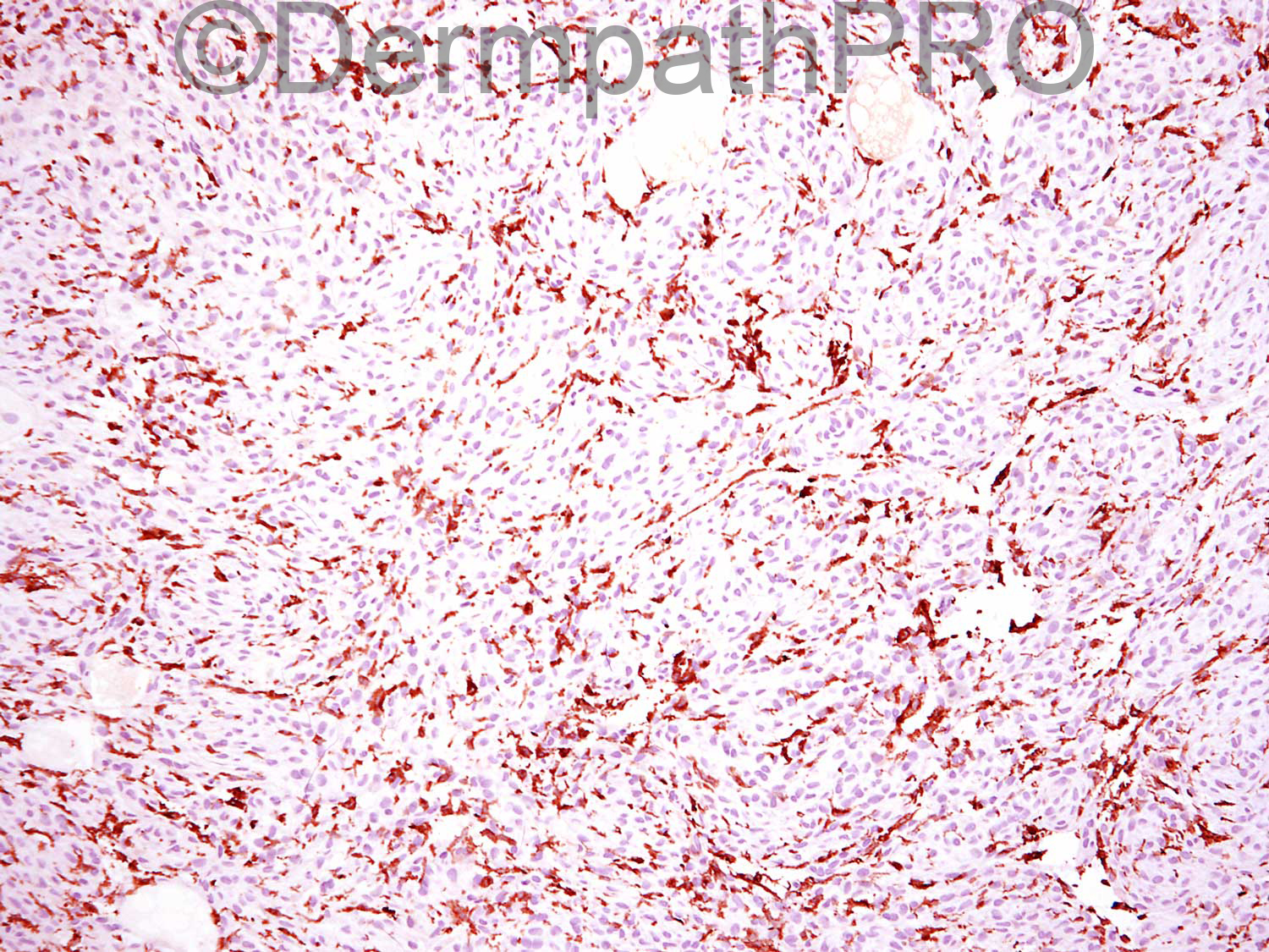
Circumscribed tumor, predominantely spindle cells, some epithelioid cells, prominent pseudo-nuclear inclusions, a storiform pattern is perceived in many areas in addition to whorled arrangements also. There are myxoid foci aalso. The Immunos are not labelled !! Based on morphology, I am thinking of perineurioma. Guessing the Immunos are CD34 (first one) and EMA (second one)!!!

Share this comment


Link to comment
share_externally

Arti Bakshi

Posted

Agree completely with Mona, first thought is perineurioma with the vague whorling/storiform pattern.
Need to know the immunos.

Share this comment


Link to comment
share_externally

My first thought, given the intranuclear inclusions, was of a melanocytic tumour, possibly a cellular blue naevus.

Share this comment


Link to comment
share_externally

Guest Romualdo

Posted

I see a very superficial lesion associated with some grade of epidermal hyperplasia. Collars of epithelia demarcate the lesion at lateral margins. Neoplastic cells are frequently epithelioid and have moderately abundant cytoplasm. Whorled arrangemens of cells around small vessels is seen in some areas. I think this is epithelioid cell dermatofibroma.

Share this comment


Link to comment
share_externally

Uma Sundram

Posted

I like to keep you guys guessing :)
Yes, perineurioma (great job Mona!). 1st immuno is CD163, showing the tumor cells are negative. 2nd immuno is EMA, strongly positive. Melanocytic markers are negative.

Share this comment


Link to comment
share_externally


Join the conversation

You can post now and register later. If you have an account, sign in now to post with your account.

Guest
Add a comment...

×   Pasted as rich text.   Paste as plain text instead

  Only 75 emoji are allowed.

×   Your link has been automatically embedded.   Display as a link instead

×   Your previous content has been restored.   Clear editor

×   You cannot paste images directly. Upload or insert images from URL.

Loading...

×
×
  • Create New...