Jump to content
In this section we have spot diagnoses posted on a daily basis since June 2010, now over 4000! You can review the archived cases and read the suggested diagnoses by users and the final comment by the contributors.
Case are uploaded each week day by 10 am UK time with the correct diagnosis will generally be posted at 8 pm UK time. Why not view the most recent spot diagnosis and proffer a diagnosis?

Case Number : 1509 - 06 April Posted By: Guest

Please read the clinical history and view the images by clicking on them before you proffer your diagnosis.
Submitted Date :
   (0 reviews)

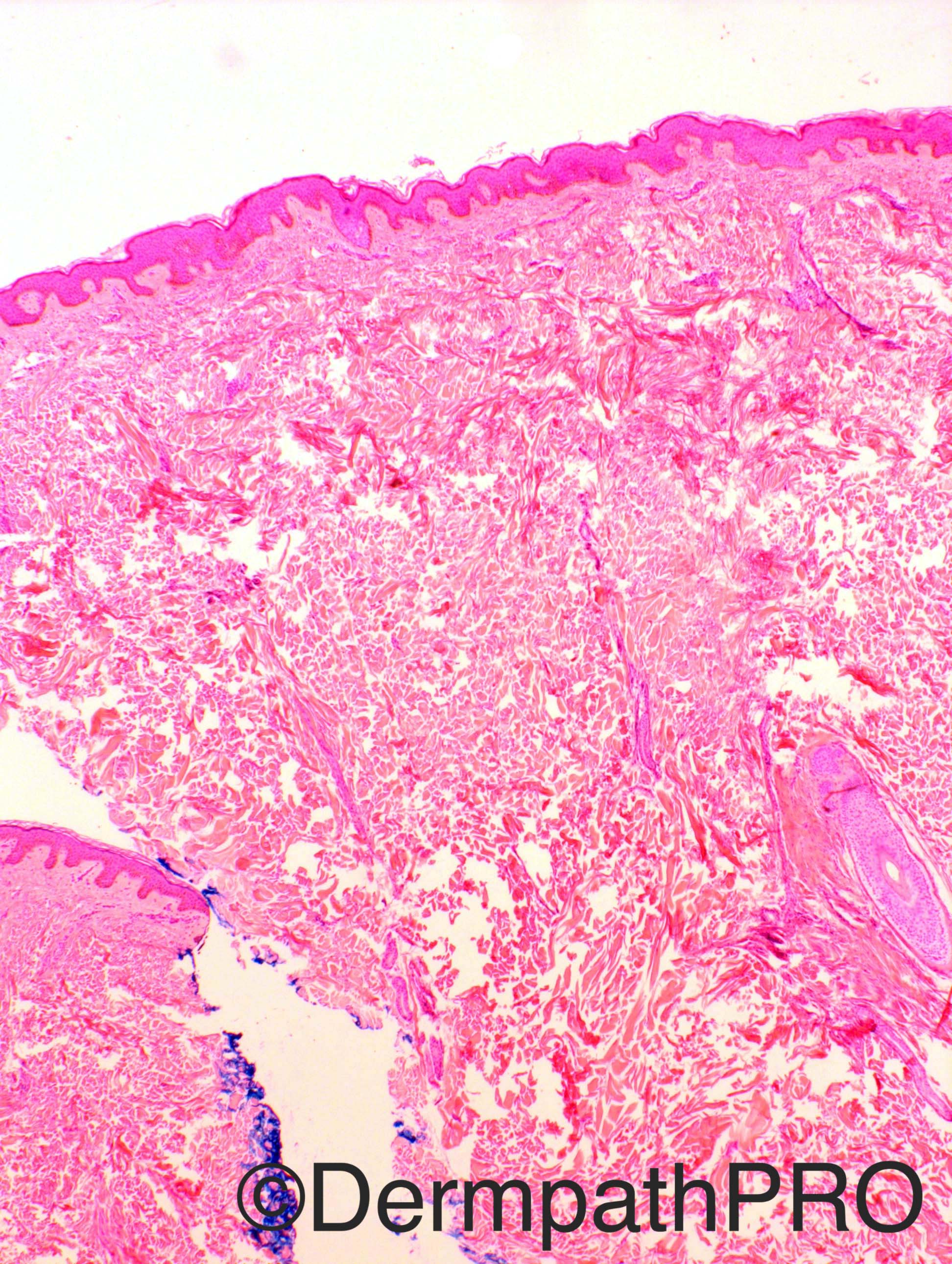
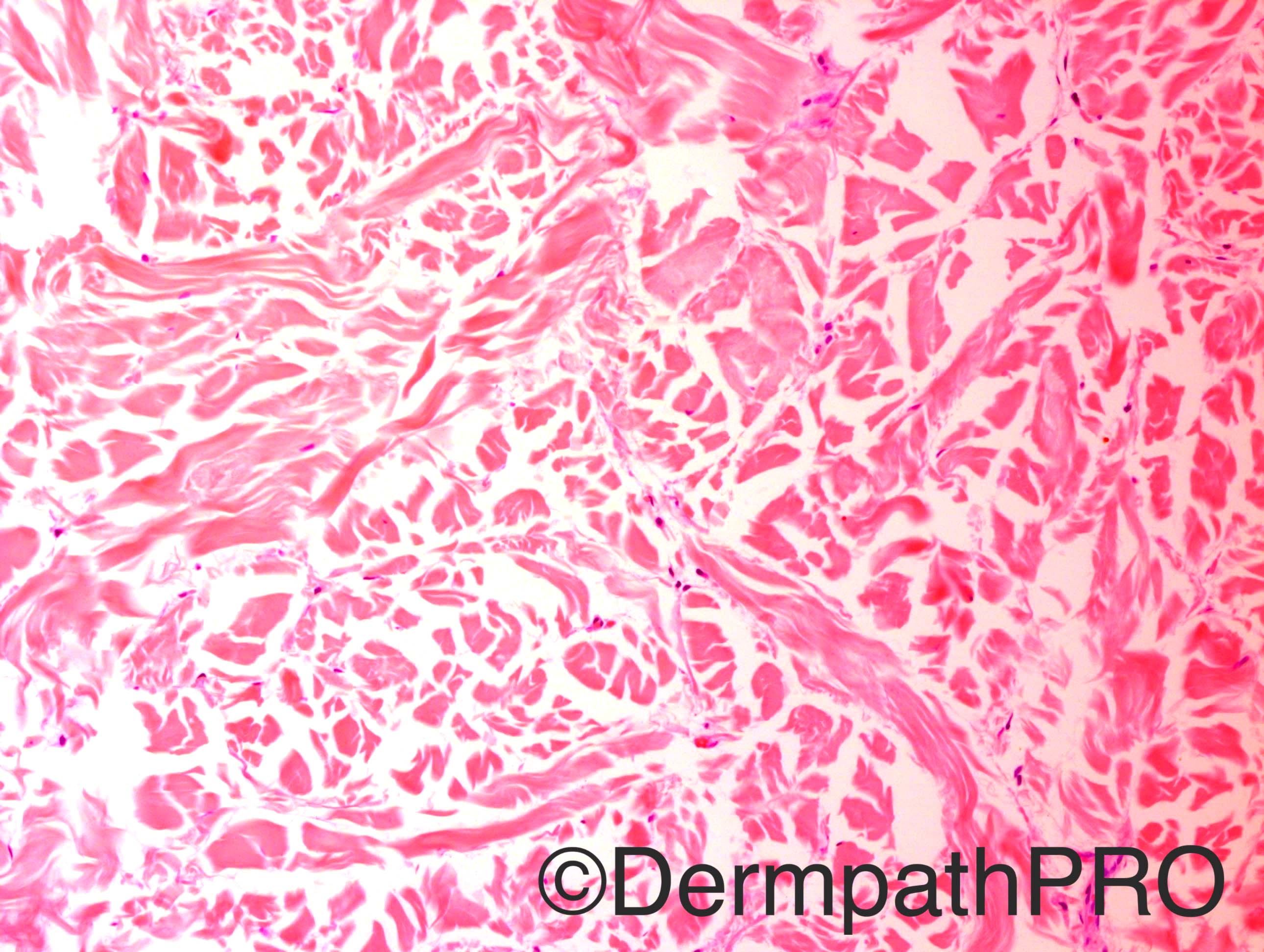
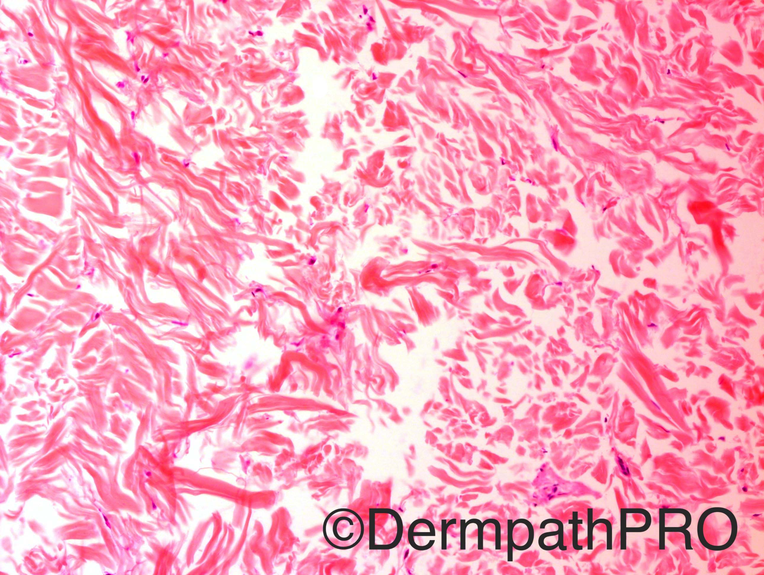
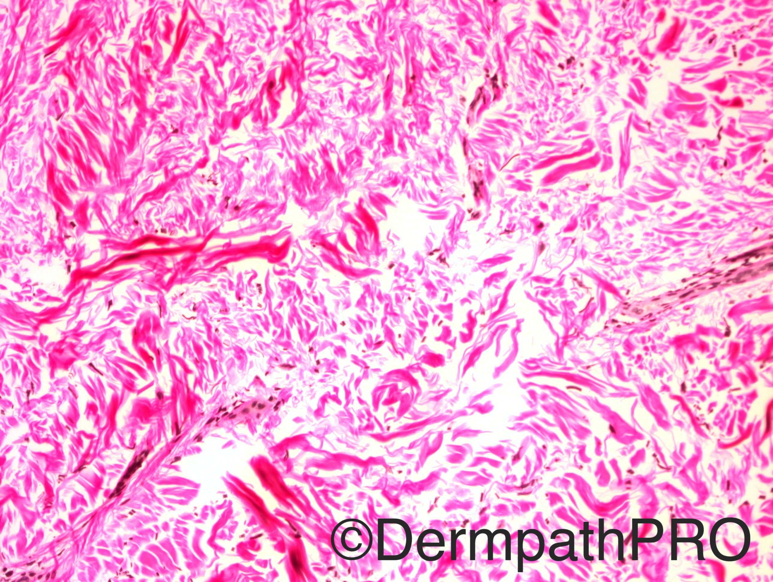
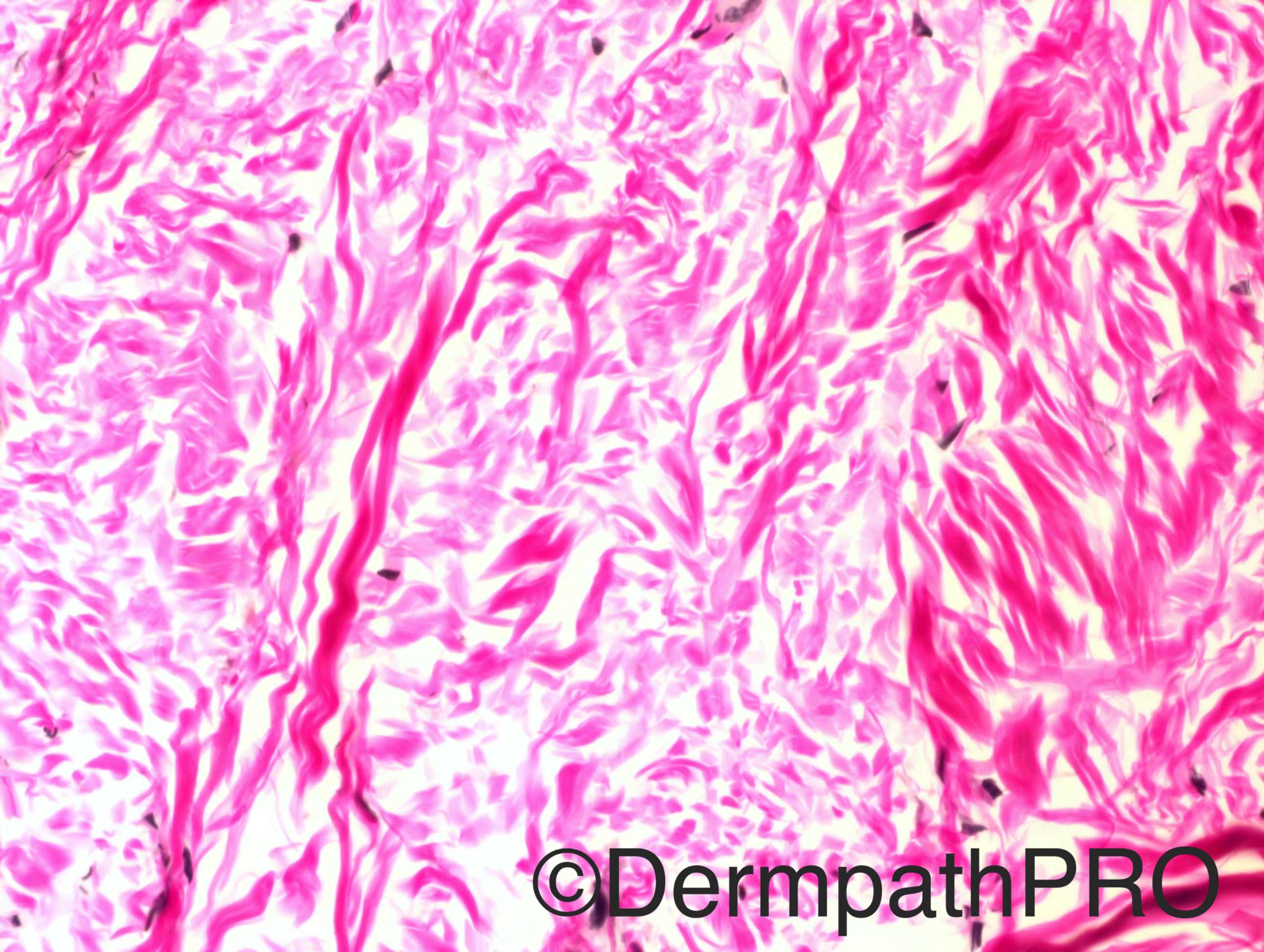
39 year-old male with 7 cm upper back mass.

Dr Hafeez Diwan.


  Report Record

User Feedback


Connective tissue nevus (collagenom)

Share this comment


Link to comment
share_externally

sorry: Connective tissue nevus (Collagenoma)

Share this comment


Link to comment
share_externally

Guest Romualdo

Posted

I think this is nuchal-type fibroma. If there is an association with FAP it should be called Gardner fibroma, but this last possibility is improbable, since Gardner fibroma tends to occur in children.

Share this comment


Link to comment
share_externally

Guest Arash Daryakarr

Posted

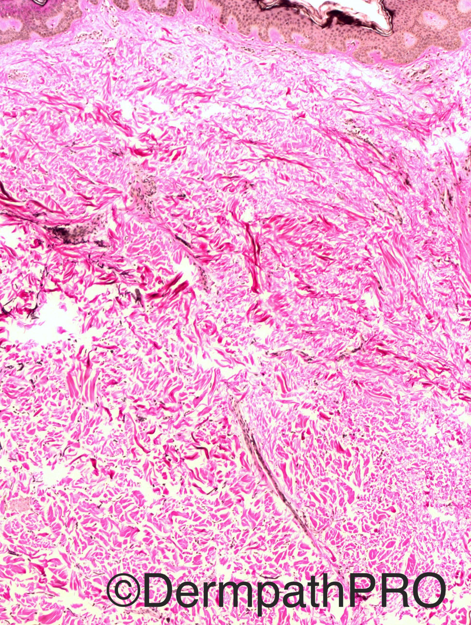
My impression is connective tissue nevus with abscence of elastic fibers(nevus anelasticus)

Share this comment


Link to comment
share_externally

Raul Perret

Posted

Favour nuchal-type fibroma due to clinical description and histology. If clinical information is not available, nuclear beta catenin expression would support Gardner fibroma

Share this comment


Link to comment
share_externally

Here are my reasons for not favouring a nuchal fibroma: 


1.  Nuchal fibromas are subcutaneous; this lesion appears to be in the dermis, albeit a thick dermis. 

2.  Continuing from the first point, nuchal fibromas usually show entrapped islands of fat, which we do not have here. 

3.  Nuchal fibromas usually show entrapped traumatic neuroma-like nerves, which we do not have here. 

4.  Nuchal fibroma is a substitutive process, with collagen bundles that are abnormal (abnormally thick and dense); this process appears to be quantitative, with collagen bundles that are normal but in number. 

5.  Lastly, my impression of nuchal fibromas is that, although they are hypocellular, they are usually more cellular than this lesion (I think). 


Of course, I may be wrong.

Share this comment


Link to comment
share_externally

Raul Perret

Posted

Abdul, nuchal fibromas are tumors that arise on the dermis and subcutaneous tissue and entrapment of adipose tissue, adnexae and nerve twigs is variable. As you said collagen bundles are usually thickenned and they usually seem to intersect, this is what we see on the third picture on the left (to my point of view). Another important feature is that they usually extend Deep entrapping skeletal muscle (a feature we dont see here). Finally I think connective tissue nevus is usually seen in patients of postpuberal age and are rarely larger than 5 cm. While nuchal type fibroma has a wider age distribution and are usually large.

Of course me too I could be wrong but I guess is good to discuss our points

Ps: There is also a frequent association between nuchal type fibroma and diabetes ( i dont know if its the case)

Share this comment


Link to comment
share_externally

Dr. Mona Abdel-Halim

Posted

My first thought was scleredema but the clinical description is strange. It presents as an area of induration rather than a mass !!

Share this comment


Link to comment
share_externally

amiratawdy

Posted

I think of elastofibroma arises usually at back

Share this comment


Link to comment
share_externally

Dr. Hafeez Diwan

Posted

Collagenoma.  The last image on both the right and left sides is of elastin (VVG),

Share this comment


Link to comment
share_externally


Join the conversation

You can post now and register later. If you have an account, sign in now to post with your account.

Guest
Add a comment...

×   Pasted as rich text.   Paste as plain text instead

  Only 75 emoji are allowed.

×   Your link has been automatically embedded.   Display as a link instead

×   Your previous content has been restored.   Clear editor

×   You cannot paste images directly. Upload or insert images from URL.

Loading...

×
×
  • Create New...