Jump to content
In this section we have spot diagnoses posted on a daily basis since June 2010, now over 4000! You can review the archived cases and read the suggested diagnoses by users and the final comment by the contributors.
Case are uploaded each week day by 10 am UK time with the correct diagnosis will generally be posted at 8 pm UK time. Why not view the most recent spot diagnosis and proffer a diagnosis?

Case Number : Case 1507 - 04 April Posted By: Guest

Please read the clinical history and view the images by clicking on them before you proffer your diagnosis.
Submitted Date :
   (0 reviews)

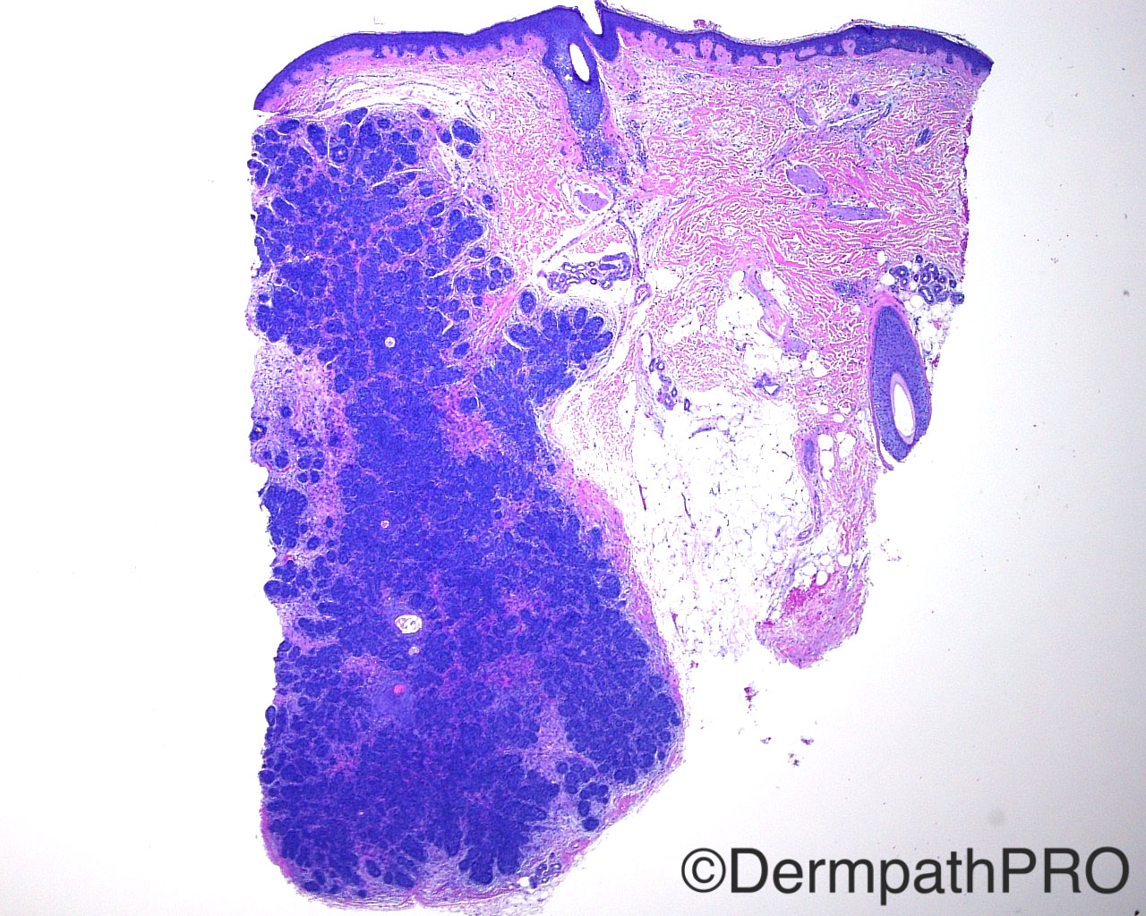
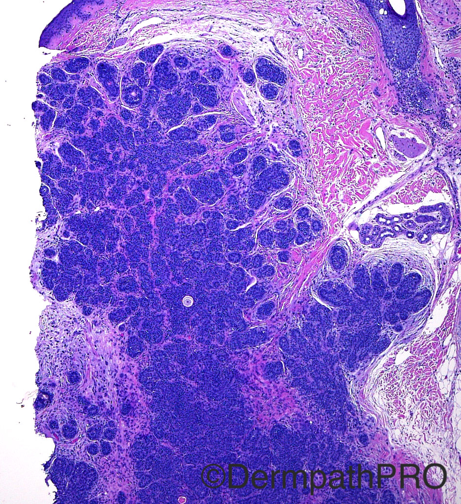
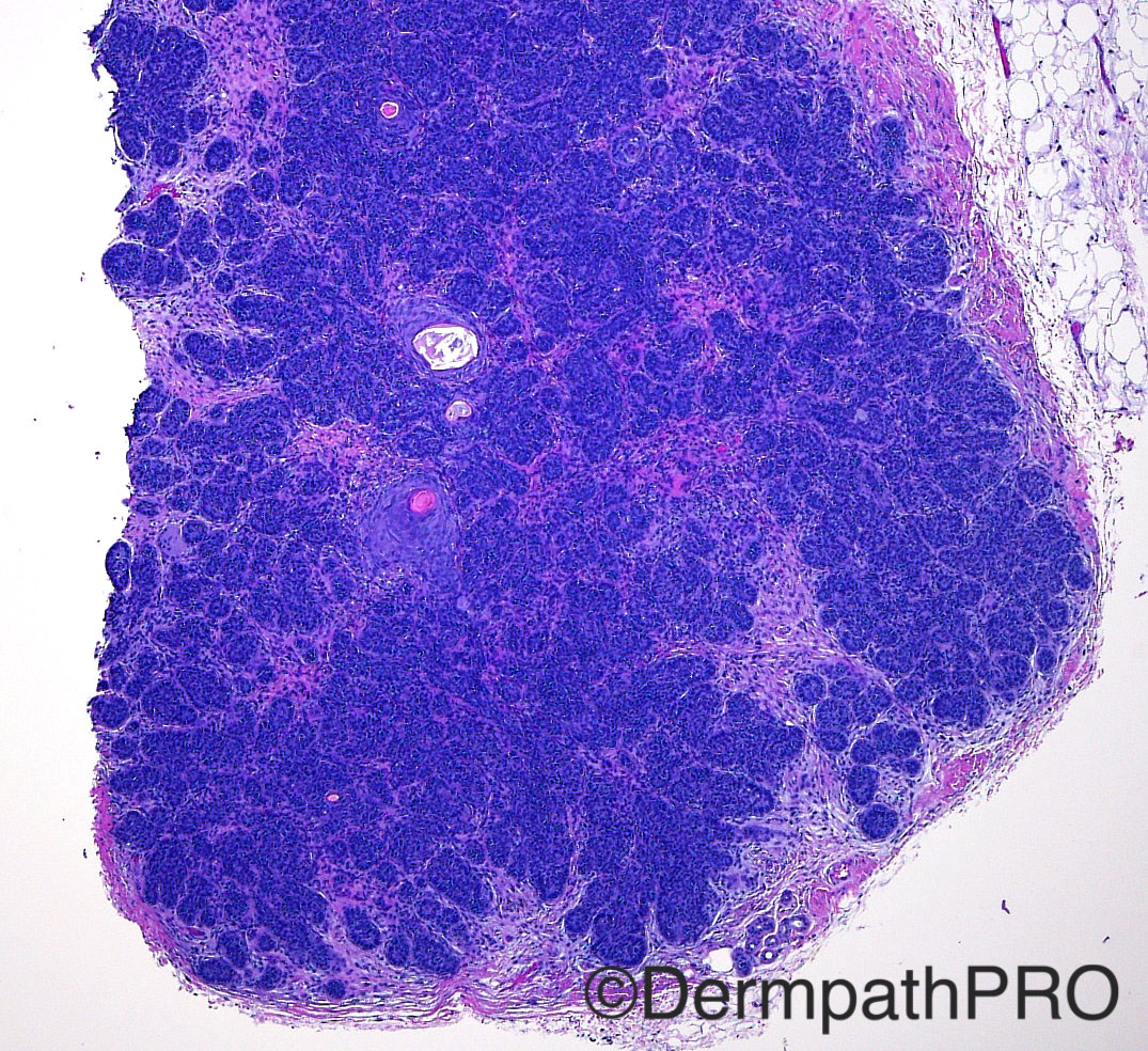
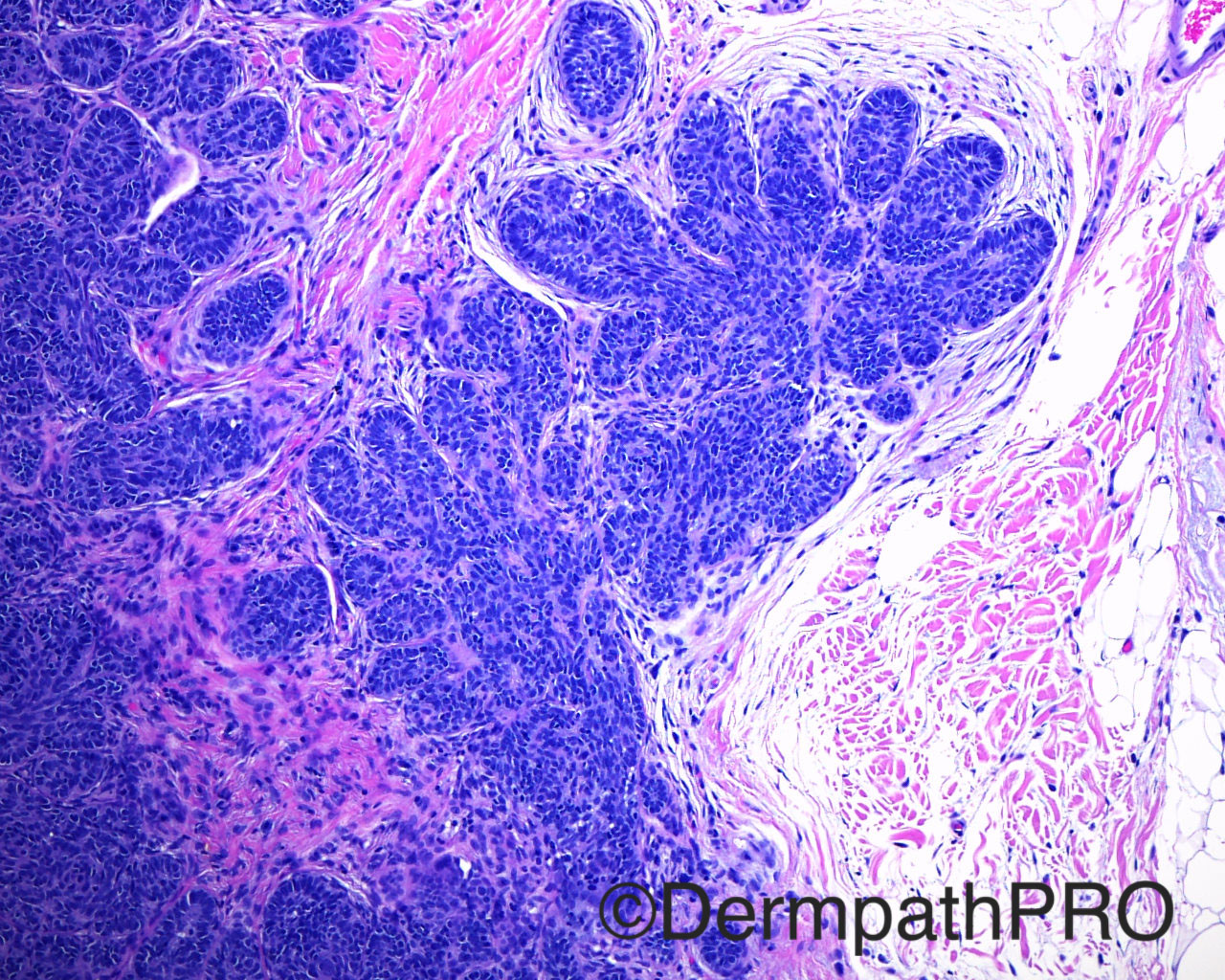
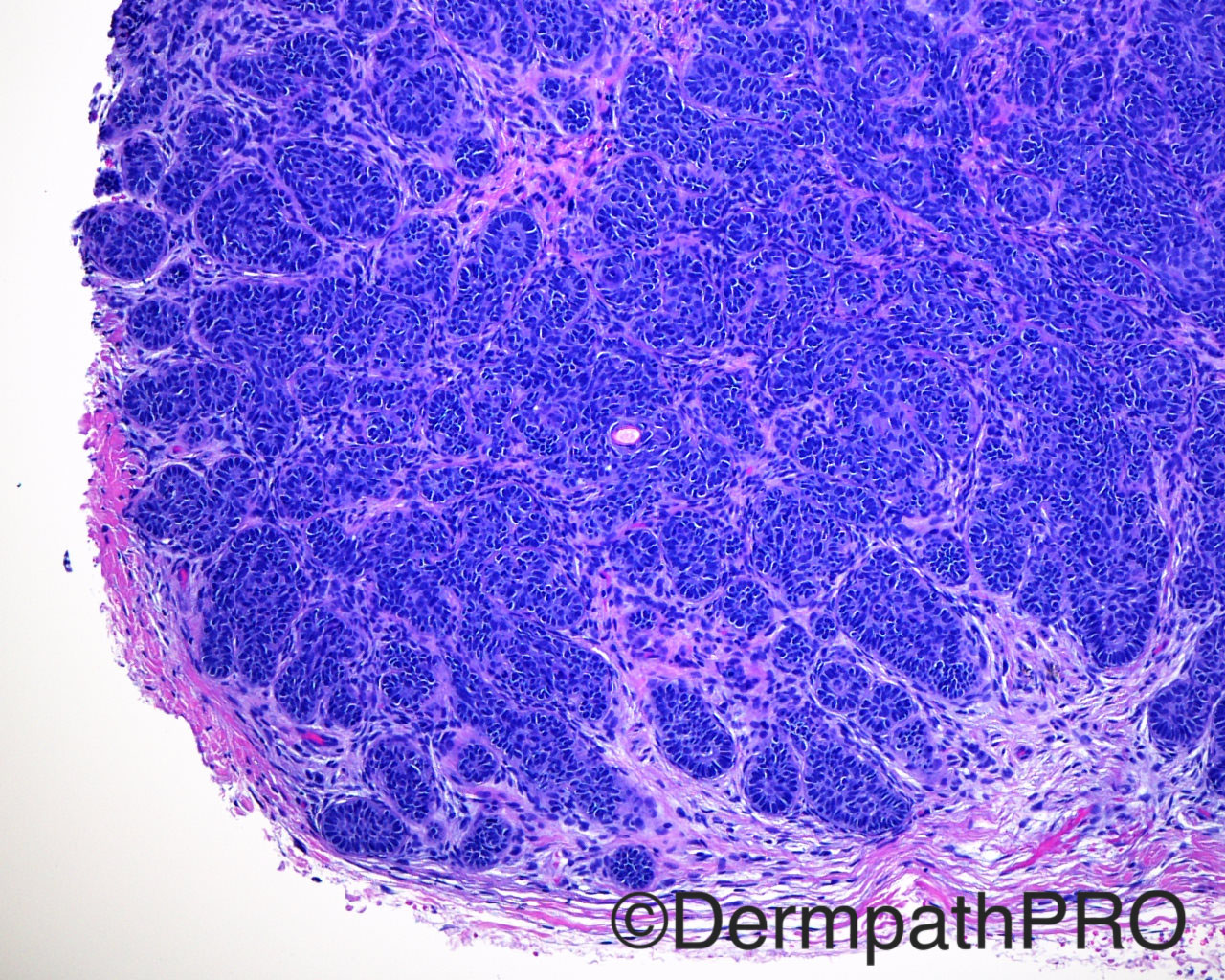
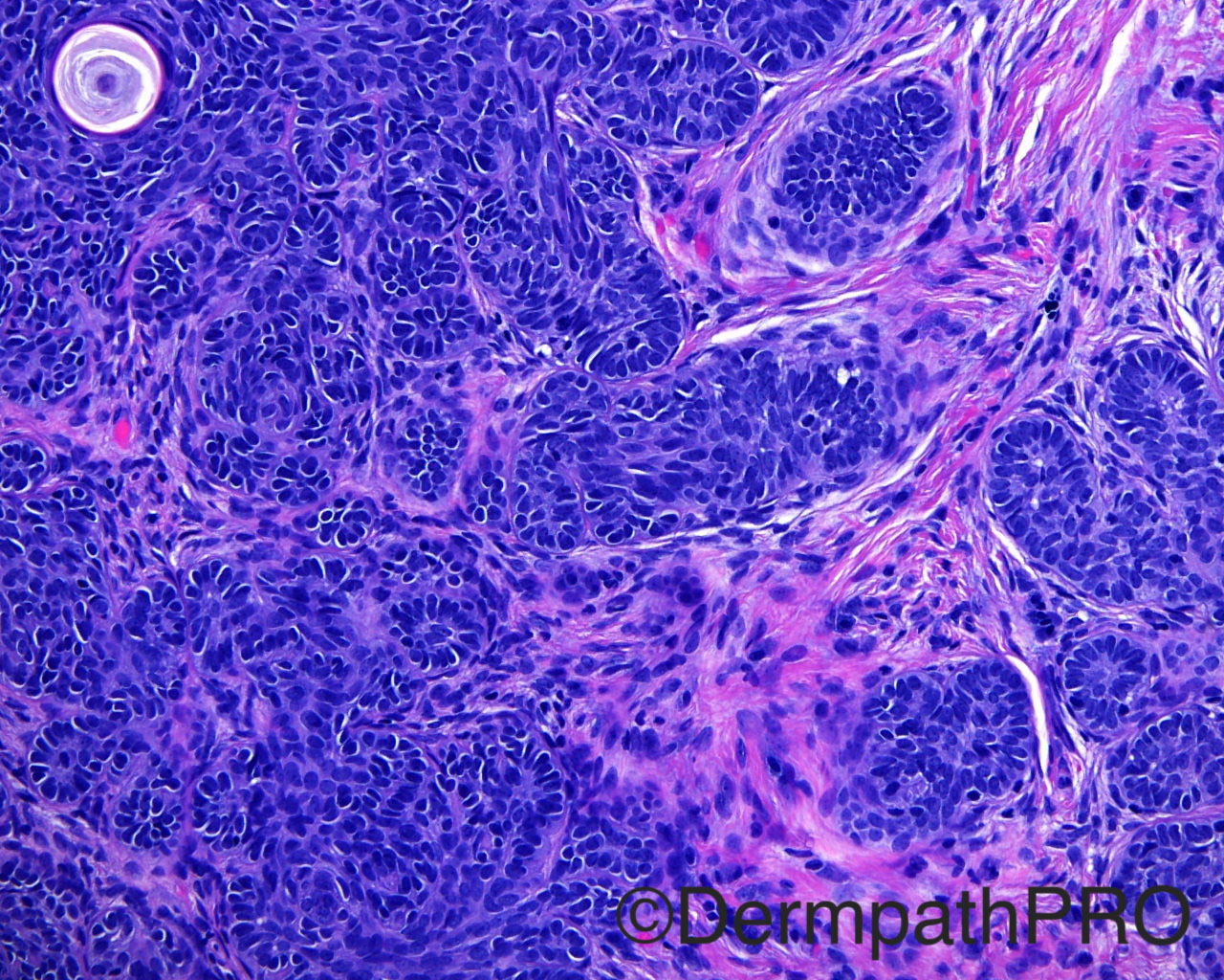
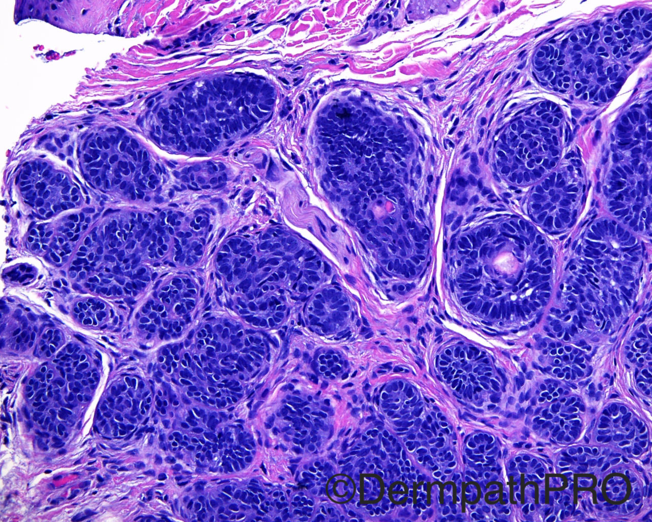
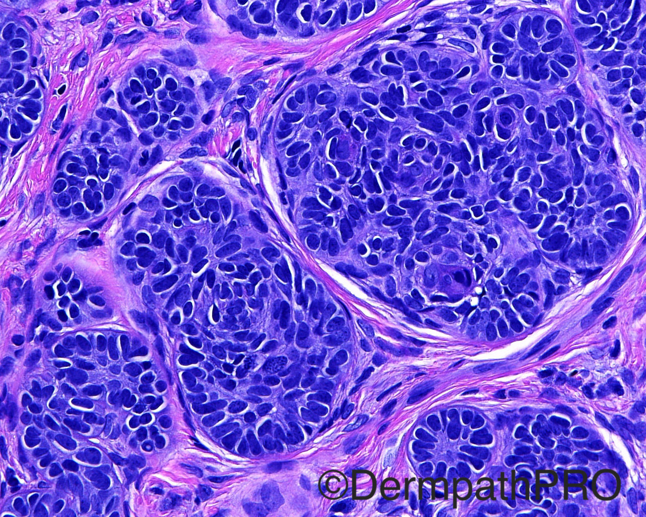
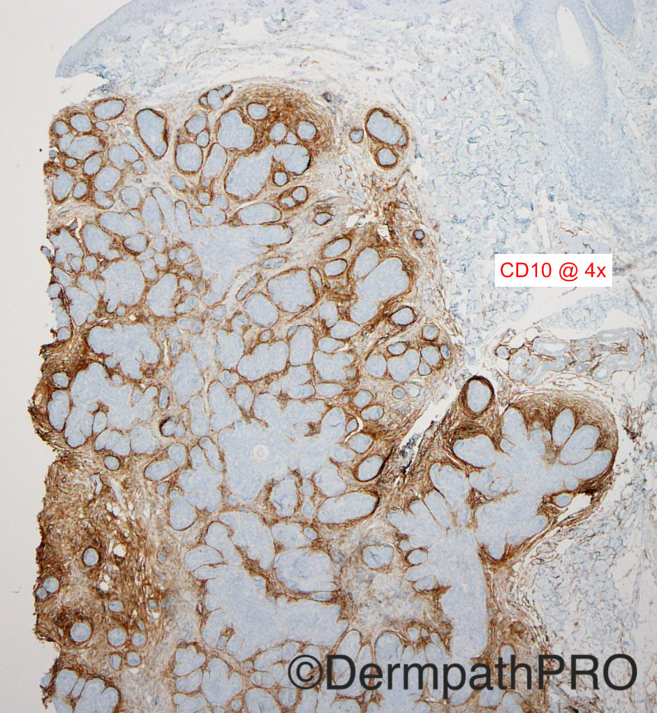
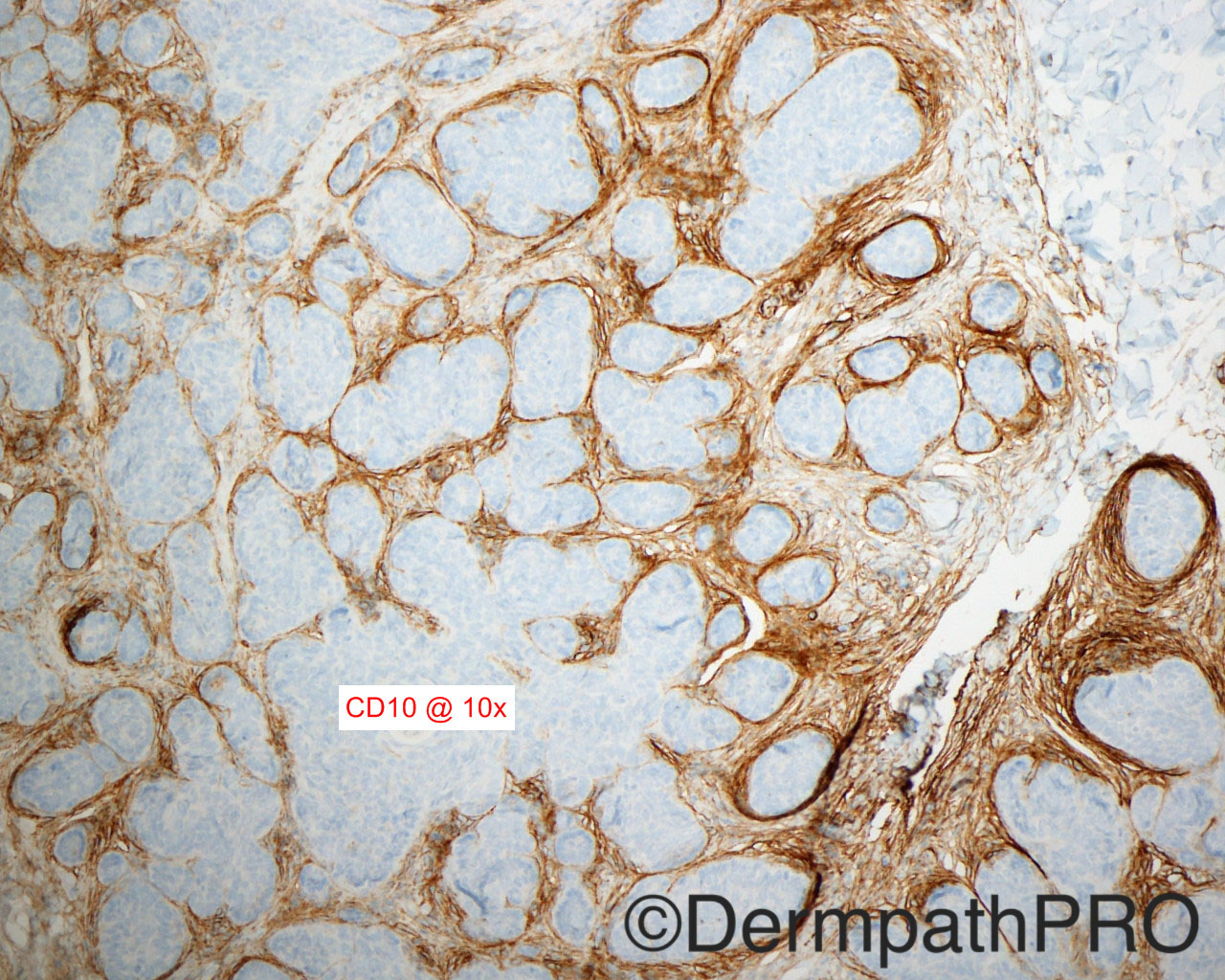
The patient is a 58 year old woman with a biopsy taken from the frontal scalp.

Dr Mark Hurt.


  Report Record

User Feedback


vincenzo polizzi

Posted

Nice CD10+ Peritumoral stromal condensation. Agree

Share this comment


Link to comment
share_externally

Dr. Mona Abdel-Halim

Posted

Trichoblastoma definitly.. Beautiful one :-)

Share this comment


Link to comment
share_externally

Guest Arash Daryakarr

Posted

Agree with trichoblastoma .stromal condesnsation is also evident together with stromal CD10 positivity.

i have a question: how can we make distinction between trichoblastoma and trichoepithelioma?

(except that trichoblastomas are bigger and deeply seated...)

Thanks in advance.

Share this comment


Link to comment
share_externally

Raul Perret

Posted

Arash: Trichoepitheliomas are usually considered variants of trichoblastoma (Mainly by Ackerman) but in particular they usually have a cribiform morphology, infundibular differentiation (usually in the form of keratocysts) whereas trichoblastomas are usually more immature (almost completely basophilic tumors). This is of course the criteria that I consider (I am not an expert) maybe the rest of the group can add other important points. I recommend you to get Kazakov's book of cutaneous adnexal tumors and check the special module's section (Dr Carr did a great review on adnexal neoplasms) 

Share this comment


Link to comment
share_externally

Arash:  Further to Raul's comment, Ackerman used trichoblastoma as a category, to include 6 types: 


- Large nodular and small nodular trichoblastomas (others limit the term "trichoblastoma" to these two types);
- cribriform, racemiform and/or retiform trichoblastomas (otherwise called trichoepitheliomas);
- and columnar trichoblastoma (otherwise called desmoplastic trichoepithelioma).  
 

To this, LeBoit adds fibroepithelioma of pinkus, as fenestrated trichoblastoma. 

Share this comment


Link to comment
share_externally

Dr. Mona Abdel-Halim

Posted

I add that TE are superficial lesions that may show focal connection to the epidermis, TB are large deep seated lesions. Otherwise they r the same, hair germ tumors with epithelial and mesenchymal components.

Share this comment


Link to comment
share_externally


Join the conversation

You can post now and register later. If you have an account, sign in now to post with your account.

Guest
Add a comment...

×   Pasted as rich text.   Paste as plain text instead

  Only 75 emoji are allowed.

×   Your link has been automatically embedded.   Display as a link instead

×   Your previous content has been restored.   Clear editor

×   You cannot paste images directly. Upload or insert images from URL.

Loading...

×
×
  • Create New...